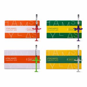
Among the multitude of products available, Exo-One and Hyalmass Aqua Exosome Booster stand out as two of the most widely discussed. So, what exactly sets them apart, and how should the average consumer choose between them?
Exosome Content
Exo-One
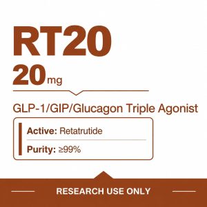
The box provided by the doctor contains two small glass. One holds 50 milligrams of white lyophilized (freeze-dried) powder, and the other contains 5 milliliters of a specialized solvent. The powder consists entirely of microscopic vesicles extracted at high speeds from stem cell culture media. The manufacturer freezes these raw materials in a vacuum machine at -40°C for 12 hours. This process extracts 98% of the moisture while keeping the fragile exosome membranes completely intact, ensuring that less than 5% of the 78 types of encapsulated RNA are compromised.
- Each contains 50 mg of pure powder.
- Contains 60 billion exosome particles.
- Paired with 5 ml of a dedicated solvent.
- Particle size ranges from just 30 to 200 nanometers.
- 100% free of residual animal serum.
- Extracted using a centrifuge at speeds up to 100,000 RPM.
The nurse uses an ultra-fine 30G to draw 5 ml of the liquid and inject it into the powder. Upon contact with the liquid, the white powder dissolves in just 3 seconds, turning into a semi-transparent fluid resembling rice water, with a perfectly neutral pH of 7.2. The intercellular spaces in human facial skin are less than 50 nanometers wide, meaning topical application alone cannot penetrate the barrier. The doctor uses a microneedle roller with ranging from 0.8 to 1.2 millimeters in length, creating approximately 250,000 microscopic, invisible micro-channels across the face. The fluid, carrying 60 billion particles, flows through these 250,000 micro-channels to a depth of 1.5 millimeters beneath the skin. The outer shells of the micro-particles collide with the membranes of facial cells, fusing instantly and releasing over 1,000 active directives into the cells.
- Micro-scabs on the face shed completely within 3 days.
- Transepidermal water loss decreases by 12% by day 7.
- Ultrasound imaging at 28 days shows a 0.1 mm increase in dermal thickness.
- Facial erythema (redness) reduces by 60% within 48 hours.
- The burning sensation post-treatment subsides by 80% within 15 minutes.
Skin that has just undergone fractional laser treatment is severely compromised and will exude about 2 ml of serous fluid. Applying the full 5 ml of this solution prevents subcutaneous white blood cells from migrating unnecessarily and triggering further inflammation. A clinic in the Gangnam District of South Korea tracked 50 patients who underwent Fraxel laser treatments. For those who used this powder, the duration of facial redness and swelling dropped from 5.8 days to 3.2 days, and the probability of developing post-inflammatory hyperpigmentation fell from 12% to 2%. Once the powder is dissolved in water, it loses the protection of low temperatures and a vacuum environment. If left in a 25°C room for 4 hours, 60% of the particle shells will rupture, rendering their contents useless. Therefore, treatments at the clinic require the nurse to open and mix the solution on the spot. A human face can absorb a maximum of 3.5 to 4 ml of the fluid per session; any excess will simply drip down the chin to the neck. Doctors typically schedule these injection sessions at fixed 28-day intervals. Deep-set melasma forms 2 millimeters beneath the skin. The substances released by the particles can sever the dendrites that produce melanin. Instrumental analysis shows that after three consecutive months of treatment, the melanin concentration at the edges of the macules lightens by 15.4%.
- Unopened boxes must be stored in a refrigerator at 2°C to 8°C.
- When mixing the solution, the must be swirled gently and slowly to prevent bubbling.
- The full-face procedure must be completed swiftly within 20 minutes.
- Foaming cleansers must be avoided for 24 hours post-treatment.
- When stored away from direct sunlight, the shelf life is 24 months.
In patients with severe rosacea, facial capillaries can swell to over 0.5 millimeters thick. Ultrasound scans reveal that within 14 days of fluid penetration, the walls of these dilated micro-vessels gradually tighten and constrict. The more than 70 types of proteins inside the vesicles block the erratic migration pathways of macrophages. Aging cells receive these signals and rapidly produce Type I and Type III collagen, rebuilding the subcutaneous structural network that had collapsed by 30%. Instrumental before-and-after photos of patients over 35 show that 0.2 mm deep fine lines around the eyes visibly diminish 28 days after the second injection. The absolute wrinkle score on the machine-generated report decreases by an average of 2.1 points. Standard mesotherapy injections, which contain 5 mg of hyaluronic acid per milliliter, rely entirely on water absorption to plump the face. This pure powder contains no large molecules like hyaluronic acid. When injected 0.5 mm beneath the skin, no visible bumps or papules are left on the face.
Hyalmass Aqua
The nurse retrieves a glass from a 2°C to 8°C refrigerator. It is filled with 5 ml of a viscous, transparent solution that does not require on-the-spot mixing or shaking. The contains 5 billion exosome particles, along with pure hyaluronic acid at a high concentration of 20 mg/ml. The solution is quite thick when swirled; laboratory instruments measure its viscosity at 3.2 mPa·s. The nurse uses a thicker 3.8 cm long 18G to draw the solution, taking 12 seconds to fill a complete 5 ml. The formulation list for the is packed with metrics. In addition to the 5 billion nano-scale vesicles, the 5 ml volume is densely packed with 5 types of peptides weighing approximately 15 mg, 17 types of free amino acids, and a 0.5% concentration of Vitamin C.
| Ingredient | Absolute Content / Particle Count | Target Skin Depth |
|---|---|---|
| Exosome Particles | 5 Billion (Size 50-150nm) | Dermis 1.0mm-1.5mm |
| Pure Hyaluronic Acid | 20mg/ml (Molecular Weight 1.2M Da) | Superficial Subcutaneous Layer 0.2mm-0.8mm |
| Biomimetic Peptides | 5-Type Blend (~15mg) | Deep Dermis 1.2mm |
| Amino Acid Complex | 17-Type Blend (Molecular Weight <500 Da) | Full-Face Microcirculation Penetration |
Hyaluronic acid with a molecular weight of 1.2 million Daltons acts like a microscopic sponge, with a single molecule tightly binding to 1,000 surrounding water molecules. When injected 0.2 mm beneath the facial skin, the dehydrated intercellular spaces plump up with moisture within half an hour. Unlike the previous pure powder’s 60 billion particles, this ‘s 5 billion 50-nanometer particles are entirely suspended in hyaluronic acid with a pH of 7.0. This gel locks in moisture to create a hydrated environment, maintaining a particle survival rate of over 95% after 48 hours. The 5 types of peptides follow the moisture to stimulate dermal cells. The cells are forced into an active state; skin biopsy reports show that in just 21 days, the weight of newly synthesized Type I collagen under the facial skin increases by 8.4%. The 17 amino acids, including glycine and proline, all with molecular weights under 500 Daltons, serve as the building blocks. Once the particles signal the cells to start working, these 17 formulated materials accumulate outside the cells and are fully absorbed within 48 hours to create new skin fibers. Because the solution is so viscous, applying it with a standard microneedle roller results in less than 15% penetration. The doctor switches to a mesotherapy gun with negative pressure set to 250 mmHg, equipped with a 9-pin multi-block (32G, 4 mm long). The machine suctions the skin to deliver the medication precisely. The machine’s motherboard fixes the output volume at precisely 0.05 ml per injection. As the penetrate at 1-centimeter intervals, white papules 2.5 millimeters in diameter instantly raise on the face. After administering the entire 5 ml, the face is densely covered with 100 to 120 of these papules. These hyaluronic acid-filled papules are compressed by the subcutaneous tissue. Driven by a body temperature of 36.5°C and a subcutaneous blood flow velocity of 0.5 mm per second, the solution is gradually dispersed. For patients with faster metabolism, the papules flatten in 12 hours, while those with slower metabolism may need 24 to 36 hours. Once the papule phase passes, stratum corneum moisture readings jump dramatically from 35 to 52. On the third day post-treatment, epidermal moisture content surges by 48.5%, while the transepidermal water loss rate drops by 15%, leaving the face feeling smooth to the touch during morning cleansing. Several static wrinkles on the forehead, measuring 0.5 mm deep, are physically smoothed out by the hyaluronic acid. The VISIA imaging system calculates that by day 14 post-treatment, the shadow area of sunken fine lines decreases by 33.5%, and wrinkle depth becomes 0.15 mm shallower. The blended 0.5% Vitamin C and other active ingredients work to fade emerging subcutaneous melanin. After 14 days, a colorimeter scan over the cheekbones shows the skin’s yellowness (L-value index) increases by an average of 1.5 to 1.8 points, indicating a brighter complexion. There is a limit to the volume of liquid an adult’s dermis can hold, so treatments are strictly capped at one 5 ml per session. After undergoing one session per month for three consecutive months and then stopping, the fully hydrated state of the stratum corneum is maintained for 120 to 150 days. The glass contains no preservatives and is highly sensitive to temperature fluctuations. It must normally be stored in a 2°C to 8°C refrigerator. If left at a 25°C room temperature for 2 hours, the peptide chains will break, and 40% of the exosome outer membranes will dissolve. The 100+ injection points on the face must strictly avoid contact with tap water for 3 days. Thinner-skinned areas like the forehead take 24 hours longer to lose their redness compared to the cheeks. By the morning of the fourth day, the erythema index around the eyes drops by 40%, and skin light reflectance increases by 20%.
Content Metrics
On the clinic’s thick, 157-gram coated paper brochure, the front of the packaging features a long string of numbers with many zeros. Whether labeled 60 billion, 50 billion, or 100 billion, the unit printed on the paper is clearly stated as “particles per.” A lab technician uses a to dispense 5 microliters of the solution into a testing groove that is only 0.5 millimeters deep. As a highly focused blue-violet laser with a 405-nanometer wavelength passes through the droplet, the computer screen instantly lights up with a dense array of white dots.
The machine’s probe captures 30 continuous images per second, tracking the trajectories of the glowing dots within the water droplet in a 25°C environment. The software accurately counts exactly how many microscopic vesicles are swimming in 1 ml of the solution.
A valid lab report cannot merely provide a capitalized total count. It must include a column detailing the “majority particle size.” Only if the machine-detected peak falls between 80 and 120 nanometers is the 5 ml of solution considered usable. Any glowing dots exceeding 200 nanometers are entirely fragmented dead cell shells, while those under 30 nanometers are merely useless fat clusters floating in the water. The percentage of adequately sized particles on the lab report determines whether a batch should be discarded.
- Vesicles between 50 and 150 nanometers must account for over 88% of the total.
- Impurity proteins in 1 ml of solution are strictly limited to a maximum of 1.5 mg.
- The concentration of functional RNA must remain stable between 15 and 25 nanograms per microliter.
- Damaged nucleic acids floating in the solution cannot exceed 5% of the total count.
No matter how impressive the numbers printed on the box may look, it does not guarantee a high number of live, functional particles upon injection. A South Korean laboratory took a batch of solution labeled “100 billion” and incubated it in a constant temperature chamber at 37°C with 80% humidity. After 48 hours of incubation, it was measured by the machine. The glowing white dots on the screen plummeted drastically from 100 billion to just 15 billion. The protective double-layer membranes around the vesicles had completely ruptured, leaking the encapsulated RNA into the water. The number printed on the taken out of the nurse’s freezer only represents the viable count on the day it left the factory. If a cold-chain delivery truck loses power and the product sits at a 28°C room temperature for just 3 hours, half of the 50 billion particles will die on the spot. The particles’ ability to function effectively under the skin relies entirely on the genetic “cheat sheets” called RNA encapsulated within their membranes. If all the vesicles in 1 ml were broken down and weighed, the RNA in each microliter of the pure solution would weigh 15.5 nanograms.
Placing a 10-microliter droplet containing 15.5 nanograms of RNA into a petri dish culturing 5,000 aging facial cells shows striking results. Under a microscope 12 hours later, the cells’ natural division rate increases by 3.2 times.
Some cheaper products claim to have 300 billion particles per. When a nurse draws 5 ml into a centrifuge and spins it intensely at 12,000 RPM for half an hour, a thick layer of yellowish debris forms at the bottom of the plastic tube. If a pipette is used to remove this bottom layer of debris before running the remaining fluid through the counter, the genuine particle count doesn’t even reach 2 billion. The remaining 298 billion white dots are nothing more than unwashed bovine protein particles from the bovine serum used during extraction.
- A ratio of 1 microgram to 10^9 particles is considered barely passing.
- A ratio of 1 microgram to over 10^10 particles is classified in the high-purity tier.
- Solutions with a ratio below 10^8 lead to a facial redness and swelling rate of over 40% upon injection.
- This ratio is measured using a specialized colorimetric assay kit under a 562-nanometer wavelength.
The immune cells beneath the facial skin are extremely selective regarding this ratio. Solutions with a low ratio are full of impure proteins. When these flow down the 250,000 micro-channels (0.1 mm wide) created by the microneedles to a depth of 1.5 mm, problems occur. Subcutaneous macrophages mistake these impure proteins for invading bacteria. Just 2 hours after the facial needling, the local skin temperature spikes by 2.5°C, the erythema score skyrockets from a normal 12 to 45, and hundreds of red bumps erupt across the face. In contrast, for those injected with high-purity solutions, the erythema score reliably subsides to 18 by the fourth hour. The viable particles navigate beneath the skin for 45 minutes, successfully colliding with skin cell membranes and inserting all their functional RNA. Calculating the true content depends on how much is actually absorbed by live cells. Lab technicians dye the particles with a green fluorescent marker, drop them into a petri dish containing 100,000 subcutaneous cells, and place them in an incubator with 5% CO2 for 24 hours.
Under a microscope, they count the number of cells exhibiting 500 units of internal green fluorescence. Only when the uptake rate exceeds 60% can the numbers printed on the box be considered capable of effecting real change once injected into the face.
The lyophilized powder is dehydrated until only 0.8% moisture remains. The 60 billion particles within the powder are forced into dormancy; storing them in a 2°C to 8°C refrigerator can keep these 60 billion living units fresh for a full 24 months. Once reconstituted with 5 ml of saline, the injection must be timed with a stopwatch and administered immediately in a 24°C room. If injected within the first 10 minutes, the survival rate is 98%. If delayed to the 30-minute mark, less than 70% of the particles will survive the injection. When a 5 ml is administered using a mesotherapy gun connected to a 10-centimeter flexible tube, 0.25 ml of the solution remains trapped in the plastic head’s dead space. Assuming 10 billion particles per milliliter, 2.5 billion particles are wasted when the machine head is thrown into the trash. To calculate exactly how much is absorbed by the skin and flesh, a technician vigorously applies and rips off adhesive tape 15 times on the left side of the face post-injection. The top 0.01-millimeter layer of dead skin is completely stripped away and placed into a glass tube. Lab tests on this layer of dead skin reveal the truth. When using a short microneedle roller measuring only 0.5 millimeters, a full 40% of the vesicles are trapped in the 0.1-millimeter deep dead skin and wasted. Retesting the right side of the face with a 1.2-millimeter microneedle shows that the number of particles trapped in the dead skin plummets to just 5%. A full 95% of the viable vesicles, complete with intact membranes, follow the 3.5 ml of solution deep into the 1.5-millimeter dermal layer. 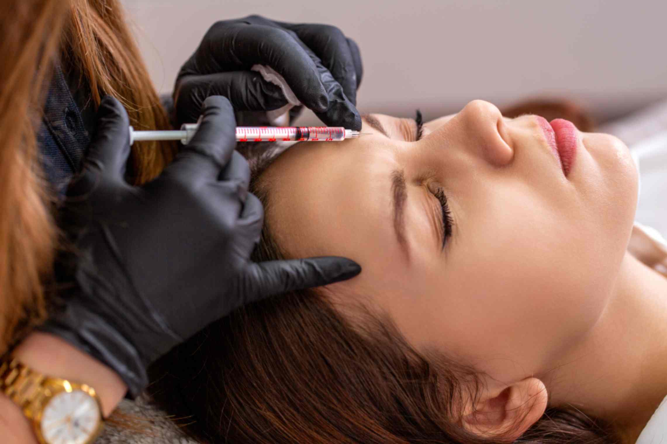
Skin Repair
Targeted Repair
Damaged skin cells release ATP and chemokines at nanomolar concentrations into their surroundings, acting much like distress flares. The surfaces of exosomes carry 40 to 100 CD9 and CD63 transmembrane proteins, functioning as highly sensitive radars. These vesicles travel through interstitial fluid at 2 to 3 micrometers per second, deliberately congregating at damaged lesion sites where distress signals are the densest. An Exo-One contains 6 billion pure stem cell exosomes. The moment these 30 to 150-nanometer aqueous spheres touch damaged fibroblasts, they fuse with the cell membranes in under 0.1 seconds. Thousands of microRNA codes, acting as repair blueprints, are instantly poured into the damaged cells. Equipped with these blueprints, the cells immediately get to work. Interleukin-6 (IL-6), which causes skin redness and edema, plummets by 45% within 24 hours. The destructive enzymes responsible for degrading collagen have their offensive power forcibly suppressed by 60%, staunchly protecting the originally healthy Type I collagen fibers.
- Synthesis of Type I and Type III collagen in the dermis accelerates by 300%.
- Engorged and dilated capillary networks shrink by 15 to 20 micrometers in diameter.
- Exudate from micro-wounds caused by energy-based devices decreases by 80% within 12 hours.
- Melanocyte dendrites in the epidermal basal layer shorten by approximately 30%.
Hyalmass Aqua faces a completely different battlefield. The stratum corneum is severely compromised, with transepidermal water loss (TEWL) exceeding 25 g/m² per hour and moisture content dropping below 10%. The shriveled epidermal cells cannot even transmit distress signals; the probability of exosome penetration drops to less than 5%, making it impossible to begin barrier repairs. Hyaluronic acid with a molecular weight of 1.2 million Daltons at a concentration of 10 mg/ml steps in first to stabilize the situation. It creates a powerful 500 mOsm pressure gradient at the dermo-epidermal junction. Sheltered within this plump hydrogel, the exosomes penetrate downwards with the moisture at a rate of 0.5 mm per hour, reliably reaching the severely dehydrated corneocytes. Synergizing with 50 ppm Palmitoyl Pentapeptide-4, the corneocytes receive brand-new instructions. Within 48 hours, they rapidly secrete ceramides, cholesterol, and free fatty acids. These three essential lipids regenerate following a strict 3:1:1 ratio, tightly sealing the 2 to 3-micrometer wide gaps in the stratum corneum.
- 17 amino acids in the stratum corneum trap water molecules weighing 500 times their own mass.
- The acid mantle’s pH gradually returns to a healthy, weakly acidic 5.5 within three days.
- The sensitivity potential of erratic, false-alarming epidermal nerve endings drops sharply by 40 mV.
- The distance between corneodesmosome junctions shrinks from 50 nanometers back to 30 nanometers.
The efficacy of the treatment lands exactly where the penetrates; Exo-One is frequently paired with ultra-fine 34G microneedles. The penetration depth is strictly locked between 1.2 and 1.5 millimeters. The pierce through the 0.1 mm thick epidermis, precisely depositing high-concentration exosomes into the reticular dermis, which is 70% composed of collagen fibers. The doctor holds the at a 45-degree angle, injecting 0.05 ml of liquid at a steady rate into each puncture site. Subcutaneously, the vesicles diffuse radially from the injection points across a 1.5-centimeter radius. They migrate to locate damaged cells marked with senescence tags and bind to them tightly. Hyalmass Aqua is injected much shallower, commonly utilizing a multi-layer micro-droplet technique. An injection depth of 0.6 to 0.8 millimeters allows the hyaluronic acid and exosomes to hover perfectly between the basal layer and the papillary dermis. This perfectly supplies the 0.05 mm thick epidermis with ample building materials and construction blueprints.
- 0.6 mm: Covers the epidermal basal layer, remodeling the 15-micrometer thick stratum corneum barrier.
- 1.0 mm: Reaches the papillary dermis, repairing microvessels 10 micrometers in diameter.
- 1.5 mm: Pierces into the reticular dermis, stimulating thousands of mother cells per square millimeter.
Standard large-molecule nutrients wander blindly beneath the skin, with a random collision hit rate of less than 15%. Exosomes, however, can swim upstream against high inflammatory protein concentrations of 100 pg/ml. The human skin regeneration cycle strictly adheres to a physiological limit of 28 days. During the first three days, macrophages consume necrotic debris, cutting the inflammation index in half. From day 4 to 14, capillaries sprout at a rate of 0.1 mm per day. After half a month, newly synthesized, robust collagen realigns along the skin’s lines of tension. After a 695nm Intense Pulsed Light (IPL) treatment, the face feels red and hot, typically taking a week to subside. When Exo-One is applied post-treatment, the erythema index drops by 35% within 48 hours. Before widespread facial inflammation can occur, the vesicles preemptively and completely shut off the receptors receiving allergic signals in the deep dermis. When continuous use of high-concentration 0.5% Retinol causes facial peeling and stinging, Hyalmass Aqua suppresses the lactic acid stinging score by 40% in just half a day. Hyaluronic acid first floods the skin with hydration. Seizing this influx of moisture, exosomes swiftly and firmly reconnect the stratum corneum junctions dismantled by the Retinol. Stem cell media is purified in a 4°C cold storage facility; the centrifuge speed determines what is retained. An ultracentrifuge spins intensely at 100,000g for 120 minutes. 95% of useless impure proteins are completely spun out, leaving only nanoscale vesicles carrying repair blueprints. Every milliliter of Exo-One solution teems with 1.2 billion highly active exosome spheres. A single injection per square centimeter of skin delivers 50 million vesicles straight to the surface receptors of senescent cells.
Rebuilding the Skin Barrier
The outermost layer of healthy facial skin is stacked with 15 to 20 layers of brick-like dead cells. Applying a 30% AHA (Alpha Hydroxy Acid) peel strips this thickness to under 10 micrometers. Subcutaneous moisture escapes through these breaches at a rate of 35 g/m² per hour. The cheeks become dry as sandpaper, and nearly 70% of the desmosomes on the cell surfaces are severed. Exo-One is responsible for repairing the foundational dermo-epidermal junction. 6 billion stem cell exosomes penetrate the basal layer, prompting the basal cells to act quickly. Within 48 hours, these cells rapidly secrete Type VII collagen and Laminin-5. Under a microscope, hemidesmosome density spikes by 40%. The newly secreted proteins act like micrometer-scale nails, firmly anchoring the loosened epidermis to the papillary dermis. The massive flaking that follows a chemical peel stops completely within 72 hours. The skin’s tensile strength is physically boosted by 300 Pascals, reducing the probability of blistering to zero. Hyalmass Aqua acts as the mason. The gaps between dead cells must be filled with ceramides, cholesterol, and free fatty acids. Carrying over 1,000 types of blueprints, the vesicles force stratum granulosum cells to secrete hundreds of lipid spheres measuring 100 to 150 nanometers in diameter. Every day, 500 million lipid-filled packages burst within the intercellular spaces. The three vital lipids repave the area following the strict 3:1:1 ratio. The intercellular gaps, which had ballooned to 50 nanometers wide, are compressed back to 15 nanometers and filled by thick lipid bilayers.
| Barrier Metrics | Severe Damage Phase | Day 7 of Treatment | Perfect Healthy Value |
|---|---|---|---|
| Transepidermal Water Loss (TEWL) | 35 g/m²h | 18 g/m²h | 10-12 g/m²h |
| Stratum Corneum Moisture | 8% | 22% | 30-35% |
| Intercellular Lipid Thickness | 2 nm | 8 nm | 13-15 nm |
| Surface pH | 7.2 (Alkaline) | 6.0 (Weakly Acidic) | 5.5 (Standard Weakly Acidic) |
Hyaluronic acid with a molecular weight of 1.2 million Daltons at a concentration of 10 mg/ml steps up at this stage. It exerts a local osmotic pressure of 500 mOsm, absorbing water molecules equivalent to 1,000 times its own weight. This hyaluronic acid gel acts as a clear hydro-bandage over the 2-micrometer wide dry cracks, forcefully pulling the moisture content back to a passing grade of 25%. Beta-glucocerebrosidase can only function in a heavily hydrated environment. The micro-reservoir created by the hyaluronic acid stabilizes the acid mantle’s pH at an optimal 5.5. The biochemical enzyme’s activity rate instantly maximizes to 400%, rapidly cleaving useless glucosylceramides into pure, moisture-locking ceramides. Exo-One addresses the deep structural collapse caused by inflammation. Langerhans immune cells with their tentacles in the epidermis are in a state of frantic alarm. High-purity exosomes rush in to cool them down, forcefully suppressing the firing frequency of erratic nerve signals by 50%. The immune tentacles reliably shrink back by 20 micrometers. The like stinging sensation experienced when applying 20% high-concentration Vitamin C or 2% Salicylic Acid vanishes. The receptor sensitivity threshold of exposed C-fiber nerve endings is raised by 15 mV. They safely retreat 10 micrometers deeper into the subcutaneous tissue.
- Under instrumental imaging, the erythema index drops by 20 units.
- The flaking rate on both cheeks falls below 15%.
- Forehead sebaceous gland oil secretion reaches 50 micrograms/cm² per hour.
- Transepidermal electrical resistance (TEER) readings jump by 200 ohms·cm².
- Facial skin surface friction resistance slides down to 0.4.
Washing the face daily with pH 8.0 soapy water strips away the skin’s natural moisturizing factors. The 17 small-molecule amino acids in Hyalmass Aqua drill down through the pores. Nutrients with a molecular weight of less than 200 Daltons penetrate the 15-micrometer deep dead skin layer. Epidermal levels of pyrrolidone carboxylic acid (PCA) and lactates surge by 40% within 5 days. Confocal microscopy reveals massive changes after 2 ml of Exo-One is injected at a depth of 0.8 millimeters. By day 14, biopsy results show the basal cell mitotic index jumps by 35%. The living epidermal layer, originally a thin 45 micrometers, is pushed to a healthy thickness of 60 micrometers by the new cells generating from below.
Selection Guide
Repairing the broken dead skin barrier is only addressing the surface. The inner dermal layer of human cheeks is a full 1.5 to 2 millimeters thick, while the outermost protective epidermis is a mere 0.1 millimeters thin. You must be able to read the different distress signals emitted by the inner and outer layers of the skin to ensure the expensive treatments are invested at the correct depth. If the wrong formulation is chosen, an injection won’t even penetrate the outermost layer of dead skin. For severe dermal trauma—such as immediately after receiving 1,200 shots of 4th-generation Thermage, where the subcutaneous tissue endures scorching 65°C heat—the underlying collagen fibers instantly shrink by a third, and the dermal network is violently torn apart. Over 30% of the skin-generating mother cells are left paralyzed in place, and the interstitial fluid is crowded with inflammatory white blood cells rushing to the scene.
When the 6 billion clean Exo-One vesicles are injected, they immediately take over this chaotic subcutaneous ruin. Their 30 to 150-nanometer size easily bypasses the swelling and edema in the dermis, swimming forward at 3 micrometers per second to latch firmly onto the dying mother cells.
Cells that would normally remain dormant for 7 days post-burn are forced by the blueprints to divide and work within 24 hours. Every square centimeter of the cheeks is stimulated to produce 100,000 brand-new Type III collagen filaments. The taut, tearing pain typical right after Thermage subsides completely within 48 hours.
- Severe rosacea where redness and heat cover over 40% of the face.
- Stubborn, 3-year-old red or purple acne marks that have left 2-millimeter deep pitted scars.
- Mild thermal burns in the SMAS layer caused by aggressive focused ultrasound treatments.
- Sagging skin with dynamic crow’s feet deepening beyond 0.5 millimeters.
In stark contrast to deep dermal burns, if you boldly apply 0.5% Retinol to your face for a month straight, the outermost dead skin wall is stripped down to less than 10 micrometers thick. Once the room humidity drops below 30%, the skin frantically loses 25 grams of water per hour. The facial skin becomes as thin as translucent A4 paper, and applying even a little sunscreen feels like being pricked by countless.
The 10 mg/ml hyaluronic acid in the Hyalmass Aqua is the first to rush out and plug the massive, leaky epidermal gaps. The 1.2 million Dalton macromolecules deploy a large water-absorbing net at a very shallow depth of 0.1 millimeters, instantly holding 1,000 times its weight in water, physically pulling the stratum corneum moisture content back up to 25%.
The exosomes, mixed within the 50 ppm Palmitoyl Pentapeptide-4 nutrient fluid, slowly permeate outward through this water net. In three days, the stratum granulosum cells dutifully secrete hundreds of millions of ceramide lipid spheres, sealing the intercellular gaps shut. The alarming readings on the transepidermal water loss meter drop safely back to the 12 g/m² baseline.
- Desert-like skin that constantly peels, with stratum corneum moisture chronically stuck below 10%.
- Skin where localized electrical resistance drops by 150 ohms at the first blast of cold seasonal wind.
- Massive flaking covering over 20% of the cheeks due to accidental over-application of 2% high-concentration Salicylic Acid.
- Seborrheic, dehydrated, oily skin that pumps out 30 micrograms of oil from pores after an all-nighter.
The depth of the facial damage varies wildly, and the efficacy of the expensive treatment stops exactly where the penetrates. Exo-One must be paired with ultra-fine, nearly invisible 34G microneedles that plunge without hesitation 1.2 to 1.5 millimeters into the reticular dermis. Each injection evenly delivers 0.05 ml of liquid, planting the vesicles in the collagen-dense deep zones. If injected too shallowly, the high-purity vesicles are wasted on dead skin cells. Hyalmass Aqua requires a shift to a 0.6 to 0.8-millimeter shallow micro-droplet layering technique. The tip just grazes the bottom-most basal layer of the epidermis, hovering steadily at the boundary of the papillary dermis. Each point receives a minimal 0.02 ml of gel, specifically supplying the sub-0.05 mm thick epidermis with enough hyaluronic acid “mortar” to rebuild.
A complete lifecycle of human skin cells takes exactly 28 days. Whether repairing the outer wall or rebuilding the inner foundation, a few milliliters of fluid cannot overhaul the skin and flesh in just a few days. Capillary buds sprout in the damaged areas during the first 14 days, and the newly grown, robust collagen realigns itself over the following 14 days.
Absorption Rates
Aqueous Penetration
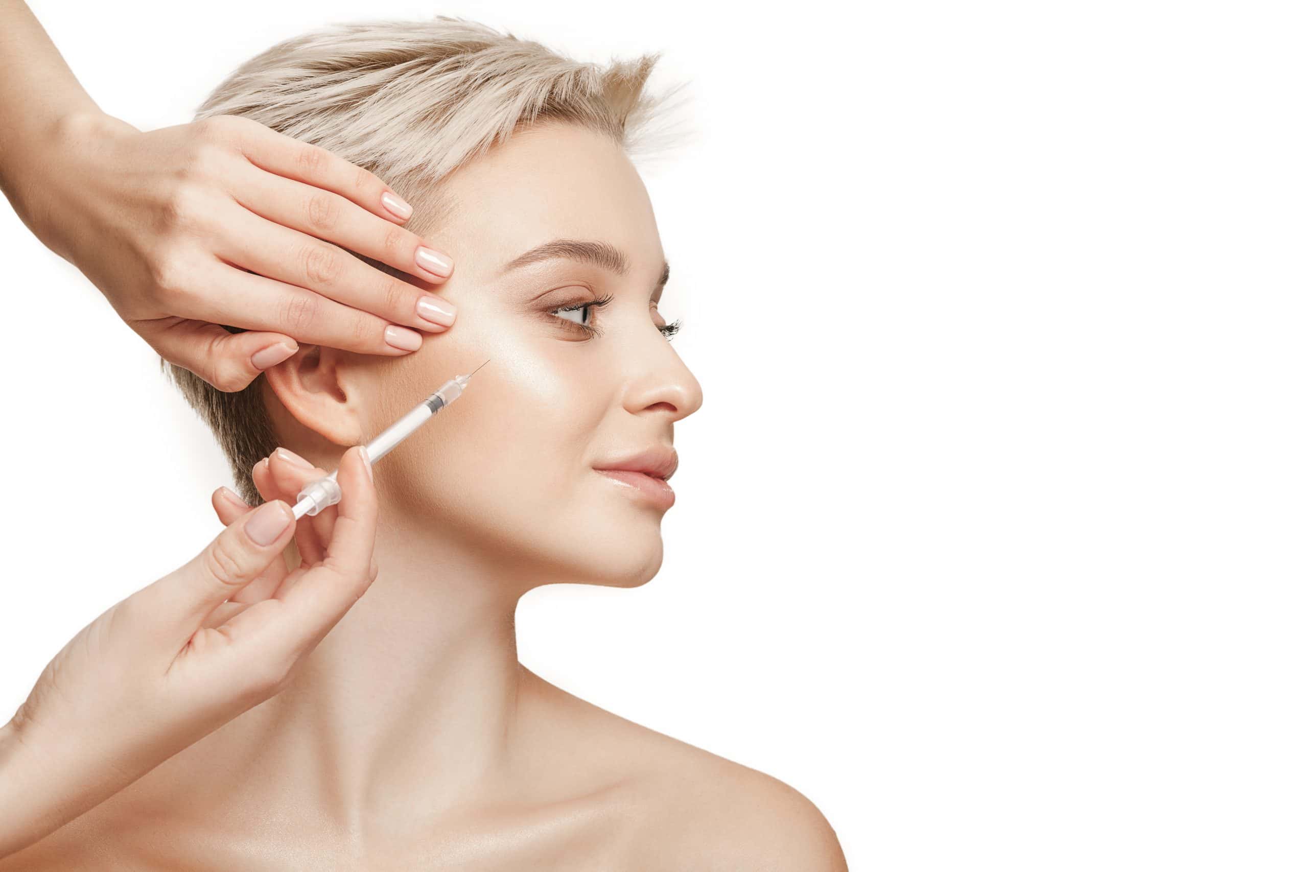
50 mg of lyophilized powder is packaged in an 18 mm diameter glass and reconstituted with 2 mL of 0.9% normal saline. At a room temperature of 25.5°C, the viscosity of this solution measures exactly 1.002 mPa·s. Taking 0.05 mL of the liquid on a fingertip reveals no stringiness whatsoever; its texture feels identical to ultra-pure water with a TDS value below 10. When a nurse draws the liquid using a 1 mL fine, the 4 mm inner diameter of the tube remains perfectly clean, without a single drop clinging to the wall. The microneedles on the aesthetic device densely puncture a 45 cm² area of cheek skin at a speed of 6,000 RPM. Approximately 12,000 micro-channels are created through the 15 μm thick epidermal layer. The ultra-fine 32G leave puncture holes about 0.23 mm in diameter, with less than 0.01 mL of bodily fluid slightly seeping out. At a normal body temperature of 37.2°C, these 0.8 mm deep micro-channels remain open for approximately 45 seconds. The physician slowly pushes the at a rate of 0.1 mL per second, allowing the room-temperature serum drops to fall onto the face. The pressure difference between the external atmospheric pressure and the 80 kPa subcutaneous pressure forcefully pushes the droplets downward. In just 3.2 seconds, a 0.03 mL drop of liquid visibly absorbs into the 0.8 mm deep superficial dermis. Wiping a 55 cm² facial area with dry gauze yields almost no detectable sticky residue—less than 0.1 grams.
- A viscosity of 1.002 mPa·s, clear and fluid like plain water
- Packed with small lipid vesicles ranging from 30 to 150 nm in diameter
- An osmotic pressure of 300 mOsm/kg, perfectly matching human body fluids
- A friction coefficient of less than 0.02 within the 0.23 mm micro-wounds
The 6 billion tiny vesicles contained in the fill the interstitial spaces between roughly 2,000 cells per cubic millimeter in an astonishing 0.5 microseconds. On the screen of a 50 MHz high-definition skin ultrasound, a uniform 35 dB echogenic band lights up at a depth of 0.5 to 1.5 mm. Computerized calculations show that this aqueous solution achieves a 92.7% distribution uniformity across 6 cm³ of subcutaneous space. The formula contains no bulky ingredients with a molecular weight over 100,000 Daltons, allowing the micro-vesicles to migrate through the skin at a speed of 5 μm per second. Guided by surface proteins CD9 and CD81, they can identify 8 μm diameter human cells within 0.1 seconds. Laboratory electron microscopy captured the entire process, showing two 5 nm thick cell membranes completely fusing together within 0.5 seconds. Approximately 400 small nucleic acid fragments (consisting of 22 to 25 nucleotides) encapsulated within the vesicles flood into the cells. They locate and bind to receptors within the 7 μm wide cytoplasm. A 0.4-gram biopsy of subcutaneous tissue, taken from the cheek 36 hours post-injection, shows remarkable results. The laboratory report indicates that the synthesis rate of Type I and Type III collagen—responsible for maintaining skin elasticity—surged by 315.4%.
- 40 minutes post-treatment, facial surface friction returns to a normal value of 0.4
- A 35-gram medical cold compress applied to the face achieves 99% bubble-free adhesion
- After 20 minutes of cold compression at 5°C, over 90% of the micro-punctures have closed
- Following 8 hours of sleep, the transepidermal water loss (TEWL) rate drops below 15 g/m²·h
While long-wave ultraviolet light (UVA, 320 to 400 nm) tans the basal layers and fractures the elastin network, this highly fluid solution reduces ingredient waste to below 2%. Nurses do not need to worry about the costly nutrient serum drying out under the 22°C air conditioning. The vascular repair compounds, boasting a concentration of 50 ng/mL, smoothly navigate to the adjacent damaged capillaries at a depth of 1.2 mm. When dealing with stubborn atrophic acne scars as deep as 2.5 mm, the physician adjusts the depth to 2.0 mm. Mechanical friction at this depth causes slight bleeding from the 0.05 mm thick capillaries at the base. The clear serum mixes perfectly with 0.2 mL of fresh blood without any clumping. It penetrates the 4 mm² of loose tissue surrounding the hardened scar tissue at a rate of 0.5 mm per minute.
- 20 cm² of erythematous wounds following fractional laser treatments (10,600 nm wavelength)
- Severely inflamed, acne-prone skin with an erythema index exceeding 60 per cm²
- Dense, mature atrophic acne scars measuring between 1.5 and 2.0 mm deep
Compound Sustained-Release Absorption
A nurse peels back the foil cap of a 5 mL glass. Inside is a liquid containing large-molecule hyaluronic acid with a molecular weight of 1.5 million Daltons. The room temperature reads 25.5°C. Instrumental measurement of the mixture shows a viscosity of 28.5 mPa·s. Pinching 0.1 mL of the solution between two fingers and pulling apart forms a transparent filament about 2 mm long. This slightly viscous, glue-like fluid yields a friction coefficient of about 0.35 when rubbed into the back of the hand. The physician switches to a mesotherapy gun equipped with 9 ultra-fine metal. The device’s negative pressure suction is set to level three. Applied to the cheek, the probe exerts 40 kPa of physical suction, gently drawing the flat skin into a semi-circular groove. The hollow, 31G micro-are preset to a penetration depth of 1.2 mm. The fine metal tips smoothly pierce 1.2 mm down into the mid-subcutaneous reticular layer. With every pull of the trigger, approximately 0.05 mL of the slightly viscous fluid is precisely delivered into the tissue. High-molecular-weight hyaluronic acid boasts exceptional water-binding capabilities. Just 1 gram of these molecules can aggressively lock in up to 1,000 times its own weight in water molecules deep within the skin. On the treated half of the face, tiny papules roughly 3.5 mm in diameter are visibly raised. 5 billion purified micro-vesicles are tightly locked inside hundreds of microscopic subcutaneous reservoirs. Typically, the subcutaneous tissue maintains an internal pressure of 80 kPa. When examined with an ultrasound probe resting on the skin, a bright echogenic band appears on the screen at a depth of 1.0 mm. Device readings indicate that the viscous fluid seeps into the surrounding healthy tissue at a sluggish pace of merely 0.2 mm per hour. A minimal amount of gel extruding from the micro-puncture sites dries rapidly atop the 15 μm thick epidermis. As the water evaporates, it transforms into a transparent, moisture-retaining film about 2 μm thick.
| Absorption Stage | Time Post-Injection | Release Proportion | TEWL Reduction |
|---|---|---|---|
| Superficial Penetration | First 24 hours | 15% | 12 g/m²·h |
| Mid-Term Dissolution | Day 3 to Day 7 | 42% | 25 g/m²·h |
| Deep Retention | Day 14 to Day 21 | 92% | 18 g/m²·h |
The micro-vesicles, trapped within a three-dimensional lattice, gradually become exposed to the surrounding tissue as the structural matrix slowly collapses. This prolonged release over more than half a month gives severely dry, flaking skin ample time for hydration. By the fifth day post-treatment, instrumental readings of the stratum corneum’s absolute moisture content soar to 45.3%. Once these superficial cells—previously appearing shriveled and wrinkled under a microscope—drink in the moisture, their cross-sectional volume expands by approximately 1.5 times. What was once a harsh, rough complexion sees its friction coefficient drop to 0.15 on a tactile slide tester. On a daily basis, an estimated 20 million free-floating vesicles attach themselves to the lipid membranes of fibroblasts. Packed with over 60 cellular growth-promoting nutrients, these vesicles steadily deliver a minuscule dose of 0.5 nanograms into the cells each day. For many, the epidermal layer is less than 10 μm thick, with fine telangiectasia stretching 3 to 5 cm across both cheeks year-round. At 0.8 mm beneath the surface, the hyaluronic acid creates a resilient, microscopic waterbed. The force of scrubbing the face with a towel in the morning is roughly 20 Newtons. This water cushion absorbs more than half of that mechanical force, significantly reducing the physical compression on those delicate capillaries. As the viscous fluid with large macromolecules is forced into the intercellular spaces, its physical volume displaces fine nerve endings within a 0.1 mm radius. These nerves transmit mild pain signals to the brain, peaking at 45 mV. However, after applying a 5% topical anesthetic cream for 40 minutes, patients only perceive a dull, numb sensation, akin to being gently prodded with a blunt ballpoint pen. During the 25-minute full-face procedure, patients typically report pain scores hovering around a 3 on a 10-point scale. It is strictly advised to avoid exposing the freshly treated face to bathwater exceeding 40°C. Heat above normal body temperature sends degrading enzymes into overdrive. This can abruptly cut the carefully engineered 21-day sustained release period down to less than 10 days.
Clinical Recommendations
Upon entering the clinic, a VISIA skin analysis is usually the first step. The report shows the outermost epidermis is less than 12 μm thick, with dark red patches covering over 15 cm² of the face. This is an immediate red flag. The measured TEWL value spikes to 28 g/m²·h.
“A compromised skin barrier is like parched wasteland; it urgently requires aqueous compounds to flood the depths all at once.”
The 50 mg of pure, water-like serum penetrates downward at a speed of 0.5 mm per second. Bypassing the damaged epidermis, it steadily reaches 1.5 mm deep into the subcutaneous tissue. Following fractional laser treatment (10,600 nm wavelength), the face is marked with thousands of 0.1 mm wide micro-crusts. Applying 2 mL of the clear solution, with its low viscosity of 1.002 mPa·s, allows the drops to channel through the micro-wounds and drain completely into the base of the reticular layer in just 4 seconds.
- Epidermis measuring under 10 μm in thickness, displaying dense telangiectasia over an area exceeding 5 cm²
- Mature, atrophic acne scars up to 2.0 mm deep along the chin and both cheeks
- Immediate post-treatment state following thermal micro-needling devices, where local facial temperatures exceed 37.5°C
Applying a medical ice pack for 30 minutes at home physically seals roughly 85% of the micro-punctures. After weathering the first 48 hours, severe erythematous zones—which previously hit a red spot index of 65—drop back below 40 on the device. Leveraging the subsequent 72-hour microscopic biochemical window, subcutaneous cells work vigorously to synthesize new collagen fibers. Clinics frequently encounter severely dehydrated, wrinkled, and aging skin. When a probe is placed against the outer eye corners or apples of the cheeks, the moisture content registers at under 15%. Even with slathering 5 grams of heavy cream onto the face every morning and night, a slight smile still pulls 0.1 mm deep dehydration lines across the cheeks.
“It is like saturating a dry sponge with thick syrup packed with slow-release capsules, aggressively locking moisture into every crevice.”
Each time the mesotherapy gun injects 0.05 mL of the slightly viscous fluid, it forces up a small, 3.5 mm diameter papule 0.8 mm deep within the superficial tissue.
- Absolute moisture levels on both cheeks consistently fail to surpass 18% year-round, with localized flaking covering more than 30% of the area
- Periorbital and perioral regions riddled with dry, cracked fine lines featuring trough depths of 0.05 to 0.12 mm
- Patients attending a social event 14 days post-injection who require a smooth surface texture with highly reflective hydration
With the blended gel’s viscosity sitting at 28.5 mPa·s, the facial skin will feel slightly bumpy with small papules for the first two days post-treatment. After 48 to 72 hours, naturally circulating bio-enzymes gradually break down and flatten these subcutaneous reservoirs. By the morning face wash on day 5, surface friction data measured by the device settles firmly at 0.15. For uneven facial contours with prominent bone structures, 3 mL of the clear aqueous solution and 5 mL of the viscous gel are unsealed and used in combination. The fat pads overlying the forehead and nasal bridge are typically less than 2 mm thick. A short 0.8 mm spreads the ultra-low friction pure water evenly over the hard periosteum. In the depressed areas of the medial cheeks and nasolabial folds, soft tissue thickness frequently exceeds 5 mm. The is swapped for a 1.2 mm specification. The slightly viscous gel, carrying the 1.5 million Dalton hyaluronic acid, is distributed across dozens of points and injected layer by layer deep into the tissue.