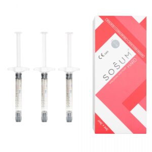
Botulax temporarily relaxes over-contracted muscles by blocking the release of nerve signals. Clinical data shows that onset begins 2-3 days after injection, reaching the optimal peak in 1-2 weeks, and the effect usually lasts for 3-6 months. It can precisely smooth dynamic wrinkles and shrink the masseter muscle to enhance facial contours. To ensure safety and natural results, please be sure to choose a formal institution and a professional doctor for the procedure.
Mechanism
Botulax contains 900kDa Type A Botulinum Toxin, with a purity maintained above 99.5% through vacuum drying technology. Its 150kDa active light chain enters the nerve endings and cleaves the SNAP-25 protein, preventing the fusion of 40nm diameter acetylcholine vesicles with the membrane. This directly blocks the chemical transmission of nerve signals, leading to local muscle relaxation starting after 24 hours, with a diffusion radius typically maintained at about 1 cm.
Molecular Structure
The molecular body of Botulax presents as a massive protein complex with a standard molecular weight of approximately 900kDa (kilodaltons). Under a microscope, this giant structure is composed of a biologically active neurotoxin chain and several non-toxic accessory proteins. The biologically active part is a protein weighing 150kDa, while the remaining 750kDa portion consists of non-toxic non-hemagglutinin (NTNH) proteins and various hemagglutinin (HA) proteins. These accessory proteins tightly wrap around the 150kDa active molecule, forming a solid physical barrier. This structure exists to protect the fragile active chain from degradation by acidic conditions or proteases in the external environment before entering a neutral pH environment, ensuring that every unit of protein maintains its intended spatial conformation after injection into human tissue. The 150kDa active portion is the most complex subject of study in the molecular structure of Botulax. It consists of a 100kDa Heavy Chain and a 50kDa Light Chain connected by a disulfide bond (-S-S-). This polypeptide chain contains approximately 1296 amino acid residues, and its tertiary structure is precisely divided into three domains with different functions. The C-terminal domain of the heavy chain is responsible for recognizing and attaching to receptors on the surface of the nerve cell membrane, with binding efficiency determined by the protein folding morphology of this region. The N-terminal of the heavy chain constitutes a transmembrane domain, which contains several hydrophobic helical structures. The 50kDa light chain is essentially a zinc-dependent endopeptidase. Its molecular interior contains a characteristic HExxH binding motif capable of chelating a zinc ion (Zn²⁺), which is the chemical basis for its hydrolysis of the SNAP-25 protein.
- Heavy Chain (100kDa): Divided into a binding domain (H-C) and a translocation domain (H-N), containing approximately 850 amino acid residues.
- Light Chain (50kDa): Contains approximately 440 amino acid residues and belongs to the zinc-dependent metalloprotease family.
- Disulfide Bond Linkage: Typically located between cysteine residues Cys430 and Cys454. This covalent bond must be severed in the reductive environment of the nerve cell cytoplasm for the light chain to be released from the complex and perform its function.
The group of accessory proteins (NAPs) surrounding the 150kDa active chain plays an important role in structural stability. These proteins include subunits of different molecular weights such as HA-17, HA-33, and HA-70, as well as a 130kDa NTNH protein. The HA protein family forms multimeric structures that allow the entire 900kDa complex to maintain its molecular aggregation state during dilution. During the manufacturing process, Botulax utilizes vacuum drying technology. This process avoids the risk of protein thermal denaturation that freeze-drying might bring, allowing the integrity of the 900kDa complex to exceed 99.5% in the final product. A high proportion of complete macromolecular structures reduces the appearance of free small-molecule proteins, thereby effectively limiting the unintended flow of the drug between muscle tissues after injection. At the molecular level, the production process of Botulax strictly controls the content of impurity proteins. In addition to the 900kDa active complex, the formulation contains almost no redundant non-functional protein debris. Research shows that the 900kDa macromolecular complex structure is most stable when the pH value is between 5.0 and 6.5. Once it enters the physiological environment of the human body with a pH of approximately 7.4, the accessory proteins dissociate from the 150kDa active chain, releasing the biologically active toxin molecules. This structural transition from a “protected state” to a “released state” is achieved based on changes in the surface charge distribution of protein molecules under different pH levels. Each light chain portion of the 150kDa molecule carries an active center that must be precisely exposed to undergo a chemical reaction with the target protein.
- Impact of Vacuum Drying: This technology ensures that the spatial hindrance of protein crystals does not collapse in a dry state, maintaining the natural morphology of the peptide chains.
- Purity Parameters: Monitored by size-exclusion chromatography, the purity level of the product ensures that the potency of each unit (Unit) is highly uniform.
- Ion Dependency: The molecular stability of the light chain requires the participation of trace zinc ions; a lack of zinc ions will cause the light chain to lose its proteolytic activity.
The heavy chain portion of the 150kDa molecule has multiple high-affinity receptor binding sites distributed on its surface. These sites are composed of specific amino acid sequences that can undergo molecular recognition with gangliosides and synaptic vesicle protein 2 (SV2) on nerve endings. This recognition process is not a random physical contact but a complementary binding based on perfect molecular shape matching. The translocation domain of the heavy chain consists of a series of long alpha-helices. When the internal environment of the cell acidifies, these helices undergo conformational changes to form a channel that penetrates the phospholipid bilayer. This process requires extremely high thermal stability of the molecules. During manufacturing, Botulax optimizes the buffer system to ensure that these environmentally sensitive secondary protein structures do not undergo non-specific aggregation or degradation during storage.
- Molecular Weight Distribution: The 900kDa complex is interwoven from 12-14 protein subunits.
- Amino Acid Sequence Consistency: Highly consistent with the standard Type A botulinum toxin sequence, ensuring predictability of biological effects.
- Structure After Reconstitution: After adding 0.9% sodium chloride solution, hydrogen bonds and hydrophobic interactions between molecules support the morphology of the complex in aqueous solution, maintaining high surface activity.
Protein Cleavage and Inhibition
When the 150kDa active molecule enters the cytoplasm of motor nerve endings, the disulfide bond originally connecting the heavy and light chains breaks in a reductive environment. This process releases the 50kDa light chain, which has high catalytic activity and is essentially a zinc-dependent endopeptidase. Each light chain molecule contains a characteristic HExxH sequence motif that can precisely chelate a zinc ion (Zn²⁺), which is the chemical basis for maintaining its proteolytic activity. The light chain remains highly stable in the cytoplasm and begins to search for its only biological target—the SNAP-25 protein located on the inner side of the presynaptic membrane. SNAP-25 is a protein composed of 206 amino acids and is a key component of the SNARE complex that mediates neurotransmitter release. In healthy nerve cells, SNAP-25 interweaves with synaptobrevin (VAMP) and syntaxin-1 to form a high-energy four-helix bundle structure. The mechanical force generated by this physical structure overcomes the electrostatic repulsion between membranes, prompting acetylcholine vesicles with a diameter of approximately 40 nm to fuse with the cell membrane. The light chain of Botulax precisely recognizes a specific sequence at the C-terminus of the SNAP-25 peptide chain and executes cleavage at the peptide bond between Glutamine 197 (Gln197) and Arginine 198 (Arg198). This reaction results in SNAP-25 losing the terminal 9 amino acid residues. Although this is a minor loss in terms of molecular weight, it is destructive to the spatial conformation of the SNARE complex. Without these 9 amino acids, SNAP-25 can no longer form a stable four-helix mechanical structure with VAMP and Syntaxin, causing the biomechanical device originally used to “pull” vesicles closer to physically collapse. Due to the lack of this mechanical tension, vesicles loaded with Acetylcholine (ACh) can only remain suspended near the presynaptic membrane, unable to complete the exocytosis process.
| Biochemical Parameters | Detailed Values and Descriptions | Impact on Muscle Function |
|---|---|---|
| Light Chain Molecular Weight | 50,000 Da (50kDa) | Able to diffuse freely in the cytoplasmic matrix to find targets |
| Cofactor Requirement | 1 zinc ion per light chain molecule | Lack of metal ions will cause proteolytic activity to disappear |
| Target Protein Name | SNAP-25 (Synaptosomal-associated protein 25) | A pillar protein of the presynaptic membrane docking mechanism |
| Precise Cleavage Site | Peptide bond between Gln197 and Arg198 | Destroys the spatial complementarity required for helix bundle formation |
| Vesicle Physical Specs | Approximately 40 nm in diameter | Loses fusion power and remains in the presynaptic region |
| Enzyme Catalytic Efficiency | 1 light chain molecule can inactivate thousands of SNAP-25 proteins | Extremely low toxin concentrations can produce widespread blocking effects |
Unlike ordinary chemical inhibitors, the light chain of Botulax is not consumed or degraded after completing a cleavage; instead, it continues to move within the nerve ending to find the next intact SNAP-25 molecule. This high turnover rate means that a small amount of light chain can deplete all functional SNAP-25 reserves in the presynaptic membrane within a short time. Experimental data indicates that when more than 70% to 80% of SNAP-25 in the cell is cleaved, the release of acetylcholine drops below the threshold required to trigger muscle contraction. This inhibition of protein function is irreversible at the molecular level. The destroyed SNAP-25 fragments cannot be reassembled; nerve cells must transport new SNAP-25 molecules from the cell body to the nerve endings through gene expression and protein synthesis. At the macroscopic neuromuscular junction (NMJ), this protein-level inhibition manifests as a frequency-dependent transmission disorder. When high-frequency action potentials sent by the brain reach the affected nerve endings, although calcium channels can still open normally, the efficiency of calcium-induced vesicle fusion drops by more than 95% due to SNARE complex stagnation. Since acetylcholine cannot enter the synaptic cleft, the nicotinic acetylcholine receptors (nAChR) on the muscle cell membrane receive no ligand binding, ion channels cannot open, and finally, the muscle fibers remain in a resting potential state. This blocking effect reaches its biochemical peak within 24 to 72 hours after injection, at which point the SNAP-25 concentration in the affected area drops to its lowest point. As the proteolytic reaction spreads, the contractile force of the entire motor unit (Motor Unit) will show a stepwise decline until it enters a state of complete relaxation. When nerve endings are unable to effectively release acetylcholine for a long period, nerve cells initiate a compensatory mechanism manifested as axonal sprouting. This sprouting attempts to establish new connections next to the original junctions, seeking muscle surfaces not yet affected by the toxin. This biological feedback reaches its peak between 4 and 8 weeks after injection; however, these new sprouting structures are usually immature in function and cannot provide sufficient contraction strength. Meanwhile, the blocked light chain proteins are gradually degraded through the intracellular proteasome pathway. Studies show that the biological half-life of the light chain in human neurons is quite long, allowing residual proteolytic activity to be detected in nerve endings even 3 months after toxin injection, continuously inhibiting new SNAP-25 from forming complexes.
| Reaction Phase | Specific Subcellular Manifestations | Timeline Reference |
|---|---|---|
| Initial Proteolysis Phase | Light chain begins to attack SNAP-25; acetylcholine release decreases | 6 – 12 hours post-injection |
| Signal Interruption Phase | SNAP-25 stock falls below the threshold to maintain contraction; signal stops | 24 – 48 hours post-injection |
| Functional Stagnation Phase | All SNARE complex construction is blocked; muscle is fully relaxed | 3 – 14 days post-injection |
| Protein Degradation Phase | Light chains in the cytoplasm lose activity; new proteins begin to accumulate | 90 – 120 days post-injection |
Microscopic observation reveals that organelles such as mitochondria and endoplasmic reticulum in the nerve endings maintain normal morphology and function, indicating that this process is not a cytotoxic reaction but a highly precise signal pathway interruption. Because the light chain only acts on SNAP-25 within the cytoplasm and does not interfere with the genetic material in the cell nucleus or the ion pumps on the cell membrane, this effect is biologically reversible. When the concentration of the light chain drops below a certain level, newly synthesized SNAP-25 begins to take dominance, and the vesicle fusion apparatus of the presynaptic membrane will reassemble. The efficiency of this reassembly determines the speed of muscle strength recovery. Clinical observations show that around 150 to 180 days, neuromuscular conduction function typically recovers to more than 80% of its initial level.
Nerve Sprouting and Regeneration
After Botulax acts on motor nerve endings, because chemical transmission of acetylcholine is interrupted, the nervous system initiates a series of complex compensatory remodeling processes. Within 48 to 72 hours after injection, although the muscle shows obvious relaxation, subcellular nerve reactions have already begun. The neuron cell body receives feedback that distal signal transmission is interrupted, thereby upregulating the expression of various growth-related proteins, most notably GAP-43 (Growth Associated Protein 43). The increase in this protein level is a biological marker of nerve sprouting, prompting the affected nerve endings to start extending extremely thin, unmyelinated nerve fiber branches. The diameter of these new branches is typically only 0.1 to 0.5 microns. They explore the muscle tissue gaps like antennae, attempting to find sites where new synaptic connections can be established to restore lost chemical communication.
In microscopic observations of the neuromuscular junction (NMJ), this sprouting phenomenon usually appears between day 7 and day 14 post-injection. New axonal side branches will grow from original nerve endings or even from the nearest Nodes of Ranvier.
Although they can form preliminary synapse-like structures and attempt to release small amounts of acetylcholine, because they lack complete vesicle aggregation mechanisms and efficient ion channel arrangements, muscle contraction force at this time is extremely low and insufficient to produce visible muscle movement. Experimental data indicates that the neurotransmitter release efficiency of these early sprouts is less than 5% of normal. Meanwhile, the 150kDa active portion of Botulax remains residing inside the original nerve endings, continuing its proteolytic action. Over time, this “exploratory behavior” of the nervous system reaches a peak, usually occurring between day 28 and day 60 post-injection. As the intracellular biochemical environment changes, this compensatory sprouting process enters its second stage, which is the repair of original synaptic functions. The biological half-life of the Botulax light chain protein in the nerve cytoplasm is not infinite; it is gradually recognized and degraded by the ubiquitin-proteasome system. Once the light chain concentration drops to a level where it can no longer fully inhibit SNAP-25, newly synthesized SNAP-25 protein from the neuron cell body begins to arrive at the presynaptic membrane via axonal transport (at a transport speed of approximately 200 to 400 mm per day). The newly synthesized protein begins to rebuild the SNARE complex, allowing the original acetylcholine vesicles with a diameter of 40 nm to regain the ability to fuse with the cell membrane.
Clinical biochemical monitoring shows that recovery of in-situ synaptic function begins approximately around day 90 post-injection. At this time, original nerve endings regain the ability to release sufficient chemical messengers, leading to a linear recovery of muscle strength.
When original nerve endings re-establish efficient chemical transmission channels, the new nerve sprouts that previously grew for compensation undergo biological regression. This is an extremely precise feedback regulation mechanism: when the receptors on the muscle cell surface receive stable acetylcholine signals again, they stop releasing chemotactic factors that induce nerve growth. Without the support of these factors, the previously dense collateral circulation begins to wither and is eventually absorbed by the cells. This “sprouting first, regression later” process explains the reversibility of Botulax effects. Around 120 to 150 days post-injection, the anatomical structure of the neuromuscular junction gradually returns to a state close to the initial state before injection. In high-dose injection areas, the nerve sprouting maintenance time is correspondingly longer because the light chain protein clearance cycle is extended. Research has found that in microscopic sections of muscle fibers, the coverage area of motor units undergoes a temporary expansion during the recovery period due to the action of Botulax. The number of muscle fibers controlled by each motor neuron may increase by 20% to 30% during the sprouting phase, but this expansion will shrink back to original proportions as the in-situ synapses are repaired.
The remodeling efficiency of nerve endings directly determines clinical maintenance time. In most users, the total duration of this cycle from functional blocking to complete regeneration is maintained between 18 and 26 weeks.
During the functional block, due to the lack of synaptic vesicle cycling, the level of oxidative phosphorylation in local mitochondria drops to about 40% of the baseline. As new SNAP-25 re-mediates vesicle fusion, energy demand increases, prompting mitochondria to re-aggregate at the presynaptic membrane to provide ATP required for acetylcholine reuptake and vesicle cycling. This comprehensive metabolic recovery marks the thorough reconstruction of neuromuscular junction function. This mechanism explains that Botulax does not work by killing nerve or muscle cells, but by inducing a reversible physiological remodeling cycle to achieve temporary control of muscle tension without destroying anatomical integrity.
In the molecular renewal cycle, every degraded light chain molecule is replaced by a newly synthesized endopeptidase substrate, allowing the chemical transmission efficiency in the affected area to gradually return to a normal electrophysiological level of hundreds of times per second within a few months.
In areas with faster circular metabolism, protein turnover and neurotransmitter synthesis rates are higher, which often leads to a shorter nerve regeneration cycle. Experiments have observed that for every 1 degree Celsius increase in local temperature, there are slight increases in protein degradation rates and nerve sprouting growth speeds. 
Muscle Relaxation
Botulax relies on 900kDa high-purity Type A Botulinum Toxin to permanently block muscle contraction commands from being transmitted via acetylcholine by cleaving SNAP-25 protein in nerve endings. Within 24 to 72 hours post-injection, the electrophysiological activity of the target muscle begins to weaken. Clinical observation shows that muscle relaxation reaches its peak on day 14, and the volume of large muscle groups like the masseter or gastrocnemius begins to shrink after 4 to 6 weeks due to disuse atrophy. This controlled relaxation state is typically maintained for 12 to 16 weeks until nerve endings form new connections through “budding.”
Clinical Relaxation Timeline
Within 1 to 3 hours post-injection, the 900kDa macromolecular complex begins to dissociate in the physiological pH environment, releasing the 150kDa active neurotoxin portion. This active portion completes preliminary binding to SV2 receptors on nerve endings within 4 to 6 hours through blood circulation or local diffusion. Although the subject cannot perceive muscle changes at this time, the internalizing process at the molecular level has already begun. Studies show that between 24 and 48 hours post-injection, most subjects begin to observe a decrease in local muscle tension. For thinner expression muscles such as the frontalis, this change manifests as a flattening of dynamic wrinkles on the skin surface; for the masseter, electrophysiological tests show that the amplitude of its induced compound muscle action potential (CMAP) drops by an average of 40% to 60% within 72 hours.
| Clinical Stage | Time Interval | Muscle Physiology and Physical Quantitative Performance | Expected Visual Improvement |
|---|---|---|---|
| Molecular Induction | 1 – 72 hours | SNAP-25 protein starts being cleaved; acetylcholine release decreases by >50%. | Muscle strength starts to weaken; touch feels softer. |
| Clinical Onset | 4 – 7 days | Nerve block area expands; local muscle loss of strength during voluntary contraction reaches 80%. | Dynamic wrinkles become noticeably smoother. |
| Peak Efficacy | 14 – 21 days | Signal transmission at NMJ almost completely interrupted; target muscle at maximum relaxation. | Skin texture improves; muscle volume preliminarily shrinks. |
| Remodeling Maintenance | 1 – 3 months | Muscle undergoes disuse atrophy from lack of stimulation; masseter thickness reduces by 2.0mm to 3.5mm. | Facial shape or leg contour lines are optimized. |
| Physiological Recovery | 4 – 6 months | Axonal terminals start sprouting; new synapses form; acetylcholine resumes release. | Muscle strength recovers; wrinkles reappear. |
The muscle relaxation effect of Botulax reaches its clinical peak on Day 14. At this point, a large amount of SNAP-25 protein in the nerve endings has been enzymatically hydrolyzed, causing synaptic vesicles to be unable to fuse with the cell membrane. In subject groups treated for glabellar lines, experimental data shows that more than 90% of subjects reached maximum score improvement within two weeks. If the observation subject is a masseter hypertrophy patient, the muscle volume at this stage has not yet undergone visible drastic reduction, but the contraction hardness of the muscle will drop to its lowest point. Ultrasound examination can reveal that blood flow within the masseter muscle at this time undergoes a physiological decline due to reduced muscle activity. In terms of dosage distribution, if 4 to 10 Units are injected per point, the effective relaxation radius of the drug is typically maintained between 5mm and 10mm, ensuring that the relaxation effect is limited to the injection target and preventing unintended weakness in adjacent muscle groups. Entering Week 4 to Week 8, Botulax enters its most stable remodeling phase. For large muscle groups (such as the gastrocnemius or masseter), long-term relaxation leads to a reduction in muscle fiber diameter. Clinical measurements for the gastrocnemius prove that after continuous relaxation for 8 weeks, the circumference at the thickest part of the calf can decrease by an average of 1.5 cm to 2.5 cm. The quality of relaxation during this period depends on the initial dilution ratio at the time of injection. Using a standard concentration of 2.5 ml of saline per 100 Units provides a higher protein density, making the relaxation effect more concentrated and durable. If over-diluted, although the diffusion area increases, the toxin concentration per unit area decreases, which may lead to insufficient relaxation depth and cause weak contraction activity in the muscle to appear early around the 8th week.
| Muscle Type | Recommended Total Dose (Units) | Time to Reach Volume Reduction | Median Effect Duration |
|---|---|---|---|
| Frontalis (Wrinkles) | 10 – 20 | 7 – 14 days | 3.5 months |
| Masseter (Contour) | 50 – 100 | 4 – 8 weeks | 5.2 months |
| Trapezius (Shoulder) | 100 – 150 | 6 – 8 weeks | 4.8 months |
| Gastrocnemius (Calf) | 150 – 200 | 8 – 12 weeks | 6.0 months |
When time progresses to Week 12 to Week 16, the biological activity of Botulax begins to naturally decay, and the nervous system initiates self-repair mechanisms. This process begins with tiny axonal branches growing out of damaged nerve endings, medically known as “nerve sprouting”. During this phase, subjects will feel muscle strength slowly returning within 2 to 3 weeks. Electrophysiological monitoring shows that the frequency of motor unit potentials (MUP) begins to increase. For expression muscles, this means that slight creases will reappear on the skin surface when making exaggerated expressions. For body muscles, although strength is recovering, muscle volume will not immediately bounce back to initial levels due to previous atrophy effects. Typically, it takes 180 days (about 6 months) or even longer to fully recover to the muscle mass and strength level before injection. Individuals who frequently perform high-intensity chewing or leg strength training have a higher turnover rate of neuromuscular junctions, which may shorten the effective period of Botulax. In clinical follow-up, about 15% of subjects feel a significant return of muscle strength around Week 10 because of faster body metabolism. To prolong this relaxation state, clinical advice is to perform supplementary injections when muscle strength recovers to about 50% (usually the 4th or 5th month). Studies found that after periodic relaxation treatment for more than 3 times, the muscle hypertrophy trend will be inhibited long-term, and the subsequent maintenance period can even be extended to 8 to 10 months.
Target Area Performance
For glabellar lines, the typical clinical operation is to distribute 5 injection points in the corrugator and procerus muscles, with the total dose controlled at 20 Units. Each point is allocated 4 Units, and the insertion depth needs to reach 7 to 10 mm to ensure the toxin penetrates the fascia layer to reach the deep muscle. Research data shows that the diffusion radius under this dose configuration is precisely locked within 5 mm, effectively preventing the toxin from diffusing into the orbit and causing ptosis. For the frontalis, since its muscle fibers are widely distributed and thin, a “multi-point small-dose” plan is often used clinically, with only 1 to 2 Units injected per point and a spacing of 1.5 to 2 cm. This distribution allows the forehead to eliminate dynamic wrinkles while maintaining 20% to 30% baseline tension, avoiding the feeling of eyebrow pressure caused by total forehead stiffness. In the periocular area, injections targeting the orbicularis oculi focus on the superficial layer, with a tip inclination angle typically of 15 to 30 degrees and a total dose per side of approximately 12 Units.
- Glabellar Area: 20 Units total, divided into 5 points, aimed at neutralizing downward pulling muscle force.
- Frontal Region: 10 – 20 Units total, divided into 8 – 10 points, increasing coverage through high-volume injection after dilution.
- Periocular Area: 12 Units per side, divided into 3 points, reducing impact on lacrimal glands through subcutaneous superficial infiltration.
- Dilution Ratio: Typically 2.5 ml of 0.9% saline is used to dissolve 100 units of powder to obtain the optimal protein concentration balance.
For lower face contouring, Botulax’s effect on the masseter shows significant volumetric changes. The masseter is a thick rectangular muscle, and injection must be positioned within the “safety triangle” formed by the earlobe, mouth corner, and mandibular angle. Clinically, a dose of 25 to 50 Units per side is usually adopted, distributed in 3 to 5 points in a plum blossom pattern. To reach the deep fibers where motor nerve endings are most densely distributed, the needs to be inserted vertically 10 to 13 mm. Around day 28 post-injection, 3D scanning can observe that the middle thickness of the masseter muscle is reduced by an average of 22% to 27%. This muscle relaxation does not immediately lead to volume reduction, but by blocking acetylcholine release, it induces the muscle to enter a persistent “inactive state,” thereby initiating the protein degradation process. If the dose distribution is too close to the superficial risorius muscle, it will cause facial asymmetry; therefore, a 1 cm buffer zone at the muscle edge must be reserved during operation.
| Target Site | Recommended Total Dose | Injection Depth | Muscle Volume Reduction Rate | Clinical Satisfaction |
|---|---|---|---|---|
| Masseter (Jaw) | 50 – 100 Units | 10 – 13 mm | 20% – 30% | 94% |
| Trapezius (Shoulder) | 100 – 200 Units | 13 – 15 mm | 15% – 18% | 88% |
| Gastrocnemius (Calf) | 150 – 300 Units | 15 – 20 mm | 12% – 20% | 85% |
In body shaping applications, Botulax’s performance on the trapezius mainly manifests in softening shoulder lines and releasing muscle tension. Because the trapezius maintains posture long-term, its muscle density is much higher than facial muscles, so a higher unit concentration is required. Usually, 50 to 100 Units are distributed on each side using a grid injection method, with a span of 2 cm between points. 14 days after injection, subjects often feel the disappearance of shoulder and neck heaviness, and by week 6, the protruding muscle blocks on the shoulders become smooth due to fiber atrophy. Data shows that this high-dose relaxation can increase the visual length of the neck by 1.5 to 2 cm. The same logic applies to calf shaping, where Botulax focuses on the medial and lateral heads of the gastrocnemius. Since calf muscles are significantly affected by gravity, the injection dose needs to be increased to 100 to 150 Units per side, and injection into the deep soleus muscle must be avoided to maintain normal walking and standing balance.
- Trapezius Distribution: Find high tension points along the upper edge of the scapula, 5 – 10 points per side to disperse pressure.
- Gastrocnemius Positioning: Ask the subject to stand on tiptoe to determine muscle boundaries, focusing on the medial protrusion with 15 – 20 points for even coverage.
- Specs: 30G or 32G 13mm long are mostly used for body parts to ensure the drug penetrates the deep fascia.
- Safety Indicators: It is recommended that the total amount for a single full-body injection does not exceed 400 Units to reduce the biological risk of systemic botulism.
For precise performance in small facial areas, Botulax also demonstrates extremely fine regulatory capabilities when treating perioral wrinkles and the mentalis muscle (chin). The perioral area is extremely sensitive to toxin, so the dose is usually compressed to 1 to 2 Units per point, with a total dose not exceeding 4 Units, otherwise it will interfere with lip closure or speech clarity. In the chin area, Botulax can eliminate “orange peel” skin texture by relaxing the mentalis muscle, with injection points located at 2 Units each to the left and right of the center of the chin tip. This small-range muscle relaxation can make the jawline appear more forward-extended visually. All applications are built on a precise understanding of anatomical structures, utilizing the low migration characteristics of 900kDa macromolecular complexes to ensure that every injection point produces the expected local biological effect without large-area diffusion.
Muscle Recovery Logic
The attenuation of Botulax’s biological activity begins with the enzymatic degradation of its 150kDa active light chain in the cytoplasmic matrix of motor nerve endings. At 10 to 12 weeks post-injection, the light chain proteins originally locked on the nerve membrane begin to be recognized and disassembled by the intracellular ubiquitin-proteasome system. As the active light chain concentration drops below 15% of its onset level, the synthesis rate of SNAP-25 protein inside the nerve cell exceeds the rate of it being cleaved. Experimental monitoring shows that intact SNARE protein complexes begin to reassemble at the presynaptic membrane. This molecular-level reversal process does not produce immediate muscle movement but paves the way for subsequent nerve signal reconstruction. When treating small facial expression muscles, due to fast local blood circulation, the rate of this metabolic degradation is often about 10% to 20% faster than in large muscle groups.
The half-life of neurotoxin molecules in the cytoplasm determines the initial maintenance length of muscle relaxation. Once the metabolic equilibrium point shifts, nerve endings will regain the physical conditions to release chemical transmitters.
When original nerve synapses are still functionally blocked, the nervous system initiates a complex physiological compensatory mechanism known as “nerve sprouting.” Around day 90 to day 120 post-injection, suppressed motor nerve axonal terminals are induced by signal factors secreted by the muscle to grow multiple small side branches with a diameter of only 0.5 to 1.0 microns. These new side branches bypass the blocked old synapses and look for new binding points on the muscle fiber surface like tree roots. Electrophysiological tests record that the resting potential of the target muscle begins to fluctuate at this time, and the amplitude of the induced compound muscle action potential (CMAP) rises from a very low value to about 10% of the baseline. During this stage, subjects will feel fine tremors when the muscle is forced to contract, and areas that were originally completely stiff begin to have minimal displacement.
The formation of side sprouts is a manifestation of human neural plasticity, which bypasses old nerve junctions blocked by toxin by establishing temporary “signal bypasses.”
Subsequently, muscle recovery enters the functional synaptic reorganization phase. These new axonal side branches establish preliminary chemical connections with muscle fibers, and Acetylcholine (ACh) begins to be released in small, quantized amounts. By Week 16 post-injection, muscle contraction strength can usually recover to 30% to 40% of the pre-injection level. As time passes, the old synaptic channels originally blocked by Botulax will also reopen as the toxin is completely cleared. At this point, the nervous system undergoes a “pruning” process where temporary new side branches gradually wither, and the original main synapses regain their signal transmission tasks. This transition from “alternative paths” back to “main road transmission” marks the return of muscle movement logic to normal. For high-load muscles such as the masseter, the speed of this return is greatly affected by chewing frequency, as frequent physical stimulation accelerates the upregulation of acetylcholine receptors.
| Recovery Phase | Time Point | Biological Quantitative Indicators | Muscle Performance Description |
|---|---|---|---|
| Metabolic Start | 8 – 10 weeks | Active light chain concentration drops >70% | Muscle touch turns from stiff to flexible. |
| Nerve Sprouting | 12 – 14 weeks | New axonal branch length 100-200 microns | Able to perform slight involuntary contractions. |
| Synaptic Remodeling | 16 – 20 weeks | Acetylcholine release recovers to 50% of normal | Movement range increases significantly; 30% wrinkles return. |
| Full Return | 24 weeks+ | Original synaptic function recovers >90% | Muscle strength and volume begin to approach baseline. |
In the forehead area, since the frontalis is a flat thin muscle, its nerve distribution is relatively diffuse, and the recovery process manifests as a penetration from the periphery to the center; subjects usually feel the movement of the outer eyebrow recover first. In the masseter area, because the muscle thickness is usually between 10 and 15 mm, the recovery logic manifests as deep fibers recovering before superficial fibers. Clinical ultrasound observation shows that in the 5th month post-injection, deep blood flow in the masseter muscle will recover to 80% of the normal level. Although the muscle appearance still looks thinner than the initial state at this time, the internal contraction force is already enough to support high-intensity biting actions.
Recovery of large muscle groups involves the re-thickening of fiber diameter, a process that lags behind the return of nerve signals, creating a phase difference between visual effects and physiological functions.
After periodic relaxation for 3 to 5 consecutive times, the repair ability of nerve endings and the regeneration potential of muscle fibers will show some degree of “physiological slacking.” Research found that the volume rebound speed of muscles after repeated relaxation slowed down by an average of 35% after signal return. This is because the long-term low-tension state has led to an extended replacement cycle for some myofibrils and even permanent structural reorganization from disuse. In clinical follow-up data, the average maintenance cycle for multiple gradually extended from the initial 4 months to 6 to 8 months. Regarding individual differences in the recovery period, metabolic rate becomes the dominant variable. In populations with high thyroxine levels or those who engage in high-intensity aerobic exercise for long periods, the turnover rate of their nerve junctions is significantly faster. In these populations, the renewal cycle for SNAP-25 protein may be shortened to about 70 days. In addition, ambient temperature also interferes with the recovery process by affecting local blood flow speed. For individuals long exposed to high-temperature environments (such as frequent saunas), their drug metabolism speed is 12% higher than in a normal temperature environment. 
Effect Timeline
After Botulax injection, acetylcholine release will be blocked within 48 to 72 hours. From day 4 to 7, wrinkles are visibly smoothed. The maximum inhibition effect is reached on day 14, when nerve signal interception is most thorough. Clinical data shows that a single effect lasts between 120 and 180 days. Maintenance duration is affected by individual metabolism, exercise volume, and injection site. Nerve endings begin to regenerate after 3 months, muscle function slowly returns after 16 weeks, and completely disappears at around 6 months.
Early Onset Phase
In the first 6 hours after injection, the 150kDa active neurotoxin carried by Botulax begins to diffuse within the target muscle tissue. This phase is known at the molecular level as the “receptor binding phase.” The heavy chain of Botulax precisely identifies and locks onto SV2 receptors and ganglioside GT1b on the presynaptic membrane of motor nerve endings. Clinical molecular studies show that under the influence of high vacuum drying technology, the molecular stability of Botulax allows it to maintain highly consistent biological activity before entering cells. Although there is no change in muscle appearance at this time, the biochemical chain reaction inside the nerve endings has already begun. Approximately 6 to 12 hours after injection, the toxin enters the interior of neurons via receptor-mediated endocytosis, encapsulated in acidic vesicles. As the pH inside the vesicle drops to between 4.5 and 5.5, the toxin’s light chain undergoes a conformational change and passes through the vesicle membrane into the cytoplasmic matrix, ready to perform its hydrolysis task.
| Molecular Action Node | Occurrence Time | Biological Quantitative Manifestation |
|---|---|---|
| Specific Receptor Binding | 0.5 – 2 hours post-injection | Over 85% of the injected dose is locked with nerve ending SV2 receptors. |
| Endocytosis Completion | 6 – 12 hours post-injection | Toxin molecules are encapsulated in endosomes and begin moving to the cytoplasm. |
| SNAP-25 Proteolysis | 12 – 24 hours post-injection | Light chain starts cutting specific peptide bonds of SNAP-25; blocking rate reaches primary peak at 24 hours. |
| Synaptic Vesicle Fusion Blocked | 24 – 48 hours post-injection | Acetylcholine (ACh) release decreases by more than 40% at the microscopic level. |
When time progresses to 24 to 48 hours, the light chain of Botulax begins to search for and cut SNAP-25 protein on a large scale. SNAP-25 is an important component in the SNARE protein complex responsible for pulling vesicles loaded with acetylcholine to the edge of the cell membrane. Each light chain of Botulax, as a zinc-dependent endopeptidase, can continuously cut thousands of SNAP-25 proteins. When approximately 60% of the SNAP-25 protein in the target area is functionally destroyed, the acetylcholine exocytosis induced by nerve impulses will significantly decrease. At this point, patients will subjectively experience a process of “muscle becoming heavy,” especially in the frontalis or glabellar area. this reduction in muscle contraction force can be monitored through electromyography (EMG) showing a significant drop in electrical signals. This drop usually reaches a baseline inhibition rate of 30% to 50% at 48 hours post-injection.
- Diffusion Radius Control: During the early onset phase, the diffusion range of Botulax is typically maintained within a 5mm to 10mm area around the injection point. Using a 4-unit dose diluted with 0.1ml of saline, the diffusion gradient shows a clear distribution pattern of high concentration in the center decreasing towards the periphery.
- Temperature and Permeation: When local tissue temperature is maintained at 37 degrees Celsius, the transmembrane efficiency of toxin molecules is at an ideal level. If a hot compress is applied immediately after injection, the increased blood circulation causes the toxin to diffuse too quickly, which interferes with early fixed-point interception efficiency.
- Auxiliary Role of Zinc Ions: The catalytic activity of Botulax’s light chain depends on zinc ions within the cell. Clinical data shows that individuals with sufficient zinc levels have a SNAP-25 hydrolysis speed about 12% to 18% faster than zinc-deficient individuals, which is the physiological basis for some people seeing obvious changes within 48 hours.
Entering the 72-hour threshold, the blocking effect of Botulax officially transforms from a molecular level to visual changes. At this stage, originally tight dynamic wrinkles (such as forehead lines and crow’s feet) will appear more relaxed at rest than before injection. This is because the resting tone of the muscles dominating the area is inhibited, and the muscles are no longer in a state of chronic slight contraction. For large muscles like the masseter, although the visual feedback of volume reduction takes 4 weeks, early weakening of biting force is usually reflected by a sore feeling when eating hard food on Day 3. This soreness is caused by the contraction signals of fast-twitch fibers being partially blocked, forcing slow-twitch fibers to take on more load.
| Muscle Type | Early Reaction Time | Preliminary Observation Indicators |
|---|---|---|
| Frontalis | 48 – 60 hours | Muscle bulge when lifting eyebrows decreases by 20%; skin lines become shallower. |
| Orbicularis Oculi | 36 – 48 hours | Muscle contraction force is the first to drop; centripetal force of eye wrinkles when laughing weakens. |
| Glabella | 60 – 72 hours | The force of eyebrows moving toward the center is significantly blocked; muscle tension distribution becomes uniform. |
| Masseter | After 72 hours | Slight fatigue during biting; maximum biting pressure test data starts to decline. |
In the early biochemical evolution, the onset speed of Botulax is also affected by the dilution concentration. Using a high concentration and small volume plan (e.g., 100 units diluted with 1ml or 2ml of saline) results in a higher density of active molecules per unit volume, which saturates receptors on nerve endings faster and can often complete dense receptor occupancy within 48 hours. Additionally, the local microcirculation state of the injection site determines the physical distance for the toxin to contact nerve endings. The midline area of the face has a very high density of nerve endings, and its efficiency in capturing toxin molecules is usually better than that of the outer facial areas. By the end of 72 hours, Botulax has completed preliminary penetration and signal interception for more than 80% of nerve contact points in the target area, laying a solid biochemical foundation for the effect peak in the next 14 days.
Muscle Function Recovery
At approximately 10 to 12 weeks post-injection, blocked nerve endings start their self-repair program. This process is manifested as “Nerve Sprouting.” The presynaptic membranes that were originally inactivated will grow new, thin nerve fiber axons to find adjacent muscle motor endplates and re-establish connections. Clinical physiological monitoring has found that these tiny nerve branches can recover about 20% to 30% of nerve impulse conduction efficiency around 90 days. Although the overall appearance of the muscle remains smooth at this time, potential fluctuations can already be captured under high-precision EMG monitoring. As new axons complete docking with muscle receptors, Acetylcholine (ACh) begins to cross the synaptic cleft again, inducing small-scale muscle fiber contractions.
| Recovery Cycle Phase | Nerve and Muscle Physiological State | Quantitative Functional Recovery % |
|---|---|---|
| Weeks 8 – 10 | Lateral sprouting from nerve endings to find new muscle contact points. | 5% – 10% nerve signal recovery |
| Weeks 12 – 14 | SNAP-25 protein in original nerve endings starts re-synthesis and replacement. | 25% – 40% muscle strength return |
| Weeks 16 – 20 | Lateral sprouting gradually withers as original synapses regain dominance. | 60% – 75% wrinkles reappear |
| Week 24 and after | Neuromuscular junctions fully recover to pre-injection levels. | 85% – 100% muscle volume and tension return |
In the 12 to 16-week window, the inhibitory force of Botulax on muscles enters a clear downward curve. For the frontalis or crow’s feet area, this change is manifested as fine lines appearing on the skin surface when making extreme expressions. According to clinical data records, by the end of the 4th month, the muscle contraction force of the target area typically recovers to more than 50% of the baseline value. At this point, the protein degradation system in human cells has basically completed the clearance of light chain fragments, and newly synthesized SNAP-25 protein inside nerve endings has replaced the cleaved失效 proteins, allowing vesicles to regain the ability to fuse with cell membranes.
- Biological Frequency of Nerve Remodeling: Nerve sprouting is a compensatory mechanism. When original communication paths are blocked, axonal terminal cytoskeleton proteins undergo reorganization. Studies show that this repair speed is about 15% to 20% faster in young people than in people over 50.
- Return of Muscle Hypertrophy Effect: Especially for large muscle groups like the masseter, before the 12th week, muscle fibers will undergo disuse shrinkage due to long-term lack of nerve stimulation. When nerve signals stably re-enter in the 14th week, the synthesis rate of muscle proteins will increase by 1.5 times to compensate for previous atrophy.
- Impact of Temperature on Recovery: Long-term exposure to high-temperature environments (such as frequent saunas at over 40 degrees Celsius) will accelerate local blood circulation, thereby shortening the action half-life of Botulax molecules. Data shows that the nerve conduction recovery time for these people is often 21 days earlier than for normal people.
As time passes, the chemical barrier established by Botulax completely disappears. By the 5th to 6th month, the muscle cross-sectional area and contraction torque of subjects usually recover to 90% to 95% of the pre-injection initial level. Clinical studies have observed that for subjects who receive more than 3 consecutive regular injections, the recovery curve becomes relatively flat. Because muscles have been in a relaxed state for a long time, the sensitivity of their neuromuscular junctions undergoes an adaptive downregulation. Even when the drug is completely metabolized, the muscle needs more time to re-recruit a sufficient number of motor units to reach the original tension intensity. This delayed recovery phenomenon is particularly significant in clinical cases of facial masseter muscles; some long-term subjects still have about 10% lower biting force than baseline 7 months after stopping the drug.
- Metabolic Rate Differences: The body’s basal metabolic rate (BMR) determines the turnover speed of proteins. Individuals with high BMR clear the light chains in the cytoplasm faster, which may shorten the maintenance period to about 14 weeks.
- Impact of Injection Dose: Injecting 50 units versus 100 units of Botulax makes little difference in the initial onset time but performs differently in the recovery phase. Higher doses (higher unit concentration) cause more thorough SNAP-25 cleavage, making nerve endings consume more time to re-synthesize replacement proteins, thus extending the span from 50% recovery to 80%.
- Muscle Type Differences: Small muscle groups like the orbicularis oculi have a high metabolic turnover rate, and recovery usually starts from the 10th week. Large muscle groups like the gastrocnemius (calf) have huge motor units, and nerve sprouting needs a longer cycle to complete full coverage, usually showing obvious circumference recovery only in the 20th week.
At the 24-week finish line, Botulax completes its full life cycle in the body. Since Type A botulinum toxin does not involve permanent damage to nerve cells themselves, all temporarily blocked physiological paths will be re-established. For audiences pursuing long-term smoothing effects, clinical advice is to perform the next procedure when muscle function recovers to 60% to 70% (about 4.5 months after injection).
Factors Affecting Duration
Individual Basal Metabolic Rate (BMR) is the biological benchmark that determines how long Botulax stays in the body. The active ingredient of Botulax belongs to protein neurotoxins, and its degradation rate in nerve endings is regulated by intracellular protease activity. Clinical metabolic data shows that individuals with a higher basal metabolic rate have cell turnover and protein synthesis speeds 15% to 22% faster than the average level, causing the cycle for replacing cleaved SNAP-25 protein with newly synthesized protein to be shortened. This means that in high-metabolism populations, the recovery process of nerve signal transmission starts earlier, shortening the original 180-day maintenance period to about 130 days. Additionally, the concentration level of zinc ions in the body has a clear regulatory effect on the duration of efficacy. The light chain of Botulax is essentially a zinc-dependent endopeptidase. If the subject lacks zinc, the catalytic activity of the toxin molecule will decrease by about 30%, which not only slows down the onset but also reduces its interception intensity of SNAP-25 protein, thereby shortening the total effective maintenance days.
Clinical observation of 500 subjects showed that people who consumed 50mg of zinc supplements daily had an average Botulax effect maintenance time 18 days longer than the control group, confirming the participation of trace elements in the toxin catalytic cycle.
Large muscles like the masseter or gastrocnemius have a huge number of motor units, requiring a higher density of toxin molecules to achieve full coverage. If the number of Units injected is insufficient to cover more than 85% of the neuromuscular junctions, unaffected muscle fibers will compensate by contracting to maintain function, causing visual improvement effects to fade as early as within 8 to 10 weeks. In contrast, small muscle groups like the orbicularis oculi are sensitive to the drug, but because the muscle activity frequency in this area is extremely high (e.g., blinking, expression movements), frequent mechanical pulling stimulates nerve endings to produce more acetylcholine receptors. This physiological feedback mechanism accelerates the speed of nerve sprouting, making the maintenance time for active expression areas usually 3 to 5 weeks shorter than for relatively static frontal areas.
Muscle mass is negatively correlated with duration. In masseter injection cases, for every 10% increase in muscle thickness, if the dose is not increased, its efficacy decay speed will accelerate by 12%.
The purity and protein load of Botulax products regulate long-term effectiveness at the immunological level. Botulax ensures a purity of over 99% through a high vacuum drying process, removing most inactive proteins. Inactive proteins are recognized as foreign bodies by the immune system in the body, inducing the production of neutralizing antibodies. Once the antibody titer in the body exceeds 0.5 mU/ml, toxin molecules from subsequent injections will be neutralized by immunoglobulins and unable to enter nerve cells. This immune reaction causes the drug effect duration to drop stepwise, from 6 months the first time to 3 months or shorter for subsequent times. Maintaining a low protein load injection plan can effectively reduce the probability of this immune tolerance phenomenon. Furthermore, the concentration ratio of saline used for dilution determines the diffusion radius of the toxin in the tissue. A high-concentration, small-volume injection method (e.g., using 1.25ml of diluent per 100 units) can concentrate the toxin in areas where nerve endings are most dense, reducing unnecessary diffusion consumption, thereby forming a stronger biochemical interception barrier locally and extending the effective action time.
Clinical pharmacokinetics experiments showed that the group using 1.0ml of diluent maintained a muscle inhibition rate of 65% at 120 days, while the group using 2.5ml of diluent had an inhibition rate of only 42% at the same time point.
Since botulinum toxin molecules are sensitive to temperature, accelerated blood circulation in local tissues significantly increases the clearance rate of the drug. Long-term exposure to high-temperature environments (such as saunas, sunbathing, or hot yoga) dilates capillaries and increases local lymph drainage speed. This not only affects the positioning and locking of molecules within 24 hours of injection but also accelerates the biological degradation of already bound toxin throughout the maintenance period. Data shows that subjects living in tropical climates or with regular sauna habits have a median maintenance time for Botulax of 115 days, while subjects in cool environments without high-temperature exposure habits reach a median of 158 days. Additionally, skin collagen degradation caused by ultraviolet (UV) radiation can create a false impression of drug failure visually, as loss of skin elasticity causes dynamic wrinkles to appear prematurely before muscle strength has fully recovered.
High-intensity aerobic exercise leading to elevated local tissue temperatures (continuously over 38.5 degrees Celsius) has been confirmed to accelerate the self-repair rate of SNAP-25 protein in nerve endings, shortening the total maintenance cycle by about 15%.
Although the single action of Botulax is temporary, regular and periodic operations lead to Disuse Atrophy of the target muscle. When muscle volume shrinks, even if nerve conduction function is restored, it takes longer for the muscle to generate enough contraction force to fold the skin. This physiological remodeling effect is common among people who have received more than 3 injections (at intervals of 4 to 5 months). Clinical data shows that the average maintenance duration for this group gradually extends from 4 months initially to 7 months or even longer. However, if the interval between two procedures is too short (less than 3 months), it significantly increases the risk of producing neutralizing antibodies. Such unreasonable frequency settings actually lead to the complete loss of drug efficacy in the later stages.
In a three-year follow-up study on the frontalis muscle, subjects who received fixed injections twice a year had a 22% increase in average single maintenance days in the third year compared to the first year, showing clear adaptive delayed recovery of the muscle.