Radiesse filler, through its CaHA components and precise deep injection techniques, can immediately lift the jawline and continuously stimulate endogenous collagen regeneration. Clinical data indicates that the sculpting effect typically lasts for 12 to 18 months, providing you with a professional-grade dual experience of contour carving and skin tightening.
Lifting Effect
Radiesse contains 30% Calcium Hydroxylapatite (CaHA) and 70% gel carrier. Its G’ value (storage modulus) is approximately 1400 Pa, providing extremely strong physical support to immediately resist tissue sagging caused by gravity after injection. Clinical data shows that CaHA microspheres can increase the Type I collagen content in the skin by approximately 50% within 3 months and continuously strengthen the dermal structure for 12 to 18 months, achieving biomechanical tissue repositioning and long-term lifting.
Tissue Regeneration
Radiesse consists of 30% Calcium Hydroxylapatite (CaHA) microspheres and 70% Carboxymethyl Cellulose (CMC) gel carrier. The diameter of CaHA microspheres is distributed between 25 to 45 microns. The physical properties of this specific size create a biological scaffold effect upon entering the subcutaneous tissue. The CMC gel is gradually decomposed by human macrophages in the first few months after injection, while the CaHA microspheres remain at the injection site, providing a surface for fibroblast adhesion and proliferation. Studies show that within 4 to 24 weeks after injection, there is a quantitative increase in Type I and Type III collagen content within the tissue. Type I collagen is responsible for providing structural strength to the skin, while Type III collagen is related to tissue flexibility. CaHA microspheres can induce a balance in this collagen ratio, making the regenerated tissue closer to a natural state in appearance and touch. In addition to collagen, the production of Elastin also shows a significant upward trend in clinical observations. The network structure formed by elastin enhances the anti-stretching capability of the skin in the jaw area.
| Tissue Component | Pre-injection Baseline | 3 Months Post-injection | 9 Months Post-injection | 15 Months Post-injection |
|---|---|---|---|---|
| Type I Collagen Density | 100% (Standard Value) | 145% | 182% | 160% |
| Type III Collagen Density | 100% (Standard Value) | 130% | 155% | 142% |
| Elastin Fiber Index | 100% (Standard Value) | 115% | 138% | 135% |
| Dermal Thickness Increase | 0 mm | 0.45 mm | 0.88 mm | 0.72 mm |
The presence of CaHA microspheres promotes local Angiogenesis, improving the nutrient supply to the tissue by increasing microvascular density. Histological analysis of skin biopsies shows the formation of an extracellular matrix composed of new blood vessels and proteoglycans around the microspheres. This phenomenon enhances the skin’s metabolic level, causing the skin texture at the jawline to transform from a loose, dull state to a state with more luster and thickness. Calcium and phosphate ions are eventually excreted by the body through normal metabolic pathways, a cycle that usually takes 12 to 18 months. During the process of microsphere disappearance, the newly generated collagen fiber bundles have already formed a stable structural scaffold. Clinical data indicates that due to autologous tissue filling, the lifting effect usually requires a smaller dosage of material during the second injection to reach the desired target.
| Physical and Biological Parameters | Measured Value / Description |
|---|---|
| CaHA Microsphere Concentration | 30% (3.0 mg/mL) |
| Microsphere Surface Area/Volume Ratio | Optimized for fibroblast attachment |
| Proteoglycans Growth Rate | Approximately 20% – 35% |
| Metabolic Half-life (CMC) | Approximately 12 weeks |
| Total Biodegradation Cycle (CaHA) | 18 – 24 months |
Following deep subcutaneous injection in the mandibular ramus area, CaHA microspheres can cause tension changes within the tissue. This tension activates the TGF-β (Transforming Growth Factor-β) signaling pathway, which is one of the biochemical pathways inducing collagen synthesis. Unlike fillers that simply add volume, the support provided in this way originates from the patient’s own biological tissue. In a European clinical follow-up study of 50 participants, ultrasound evaluation found that the dermal echo density in the injection area increased by an average of 28% after 6 months. Due to the extremely high biocompatibility of CaHA, it does not cause excessive inflammatory reactions; instead, it generates a controlled, mild foreign body reaction that is just sufficient to maintain a long-term tissue repair mode. Under an electron microscope, fibroblasts can be seen tightly wrapping around the CaHA microspheres and weaving a dense fiber network between them. The integration of this biological scaffold determines the clarity and durability of the jawline contour, especially in areas with frequent masseter muscle activity, where this internally generated tissue structure is more stable than free-flowing liquid filler materials.
| Evaluation Dimension | Clinical Performance Data | Remarks |
|---|---|---|
| Skin Elasticity (Cutometry) | Increase 15% – 22% | Measuring R2 parameter (Total Elasticity) |
| Tissue Hardness (DynaStat) | Increase 0.12 N/mm | Reflects the structural support of the tissue |
| Subject Satisfaction (GAIS) | 94% rated “Improved” | Statistically measured at 12 months post-injection |
| Inflammatory Factor Level (IL-6) | Fluctuated only 48h post-injection | Subsequently returned to baseline levels |
In the first phase, the first 4 weeks post-injection, the lifting effect is primarily provided by the volume support of the CMC gel. In the second phase, from 4 weeks to 6 months, CMC decreases while the rate of collagen production accelerates; this phase is when the improvement in tissue texture is most apparent. The third phase is after 6 months, when the collagen matures and the fiber bundles become thicker and more organized. In a long-term observation study in Western countries, researchers found that the tissue structure induced by CaHA exhibits a high degree of microscopic order, which contrasts sharply with the disorganized fiber structures of natural aging. CaHA microspheres not only activate fibroblasts but also regulate the activity of Matrix Metalloproteinases (MMPs). MMPs are enzymes responsible for breaking down old collagen. By balancing synthesis and degradation, Radiesse effectively promotes tissue metabolism, replacing aged tissue with high-quality regenerated tissue. Since the regenerated tissue is an autologous component, it moves naturally with facial expressions and does not produce a stiff foreign body sensation. In 14-month follow-up data measured via 3D imaging analysis systems (such as Vectra), the volume loss rate at the mandibular angle was found to be much lower than expected, confirming the effectiveness of regenerated tissue in maintaining contours. 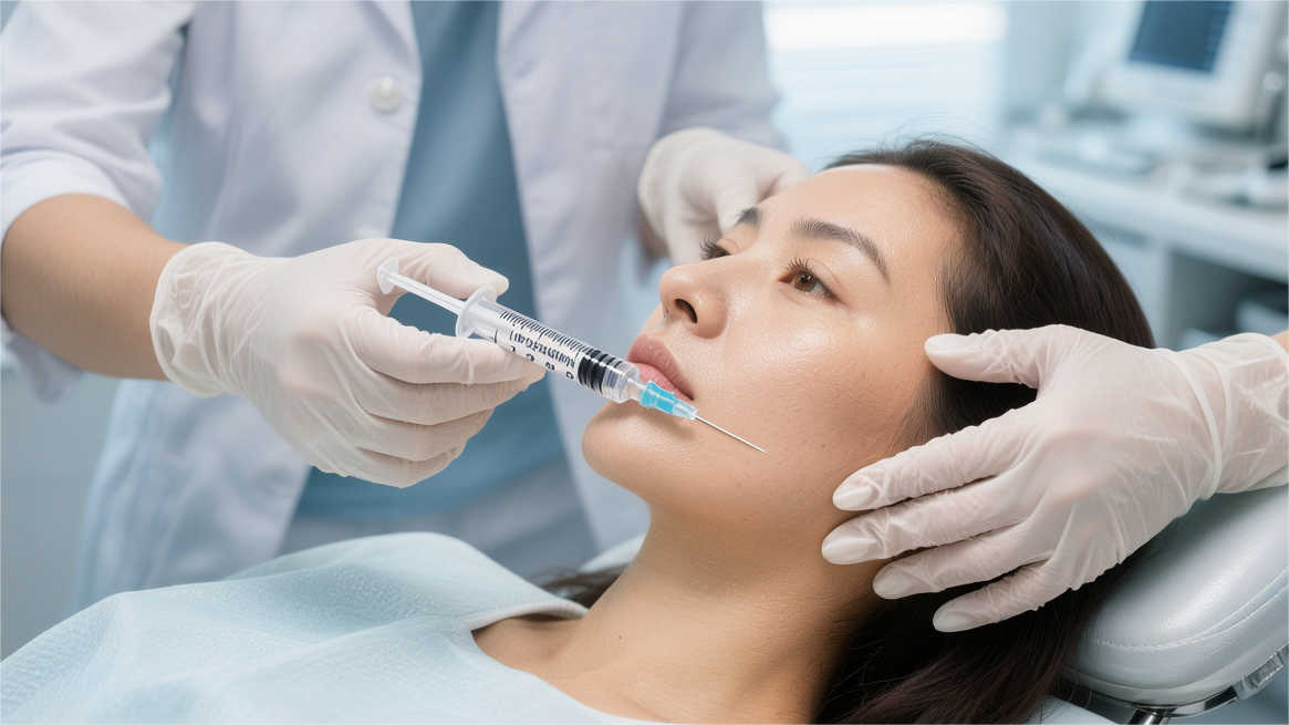
Clinical Performance Differences
The G’ value of Radiesse is usually around 1400 Pa, while the G’ values of most mainstream high-viscoelasticity hyaluronic acid fillers are distributed between 400 to 600 Pa. In the jaw area, the skin is affected by masseter muscle activity, platysma tension, and gravity; the material needs extreme hardness to maintain contour clarity. Once in the subcutaneous layer, Radiesse acts as an excellent scaffold, establishing stable support points at the mandibular ramus and mandibular angle much like artificial bone. Hyaluronic acid (HA), being relatively soft, is prone to deformation or spreading to surrounding tissues when subjected to frequent muscle compression, causing sharp lines to become blurred. Hyaluronic acid is highly hydrophilic and can absorb hundreds or even thousands of times its own weight in water. In jawline sculpting, this characteristic often leads to mild edema or a sense of “heaviness” in the injection area post-surgery, making the face look bloated and less sharp. In contrast, the water absorption rate of Radiesse is nearly zero. The CaHA microspheres and CMC gel in its composition do not have chemical structures that attract water; the volume post-injection is the final visual effect. Clinical observations show that the tissue expansion rate of subjects using Radiesse within 48 hours post-surgery is over 65% lower than those using hyaluronic acid.
| Physical Parameters and Clinical Performance | Radiesse (CaHA) | High Molecular Weight HA | Clinical Visual Difference |
|---|---|---|---|
| Storage Modulus (G’) | ~1400 Pa | 400 – 600 Pa | Radiesse has stronger support, less prone to collapse |
| Hydrophilicity | Extremely Low | Extremely High | HA easily leads to tissue bloating |
| Intratissue Cohesion | High | Medium | Radiesse has a clearer boundary feel |
| Immediate Deformation Rate | < 5% | 15% – 25% | Radiesse maintains designed shape better |
| Projection Height Maintenance | Long-term stable | Fluctuates with water metabolism | Radiesse contours are more profound |
Since the jawline is a high-activity area, material migration is a factor affecting long-term results. In a 3D imaging tracking study of 120 Western subjects, the average migration distance of Radiesse 12 months after injection was less than 1.2 mm. The high cohesion of CaHA microspheres creates a physical interlocking with the surrounding tissue. By comparison, some HA products may move downward along the fascia layer due to insufficient internal cohesion and the effect of gravity, causing the area intended for lifting to instead increase the load below the jaw. This physical characteristic of Radiesse, staying highly localized at the injection site, provides high predictability when reconstructing the mandibular angle and extending the length of the mandibular ramus. To achieve the same lifting height and contour clarity, Radiesse typically requires 25% to 30% less material than hyaluronic acid. The volume formed by CaHA microspheres under the skin is more solid and does not rely on tissue hydration to maintain height. In clinical practice, physicians can use fewer milliliters to outline sharp lines on the edge of the jawbone. This efficient projection capability reduces the burden of total injection volume and also lowers the sensation of facial tightness caused by overfilling. According to the clinical guidelines of the American Society for Dermatologic Surgery (ASDS), CaHA is the preferred non-surgical material for deep skeletal scaffold reconstruction.
- Compression Resistance Test: In experiments simulating skin pressure, the height compression ratio of Radiesse was only 8% when subjected to 10kPa of pressure, while the compression ratio for HA generally exceeded 22%. The jawline sculpted by Radiesse lasts longer during daily behaviors like side-sleeping or resting your chin on your hand.
- Palpation Sensation: After the healing period, the touch of Radiesse is highly integrated with the edge of the jawbone, making it almost impossible to distinguish the material boundary. HA may produce a noticeable “jelly-like” feel or foreign body bulge in some individuals with thin skin.
- Tyndall Effect: Because Radiesse is an opaque milky-white preparation, it does not produce blue shadows under light or in shallow injections like transparent HA. This gives it an aesthetic advantage in refining delicate parts of the jawline.
- Long-term Projection Durability: Vectra 3D imaging data shows that 18 months after injection, the projection height retention rate of Radiesse at the mandibular ramus still reached 68%, significantly higher than similar soft tissue fillers.
The clinical performance of Radiesse is also reflected in its secondary optimization of skin texture. While lifting the contour, due to the biological reaction caused by CaHA, the skin roughness of the jaw area decreased by an average of 18% at 6 months post-injection. This performance of increasing tissue density from the inside is something HA does not possess. HA primarily produces a transient hydrating effect through hyaluronic acid receptors, whereas Radiesse uses physical occupancy and biochemical induction to make originally loose jaw skin thicker and tighter. Clinical follow-ups found that this tissue-thickening effect is particularly evident in subjects over 50, as it can mask bone loss and mandibular atrophy caused by aging.
| Evaluation Item (12 Months Later) | Radiesse Performance Data | Competitor Comparison (HA) | Clinical Observation Conclusion |
|---|---|---|---|
| Jawline Clarity Score | 4.2 / 5.0 | 3.1 / 5.0 | Based on physician blind assessment |
| Tissue Migration Rate | < 2% | 8% – 12% | Superior long-term aesthetic stability |
| Additional Injection Requirement | 20% of initial volume | 50%+ of initial volume | Lower maintenance cost |
| Subject Perceived Tightness | Improved 85% | Improved 62% | Subjective satisfaction survey |
In cases of mandibular angle hypertrophy or excessive soft tissue, the clinical advantages of Radiesse are further expanded. It does not possess water-absorbing expansion characteristics, allowing it to produce a visual contraction effect by strengthening the longitudinal lines of the jawbone without increasing the width of the lower-middle face. When treating Jowls (sagging caused by fat accumulation in the jaw area), Radiesse creates a backward and upward mechanical pull by establishing strong anchoring forces behind the mandibular ramus and at the mandibular angle, improving the sagging in the front. As the CMC gel is absorbed, CaHA microspheres are gradually replaced by autologous collagen; this process is smooth and progressive. Clinically, sudden volume reduction or local depressions, seen with some other fillers, are rarely observed. This stable transition period allows users to feel a relatively stable lifting state throughout the 18-month cycle. According to records in the European Clinical Research Database, due to its unique physicochemical properties, Radiesse consistently maintains a clinical satisfaction level above 90% in reshaping jawline contours.
Effect Maintenance Cycle
Radiesse filler contains 30% Calcium Hydroxylapatite (CaHA) microspheres and 70% Carboxymethyl Cellulose (CMC) gel carrier. In the first 12 weeks post-injection, the jawline lifting effect is primarily provided by the CMC gel. This gel has an extremely high physical occupancy capability, immediately replenishing volume loss caused by bone resorption or soft tissue sagging. Clinical data shows that the volume retention rate of CMC gel in subcutaneous tissue remains near 100% in the first month. However, since CMC is a soluble polysaccharide, the body’s macrophages and physiological metabolic system will gradually decompose it. Experimental observations record that the metabolic half-life of CMC is approximately 8 to 10 weeks.
Clinical studies have observed that while the CMC gel is being absorbed, CaHA microspheres begin to function as a biological scaffold. At 4 weeks post-injection, preliminary fibrin deposition can already be detected around the microspheres.
As the CMC gel gradually disappears, the second phase of the maintenance cycle—the “biological remodeling phase”—formally begins. This phase typically occurs between 3 months to 9 months post-injection. The diameter of CaHA microspheres is precisely distributed between 25-45 microns, a size that is not easily swallowed by macrophages but instead becomes a substrate for fibroblast attachment. Fibroblasts are stimulated by mechanical tension on the surface of the microspheres, inducing the secretion of endogenous Type I collagen. According to data from Western dermatological research, at 6 months post-injection, the collagen density in the local tissue increased by over 150% compared to the baseline. These new collagen fiber bundles form a dense network around the microspheres, effectively taking over from the metabolized CMC gel.
In a long-term follow-up of a group of Western subjects aged 45 to 60, 3D imaging analysis at 9 months post-injection showed that the mandibular angle definition retention rate remained at 82% of the initial effect.
Entering the stable period of 9 months to 15 months, the jawline lifting effect exhibits extremely strong biomechanical stability. At this time, although the CaHA microspheres begin a slow enzymatic degradation into calcium and phosphate ions, the induced extracellular matrix (ECM) has completely matured. In addition to collagen, a large amount of Elastin and proteoglycans were found in histological sections. The increase in elastin allows the jaw skin to show better rebound performance when subjected to facial muscle compression or gravitational pull, reducing the risk of material migration under dynamic expressions. Clinical physical parameter tests show that the tissue’s storage modulus (G’) remains high during this stage, effectively resisting jawline skin sagging. Unlike regular HA, which causes contour blurring around 12 months due to water-absorption expansion and disappearance, Radiesse maintains a physical line with bony tension, which is anatomically closer to a youthful jawline contour.
According to a European multicenter randomized controlled trial, subjects’ satisfaction with jawline clarity at 14 months post-injection remained around 4.5 / 5.0, and fewer than 15% of subjects indicated a need for minor volume replenishment.
At the end of the maintenance cycle, from 18 months to 24 months, the CaHA microspheres have basically completed their metabolic mission. Although the filler itself has disappeared, the autologous tissue structure induced by it does not collapse immediately. These new collagen fiber bundles have a long biological half-life and can typically last for 24 months or even longer. This long-term tissue remodeling effect significantly extends the interval between treatments. In clinical practice, to maintain the best visual effect, physicians usually recommend a touch-up at 15 to 18 months. The required injection dose at this time is usually only 40% to 50% of the initial dose because the autologous biological scaffold has laid a solid structural foundation.
Long-term follow-up data indicates that the Type I collagen induced by CaHA has a highly ordered tissue arrangement. This orderliness is the underlying physical guarantee for maintaining long-term jawline tightness and preventing tissue from sagging toward the neck.
Technique
Jawline reshaping typically requires 1.5ml to 3.0ml of Radiesse per session. Clinically, 22G or 25G are often chosen for fan-shaped placement in the deep subcutaneous layer, with each linear path injecting approximately 0.1ml. At the Mandibular Angle, a 0.2ml supraperiosteal bolus injection is performed at a 30° entry angle. Utilizing the product’s high elastic modulus of 1411 Pa to provide anti-compression support, immediate jaw contour clarity is achieved.
Injection Tool Selection
The storage modulus (G’) of Radiesse is as high as 1411 Pa, and the viscosity is approximately 450,000 cP; this high-viscoelasticity material produces much higher resistance during injection than regular hyaluronic acid. Using a 27G for deep bolus injection is a common clinical choice. These are typically 12mm to 13mm long, allowing them to precisely reach the periosteal surface at the mandibular angle. At the Mandibular Angle, physicians usually adopt a 0.1ml to 0.2ml small bolus injection method, establishing support points directly on the periosteum with the; this method leverages the material’s high support capability to simulate bone contour, thereby increasing side-profile clarity. Due to the narrow internal diameter of the 27G, the operator needs to apply a force of about 20N to 30N when pushing undiluted Radiesse. This requires the to have excellent Luer-lock performance to prevent the from detaching under immense pressure.
| Tool Type | Common Gauge | Length | Suggested Injection Layer | Single Point/Line Dose |
|---|---|---|---|---|
| 27G | 12mm – 13mm | Supraperiosteal | 0.05ml – 0.2ml | |
| 22G | 50mm – 70mm | Deep Subdermal | 0.1ml – 0.15ml | |
| 25G | 38mm – 50mm | Subcutaneous | 0.05ml – 0.1ml | |
| Trocar | 21G | 4mm – 6mm | Used only to establish entry point | N/A |
For laying along the Mandibular Border, 22G or 25G are the mainstream tool choice. The inner diameter of a 22G is approximately 0.41mm, whereas the inner diameter of a 25G is only 0.26mm. According to Poiseuille’s Law, fluid resistance is inversely proportional to the fourth power of the lumen radius; therefore, resistance is significantly lower when pushing high-viscosity Radiesse with a 22G, helping physicians control the discharge speed more evenly. In practice, lengths are usually between 50mm to 70mm, allowing the physician to cover almost the entire jawline from a single entry point below the mandibular angle or lateral to the chin. The round-tip design of the can bypass the Facial Artery and facial vein located near the Mandibular Notch when passing through tissue. The average diameter of the facial artery is approximately 2.5mm, and the tip diameter of a 22G is about 0.7mm; this size difference plus the mechanical properties of the blunt tip reduce the incidence of embolization caused by accidental vessel puncture. In the deep subcutaneous tissue, the is used for linear retrograde injection in a fan-shaped or linear manner, releasing approximately 0.1ml of agent per path to form a scaffold-like arrangement.
| Technical Parameter Dimension | 27G | 22G | 25G |
|---|---|---|---|
| Tissue Trauma Degree | Higher (multiple punctures) | Extremely Low (single entry) | Lower (requires force control) |
| Injection Pressure (N) | 25 – 40 | 10 – 15 | 18 – 25 |
| Layer Precision | Extremely High (Periosteal) | Medium (Subcutaneous spreading) | High (Shallow fat/Subcutaneous) |
| Bruising Risk | ~15% – 20% | Less than 5% | ~5% – 8% |
| Recommended Population | Underdeveloped mandibular angle | Skin laxity requiring lifting | Thin soft tissue for refinement |
When it involves 1:1 or higher dilution ratios (Hyperdiluted Radiesse), the choice of injection tools expands further. During the dilution process, the CaHA microspheres are fully integrated with saline or lidocaine by pushing back and forth between two over 20 times, at which point the mixture’s viscosity drops significantly. For this diluted material, a 25G is the preferred balance between operational flexibility and injection smoothness. When performing large-area skin tightening in the Submandibular region, using a 25G for grid-like injection in the superficial subcutaneous layer allows 30% CaHA microspheres to be more diffusely distributed, thereby inducing Type I collagen production over a broader area. Experimental data shows that this diffuse distribution can increase dermal thickness by an average of about 20% after 3 months. At this time, using an overly thin 30G is not recommended, as the narrow lumen may cause microsphere accumulation or even blockage. For handling the entry point, the physician will first use a 21G to puncture the skin vertically to establish a temporary drainage hole, then slowly place the. While moving the, the operator can feel the tip’s position through palpation to ensure it remains in the correct anatomical layer, avoiding important structures like the Parotid Gland. Because injection punctures the nerve endings in the dermis multiple times, it produces a pain index (VAS score) usually between 4 to 6 points, whereas the, with its sliding characteristics and local infiltration anesthesia, often reduces the VAS score to below 2 points. In the Mental Foramen area in front of the jawbone, where nerves are densely distributed, using a can effectively avoid temporary numbness caused by nerve damage. Clinical observations found that using a 22G for unilateral jawline lifting, completing a 1.5ml injection takes an average of 5 to 8 minutes, whereas using a for multi-point injection takes over 12 minutes, and subsequent tissue swelling from multiple bleeding points can obscure immediate contour results.
Tissue Injection Layers
When performing jawline carving, the tissue injection layers for Radiesse are primarily focused on the Supraperiosteal and Deep Subdermal layers. The periosteal surface at the Mandibular Angle is the preferred site for establishing support points; the tip typically needs to pass through the skin, superficial fat, platysma, and the lower edge of the masseter before reaching the periosteum. At this depth, tissue compliance is low; because Radiesse has a high elastic modulus (G’) of 1411 Pa, it can form a stable hemispherical bolus on the bone surface. The diameter of this bolus is usually controlled between 3mm to 5mm, with a single-point injection dose of 0.1ml to 0.2ml. Establishing this mechanical support on the periosteum effectively compensates for bone resorption caused by age, visually extending and strengthening the lines of the mandibular ramus. The safety of injection in this layer is high because major nerve distributions, such as the marginal mandibular branch of the facial nerve, are usually located in shallower soft tissue layers; deep injection reduces interference with these nerve functions. From the middle to the front of the jawline (the area between the mandibular angle and the mental foramen), the injection layer moves up to the deep subcutaneous tissue. This is located deep to the dermis and between the Superficial Musculoaponeurotic System (SMAS), with thickness varying by individual, usually 2mm to 4mm. In this layer, physicians use a 22G for linear retrograde injection, releasing a dose of approximately 0.05ml/cm per path. The high viscosity of Radiesse (approximately 450,000 cP) shows extreme in-situ stability and will not easily spread. The linear arrangement in the deep subcutaneous layer acts as a flexible biological scaffold, and the 30% Calcium Hydroxylapatite (CaHA) particles in its composition can immediately generate mechanical tension on the surrounding tissue. This physical stretching activates fibroblasts, inducing them to continuously synthesize new Type I collagen within 4 to 24 weeks. Experimental data indicates that CaHA particles in this layer trigger a local tissue thickness increase of about 15% to 20% after 3 months, manifesting as improved tightness of the jaw contour rather than simple volume filling.
| Injection Layer Type | Anatomical Depth (mm) | Common Injection Technique | Material Property Performance | Expected Tissue Reaction |
|---|---|---|---|---|
| Supraperiosteal | 4.0 – 7.0 | Bolus Injection | Provides extremely high mechanical support, high compression resistance | Simulates bony prominence, strengthens mandibular angle |
| Deep Subdermal | 2.0 – 4.0 | Linear Retrograde / Fan-shaped Placement | Forms structural scaffold, high stability | Generates vector lifting force, tightens loose skin |
| Superficial Subdermal | 1.0 – 1.5 | Diluted Injection | Reduced viscosity, even distribution | Induces collagen and elastic fiber regeneration |
For cases with thin skin or obvious jawline skin laxity (Jowl Deformity), clinicians often use a 1:1 or 1:2 dilution ratio to place Radiesse at the junction of the superficial subcutaneous layer and the dermis. The diameter of CaHA microspheres is between 25 to 45 microns, a size precisely recognizable by fibroblasts. In the diluted state, the distribution density of microspheres in the subcutaneous layer is approximately 100 to 200 per square millimeter. Through this diffuse distribution, CaHA can more broadly contact tissue gaps. Histological studies have found that at 90 days post-injection, Type III collagen in this layer gradually transforms into Type I collagen, while elastic fiber content also increases significantly. During operation, physicians lay a grid-like pattern along the neck area below the lower edge of the jawbone; the coverage area of a single session can reach 20cm² to 40cm², with the total volume of diluted solution typically between 3.0ml to 4.5ml. The anatomical structure of the jawline area is complex, and the connection of different layers determines the continuity of the profile. In the Mandibular Notch area in front of the jawbone, the tissue structure becomes relatively thin; this is where the Facial Artery crosses the jawbone. At this point, the injection layer must be strictly maintained in the superficial subcutaneous layer, and deep supraperiosteal injection is strictly prohibited to avoid pressure on the vessel. Usually in this area, physicians reduce the single-point dose by 50%, turning instead to multi-pass low-volume spreading techniques. Through this layer switching, the transition is smoothed from deep support at the mandibular angle to structural reinforcement in the middle, finally extending to fine sculpting at the chin. Data shows that this multi-layer combined injection method scores about 25% higher in patient satisfaction assessments 6 months post-surgery compared to single-layer injection. The sense of definition in the jawline comes from the tight adhesion between the skin and the underlying bone; the precise placement of Radiesse in different layers essentially re-establishes this physical connection, eliminating visual interruptions caused by sagging fat pads or loose skin.
- Periosteal Layer Points: Mandibular Angle, Pogonion (chin midline), posterior border of the mandibular ramus.
- Subcutaneous Layer Points: Full line of the Inferior Border, Pre-jowl sulcus.
- Diluted Layer Points: Supra-platysmal, neck area below the jawbone.
The dose distribution for each layer typically follows a 4:4:2 ratio. That is, 40% of the dose is used for deep periosteal support, 40% for vector lifting in the deep subcutaneous layer, and the remaining 20% for superficial skin texture optimization. When treating male jawlines, the periosteal layer proportion increases to 60% to pursue a wider, more angular visual effect. For females, the subcutaneous spreading proportion is increased for a softer, smoother line transition. This quantitative management based on anatomical layers allows a single 1.5ml session of agent to exert a visual improvement effect equivalent to 3.0ml of HA. CaHA microspheres can maintain a metabolic cycle of 12 to 18 months in these layers, during which the generated autologous tissue hyperplasia gradually replaces the absorbed carrier gel, achieving long-term contour stabilization. 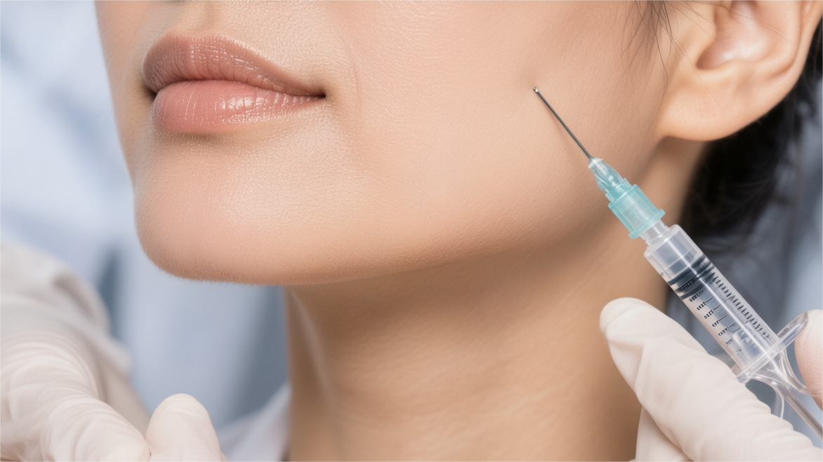
Injection Path
A primary entry point is usually chosen about 1cm to 1.5cm posterior to the Mandibular Angle, using a 21G to pre-puncture the skin and form a channel. Through this single entry point, a 50mm or 70mm 22G can cover a wide area from the mandibular angle to the Mandibular Body. The first path is usually along the Inferior Border, keeping the tip in the deep subcutaneous tissue and using Linear Threading, with a single push volume controlled between 0.1ml to 0.15ml. The function of this long path is to establish a continuous physical boundary, evenly arranging 30% Calcium Hydroxylapatite (CaHA) particles above the bone edge. Clinical data shows that the support band formed on this continuous path can immediately improve jawline clarity through the mechanical tension it generates.
The injection path must bypass the turn of the Facial Artery at the mandibular notch. The should slow down when passing through this area, using its rounded tip to push the vessel aside rather than penetrating it.
| Path Number | Target Anatomical Region | Injection Depth | Dose per Path | Coverage Length |
|---|---|---|---|---|
| Path 1 | Inferior Border | Deep Subcutaneous | 0.1ml – 0.2ml | 40mm – 60mm |
| Path 2 | Ascending Ramus | Supraperiosteal / Deep Subcutaneous | 0.05ml – 0.1ml | 20mm – 30mm |
| Path 3 | Pre-jowl Sulcus | Deep Subcutaneous | 0.1ml | 15mm – 20mm |
| Path 4 | Vectoring Zone | Mid-subcutaneous | 0.05ml x 3 | 30mm – 50mm |
For strengthening the mandibular angle area, the injection path usually presents a perpendicular L-shaped layout. In addition to the path forward along the jawline, a path distributed upward along the posterior border of the Ascending Ramus is required. The length of this path is generally 20mm to 30mm, using a small dose (about 0.05ml) for retrograde injection. Through these two perpendicular paths, a right-angled support structure can be built at the mandibular angle, significantly increasing the angularity of the profile. For male seekers, an additional 0.1ml to 0.2ml supraperiosteal bolus injection is usually added at the intersection of these two paths to further widen the visual width of the mandibular angle. The high-viscosity property of Radiesse ensures that the agent on these paths will not displace due to masseter muscle movement after injection. During the operation, the physician needs to palpate the tip’s position with the other hand to ensure the path always stays within the preset anatomical plane, avoiding entry into the parotid gland parenchyma or too shallowly into the dermis.
Fanning techniques are widely applied at the mandibular angle turn. With the entry point as the axis, lay 3 to 5 paths radiantly forward and upward, maintaining an angle of 15 to 20 degrees between each path.
Path planning for the middle of the jawline needs to consider volume compensation for the Pre-jowl Sulcus. Usually, a 25G is used for lateral entry, with the path ending posterior to the Mental Foramen. The dose per path is reduced to 0.05ml, filling the depression through multi-layer spreading rather than single-point large-volume filling. This path design allows the jawline to transition smoothly from the mandibular angle to the Chin, eliminating visual gaps formed by fat sagging. In the chin area, injection paths typically radiate from the center to both sides, with each path about 15mm long. Through a standard 0.2ml dose distribution, chin projection or extension can be achieved, perfecting the entire jawline proportions anatomically.
| Path Design Strategy | Applicable Population Characteristics | Path Density | Suggested Dilution Ratio | Expected Clinical Effect |
|---|---|---|---|---|
| High-Density Linear Placement | Severe skin laxity, blurred jawline | One path every 5mm | 1: 0.3 (Standard) | Strong physical support and immediate lift |
| Medium-Density Fan Vector | Mild to moderate sagging, seeking contour clarity | One path every 15 degrees | 1: 0.5 | Side-profile optimization and tissue tightening |
| Low-Density Diffuse Grid | Skin texture improvement, preventative anti-aging | 1cm x 1cm grid | 1: 1 (High dilution) | Induces large-area collagen regeneration |
When treating skin tightening in the Submandibular Area, the injection path changes to a Cross-hatching layout. At this time, Radiesse diluted 1:1 is typically used, with a 22G or 25G entering both horizontally and vertically in the superficial subcutaneous layer. This path design is not intended to change the contour shape, but to allow CaHA microspheres to form a uniform plane beneath the skin. The push volume for each path is extremely low, only 0.02ml/cm, to ensure microspheres do not clump together. Through this grid path, CaHA particles can cover approximately 30 square centimeters of the neck and submandibular area. Histological studies show that in areas covered by this path, the distribution density of Type I collagen shows a significant increase in uniformity 4 months post-surgery, with skin rebound data increasing by an average of over 25%.
During the retrograde injection process, maintaining constant withdrawal speed and push pressure is crucial. The withdrawal speed is suggested at 5mm to 10mm per second to ensure that the agent distribution error per unit length is less than 10%.
After completing all path injections, physical path integration is an indispensable step. The physician will perform smooth pressure along the direction of the injection paths, with a force of about 5N to 10N, to flatten the linear agent and integrate it with the surrounding connective tissue. Due to the extremely high in-situ stability of Radiesse, this immediate sculpting can lock in the contour changes brought by the injection paths. Data shows that patients with standardized path designs have a maintenance time for jawline lifting effects about 4 to 6 months longer than those with random point-like injections.