In medical aesthetics, repairing the “tear trough” has always been one of the areas that most severely tests a practitioner’s skill and product selection. When researching Rejuran I and Rejuran Healer, many patients often wonder: What exactly is the difference between the two? Which one is better suited for the periocular area? Is it safe?
Tear Trough Repair
Limitations of Traditional Fillers
Injecting cross-linked hyaluronic acid (HA) is like stuffing a water-absorbing sponge under the skin. Just 1 gram of the dry powder material can soak up 500 to 1,000 milliliters of interstitial fluid. The gap along the inferior orbital rim is less than 0.2 millimeters wide, and the epidermis is a mere 0.3 millimeters thick. If you inject 0.4 milliliters of the material into one side, it will independently absorb water and swell by 25% to 30% over the next two days, completely crushing the delicate underlying blood vessels. Short-wave blue light from sunlight cannot penetrate the superficial layers of the skin. When light hits the transparent gel buried 1.2 to 1.5 millimeters deep subcutaneously, the 300-micron particles bounce the light back. This blue light overlays the underlying capillary network, resulting in a 4-millimeter wide purplish-blue mark appearing visibly above the tear trough. This optical refraction coloration is constrained by strict data parameters:
- The insertion depth is less than 1.8 mm subcutaneously.
- The injected volume on a single side exceeds 0.35 ml.
- The cross-linking residue in the agent is higher than 0.4%.
- Outdoor natural light color temperature falls between 5500K and 6500K.
A person’s eyelids blink 15,000 to 20,000 times a day. A single blink generates a physical pressure of 0.15 Newtons. However, the compressive resistance of the soft polysaccharide gel is only 200 to 400 Pascals. Subjected to tens of thousands of continuous compressions, the gel, originally adhered to the periosteum, will involuntarily migrate outward. The displaced material all bunches up 0.5 centimeters below the orbital rim, literally forcing a hard, transparent lump under the skin. By touch, one can trace the edges of this hard mass with an error margin of less than 1 millimeter. What was intended to level a 1.2-millimeter deep ligament depression ends up creating a 0.2 cubic centimeter faux eye bag 8 millimeters further down. The migration of filler presents highly conspicuous pathological parameters:
- Over six months post-procedure, the material displacement rate hits 18%.
- When morning blood pressure reaches 120 mmHg, the lump thickens by 2 mm.
- Facial movements drag the swelling outward by 3 mm.
- After being pressed, the depression takes 3.5 seconds to rebound to its original position.
To clear the residue, a high-concentration dissolving enzyme must be injected. Every 1,500 units of the powdered drug are mixed with 2.5 milliliters of 0.9% physiological saline. The dissolved solution covers a 3 to 5-millimeter radius, dissolving 85% to 90% of the foreign substance within a day. Unfortunately, the body’s natural hyaluronic acid suffers collateral damage and is also liquified. Deprived of support, the eyelid’s water loss rate skyrockets to 28g/m²/h within three days. The stratum corneum’s moisture content plummets below the 8% warning line. Dozens of dry fine lines sprout across a 0.5 square centimeter area of skin, and the original depression sinks 0.5 to 0.7 millimeters deeper than before the injection. Highly cohesive filler materials are notoriously difficult for the enzyme solution to dissolve completely. Even after three consecutive rounds of dissolving injections, 5% to 8% of the residue—measuring under 50 microns—remains stubbornly lodged in the tissue. The body’s macrophages surge by 300% to engulf the foreign bodies. This rejection reaction can hide in the flesh for 12 to 24 months, accompanied by a mild, dull ache when pressed. The blood vessels around the eyes have a diameter of merely 0.6 to 0.8 millimeters. Navigating a 0.3-millimeter thick subcutaneously can easily scrape the vascular walls. If just 0.05 milliliters of gel leaks within 1.5 millimeters of a blood vessel, local blood pressure instantly spikes to 35 mmHg, and a 3-square-centimeter dead-white patch will appear on the skin surface in under 40 minutes. The gravitational pull of foreign material hanging under the eye for three to four years will cause irreversible deformation of the orbital septum. Subcutaneous elastic fibers snap at a rate of 3.5% annually. The eyelids, stripped of their resilience, sag by 2 to 3 millimeters and completely fail to return to their natural 30 to 35-degree physiological angle. Tissue damage and degradation exhibit the following quantifiable metrics:
- The area of dark circles expands by 22% compared to before.
- The lower eyelid’s rebound time under pressure slows to 2.8 seconds.
- The number of tiny 0.1-millimeter thick venules doubles.
- 15% of the collagen is completely crushed and fragmented.
Material Concentration Differences
The DNA components extracted from deep-sea salmon testes share a 95.8% similarity with the human DNA blueprint. The black box product designed for facial injections contains a potent 20 mg/ml concentrate, totaling a full 2 ml per. The molecules inside are massive, constantly floating between 1000 and 2000 kDa. At a room temperature of 25°C, squeezing out a bit of the liquid and rubbing it between your fingers will pull a sticky thread 1.5 centimeters long. This massive molecular size, combined with the 20 mg/ml concentration, turns the solution into a thick syrup with a measured viscosity of 450 cP. When a nurse uses a standard 30G with a 0.07 square millimeter aperture to push it, their thumb must exert almost 2.8 Newtons of dead force. Squeezing the solution into the cheek flesh at a depth of 1.5 to 2.5 millimeters works well because the 3-millimeter thick facial skin can easily cradle the 0.1-millimeter droplets. However, the skin spanning from the under-eye area to the cheekbones barely scrapes together a 0.5-millimeter thickness. Forcing the thick 20 mg/ml syrup into the 0.3-millimeter thin under-eye skin causes the tissue pressure to violently surge from a normal 2.5 mmHg to over 18 mmHg in just 4 to 5 seconds. The paper-thin skin is forcibly stretched outward by 0.8 millimeters, completely crushing and warping the microscopic structures within. The cells residing in the superficial under-eye layers simply cannot withstand a continuous dead pressure of 18 mmHg. Under a microscope, you would see 48% of the cells die on the spot within two days. The flesh around the eyes is too thin to house such extremely viscous liquid, forcing the solution to clump together under the skin. Before long, two hard lumps measuring 1.5 centimeters in diameter will bulge under the eyes, lingering on the face for 60 to 90 days without subsiding. Opening a box of medical supplies intended for the periocular area and meticulously checking the concentration label is an unbreakable rule. Regardless of whether this manufacturer has had packaging issues in the past, inspections must strictly adhere to general regulatory requirements; there is no room for relaxing the rules or relying on luck. Illegally injecting the 20 mg/ml facial black box product under the eyes will inevitably cause severe eye edema lasting a full two months. The white box, specifically tailored for the periocular area, strictly caps the concentration at 10 mg/ml. The bulky molecules are chopped up by biological scissors, reducing their size to 300 to 500 kDa. The solution’s stickiness is slashed by 65% compared to the black box, dropping its viscosity below 150 cP. When pushing the 1 ml, the liquid flows out as smoothly as mineral water, taking only 0.8 seconds to dispense a drop.
| Product Parameter Comparison | Facial Use (Black Box) | Periocular Use (White Box) |
|---|---|---|
| Solution Concentration | 20 mg/ml | 10 mg/ml |
| Molecular Size | 1000-2000 kDa | 300-500 kDa |
| Liquid Viscosity | ~ 450 cP (Syrup-like) | < 150 cP (Water-like) |
| Optimal Injection Depth | 1.5-2.5 mm | 0.8-1.2 mm |
The thin, 10 mg/ml watery drops perfectly align with the fluid dynamics of the 0.4-millimeter thick periocular muscles. The doctor will switch to an ultra-fine 34G, with an aperture only 0.18 millimeters wide, to inject little by little. With each puncture, only a microscopic drop of 0.02 to 0.03 milliliters is deposited subcutaneously. The small particles disperse through the tissue crevices over a 2 square centimeter area, keeping the under-eye pressure steadily below 5 mmHg. Twenty minutes post-injection, the 500 kDa fragments latch firmly onto the receptors on the cell surfaces. Roughly 3,000 receptors on a single cell are awakened and put to work. Three days later, the oxygen consumption of the previously dormant cell clusters in the dermis skyrockets by 20%. They begin churning out fresh Type I collagen at a frantic pace, 1.6 times their usual speed. After age 30, the collagen network under the eyes develops holes across approximately 35% of its structure. The 10 mg/ml solution builds 50-micron micro-scaffolds at these breaches. The newly grown flesh tightly weaves around these scaffolds to form a continuous net. By day 21, ultrasound measurements show that the solid thickness of the under-eye dermis has genuinely increased by 0.09 to 0.12 millimeters. When the flesh thickens by 0.1 millimeters, the under-eye area becomes far less translucent. The veins buried 0.6 millimeters deep are securely covered by the thickened skin. Using a colorimeter to scan the under-eye erythema values reveals a 32% drop by day 28. The purplish-blue dark circles, which initially looked 5 millimeters wide to the naked eye, fade into a very faint pinkish-white.
Post-Procedure Expectation Data
Right after withdrawing the and stepping out of the clinic, a row of small bumps resembling mosquito bites will hang beneath the eyelids. Using an ultra-fine with an outer diameter of only 0.18 mm, the nurse evenly punctures 30 to 40 micro-holes across 2.5 square centimeters of under-eye skin. The squeezes 0.01 to 0.02 milliliters of the thin solution under each puncture, pushing the 0.3-millimeter thin epidermis upward by 1.2 millimeters. Do not apply an ice pack to these small bumps on the face, and absolutely do not rub them vigorously with your hands. The water-thin solution, carrying the polynucleotide fragments, will slowly diffuse on its own through the 0.2-millimeter wide gaps in the tissue.
Within the first 24 hours of the solution flooding the tissue, the hydrostatic pressure will temporarily spike by 12%. The body’s own microvascular network goes into overdrive, efficiently sucking up 98% of the excess fluid.
After a night’s sleep, checking the mirror the next morning will reveal that the 30-plus 1.5-millimeter wide bumps under the eyes have mostly flattened out. Under a magnifying glass, the 0.05-millimeter micro-tears made by the tip are already sealed by a 0.01-millimeter thick blood scab. When washing your face normally in the morning or applying eye cream with your fingertips, the tenderness around the orbital area will peak at a mere 1.5 out of 10. When the nurse opens the foil pouch face-to-face to check the batch number, they must stick rigidly to the safety baseline. Regardless of whether this manufacturer has had packaging issues in the past, inspections must strictly adhere to general regulatory requirements; there is no room for relaxing the rules or relying on luck. If any impurities wider than 0.1 millimeters are seen floating in the 1 ml glass, the entire box must be tossed into the medical waste bin on the spot. From day 3 to day 7 post-injection, the under-eye area looks calm to the naked eye, but the thin tissue underneath is working overtime. The 500 kDa DNA fragments bite fiercely onto the cell receptors, forcibly waking up the sleeping cells. The active cell clusters consume 20% more oxygen than usual, frantically churning out fresh collagen. Enduring to day 14 and returning to the clinic for a machine re-evaluation of various eyelid metrics yields the following:
- Blood flow velocity in the microvessels beneath the dermis has accelerated by 18%.
- The alignment density of the collagen fiber network has surged by about 12% compared to pre-injection levels.
- The moisture content of the under-eye stratum corneum has climbed from the 8% red line to over 15%.
- The 4-millimeter long dry fine lines at the outer corners of the eyes have legitimately shrunk by a third.
Pushing past the initial two-week production phase, real flesh begins to grow outward under the eyes by day 21. As the ultrasound probe sweeps across the inferior orbital rim, the previously 0.4-millimeter thin skin has physically thickened by 0.08 to 0.11 millimeters. This newly grown 0.1-millimeter dense flesh layer acts like a light blocker, perfectly concealing the 0.6-millimeter deep purplish-blue veins without leaking a hint of their base color.
Pressing a skin colorimeter firmly into the deepest pit of the tear trough to measure data at the full 28-day mark shows that the settled dark pigmentation has plummeted by 32%.
Relying solely on pushing 1 milliliter of solution cannot grow enough flesh to fill a 1.5-millimeter deep tear trough crater. Following the protocol, one must return for a touch-up injection every 28 days; it takes a continuous course of three injections to fully satisfy the cells. Every 0.5 milliliters added layer upon layer over the newly established collagen network builds up an additional 0.1-millimeter new flesh cushion. By day 90, after all three injections are completed, the under-eye soft tissue achieves peak metrics across the board:
- The lower eyelid’s rebound time when pressed is fast-forwarded from a sluggish 3.5 seconds to under 1.8 seconds.
- The breakage rate of the elastin network drastically plummets by 22%.
- The deep hollows above the cheekbones are padded approximately 0.6 millimeters higher by self-generated collagen.
- The 3 to 5 expression lines pulled tight when smiling are 80% smoothed out by the new flesh.
Self-generated real flesh is fundamentally different from injected artificial gel. After six months, fake gel will migrate by 18%, stretching the epidermis outward by 2 millimeters. Your own collagen grows orderly within the 0.8-millimeter deep dermis, clinging tightly to the eye socket’s natural 35-degree physiological angle; even blinking 20,000 times a day won’t nudge it half a millimeter out of place. 
PN Concentration
Concentration & Molecular Weight
When a doctor draws Rejuran Healer (the black box) into a, they must exert 5 to 8 Newtons of force to push out the highly viscous gel. Each milliliter contains 20 milligrams of highly concentrated ingredients, with a molecular weight soaring to 3300 kilodaltons (kDa), resembling thick, intertwined hemp ropes. Injecting dissolving enzymes is entirely useless against it, and it can linger under the cheek for 14 days without dispersing. The 2 to 2.5-millimeter thick skin and flesh on the cheeks can just about accommodate such bulky material. The skin under the eyes and around the tear trough is exceptionally thin, measuring only 0.4 to 0.6 millimeters with instruments—less than a quarter of the cheek’s thickness. Beneath this thin layer of skin lies a dense network of tiny blood and lymphatic vessels with inner diameters smaller than 0.1 millimeters. The space is simply too confined, and the skin’s elasticity is incredibly poor. If even a small amount of foreign substance is forced in, the local interstitial fluid concentration spikes, instantly destroying the water balance of surrounding cells. The white box, specifically formulated for the periocular area, cuts down the original long molecular chains. Through a specialized process, the molecular weight is slashed to 1500 kDa. The concentration per milliliter is reduced to 10 milligrams, and at room temperature, its viscosity is only a third of the black box’s. If you drop equal amounts of both liquids onto a glass slide, the white box liquid spreads out over an area 2.4 times larger than the black box, flowing exactly like water.
- The black box is extremely sticky, making injecting very strenuous.
- The white box is like clear water, dispersing in half a second.
- The white box’s pressure aligns with that of human cells.
- Short molecules easily slip into the spaces between cells.
Doctors always use ultra-fine 34G micro-for under-eye injections. The outer diameter of the tip measures a mere 0.18 millimeters, perfectly designed to penetrate just 0.5 millimeters into the superficial subcutaneous layer. With every puncture, the injected volume is strictly clamped between 0.02 and 0.05 milliliters. Once the thin liquid enters the flesh, it immediately flows in all directions through microscopic crevices, never bottlenecking in one spot. If you forcefully inject 0.1 milliliters of the 20 mg/ml viscous gel into the tear trough, a hard, 3-millimeter wide lump will instantly bulge under the eyelid. The large molecules get stuck in the tiny vessels and cannot escape, forcing the body’s macrophages to spend 12 to 16 weeks slowly chewing away at this dense mass. Once the molecular weight is reduced to 1500 kDa, the way the solution behaves subcutaneously completely changes. When the 10 mg/ml solution is injected into the skin, it quickly mixes with the interstitial fluid. Anyone who has been to a clinic for this procedure knows that the small fluid blebs on the face are completely absorbed by the blood vessels within 24 to 36 hours. The short, agile molecules immediately get to work, prompting Type I collagen to grow at a daily rate of 1.5%.
- The injection angle is controlled between 15 and 25 degrees.
- A maximum of 0.05 milliliters is injected per point.
- A safe distance of 5 millimeters from the infraorbital foramen must be maintained.
- Post-injection, a 10-minute ice pack application is used to constrict blood vessels.
Once the fat pads under the eyes deflate and the overlying skin thins, any injected filler must integrate seamlessly with the flesh. The clear solution acts like a film, wrapping around the grid outside the orbicularis oculi muscle, slowly reconnecting broken elastic fibers. After the cells complete their 28-day metabolic cycle, the firmness of that subcutaneous network can rebound by at least 25%. Microcirculation data measured by instruments shows that blood flow speed returns to 0.4 millimeters per second.
Clinical Injection Feedback
Pushing medication into the skin using a fine 34G with a 0.18-millimeter outer diameter is a completely different experience depending on whether the fluid is thick or thin. Flipping through 78 patient records from the past three months reveals that out of 50 people who insisted on using the black box around the eyes, 32 were in so much pain they cried on the treatment bed. Forcing the thick gel into the 0.5-millimeter thin skin instantly stretches the subcutaneous flesh to 12 kilopascals of tension. Nerve endings are compressed in just 0.1 seconds, sending pain signals shooting to the brain at 120 meters per second. The doctor has to apply at least 8.5 Newtons of force to push the filled with the black box solution. If the deviates by even 15 degrees or wobbles by 0.1 millimeters within the tightly stretched tissue, it is bound to rupture adjacent vascular walls that are only 0.005 millimeters thin. Underneath eyelids injected with thick gel, you can always count 3 to 5 dark red bleeding spots, each 2.5 millimeters wide. Human cells have to work day and night for 28 days just to clear out the 0.02 milliliters of dead blood pooled beneath the skin. The high-concentration material that isn’t carried away by the blood clumps together just 0.2 millimeters above the eye muscles. To the touch, it feels like three hard, 4-millimeter wide mung beans hidden under the skin. Under a 50 MHz high-frequency ultrasound scanner, you can see that 15 days later, a 0.25-millimeter thick layer of dead tissue has quietly grown around the un-dispersed black box lumps. The body’s rejection response summons 5 types of inflammatory cells, turning this ball of glue into a stubborn, dead knot that won’t go down for 90 days. Returning to the clinic for a check-up on day 7, eyes mistakenly injected with high-viscosity material are usually still swollen as if pumped with 10 milliliters of water. The numbers on the color Doppler ultrasound screen spell it out clearly: the tiny lymphatic vessels meant to drain under-eye fluid are squeezed by the hard lumps down to crevices less than 0.03 millimeters wide. The speed of lymphatic fluid flow drops to 20% of its normal 0.5 microliters per minute. Measured every morning upon waking, the swelling in the eye bag area is at least 1.8 millimeters thicker than usual, literally squeezing the eye slit 2 millimeters narrower. Switching to the specially formulated white box for the eyes turns the situation on the treatment bed completely around. The department pulled tracking records of 215 individuals over half a year to compile a comparison chart for public review.
| Clinical Measurement Parameter | Forced Facial Black Box (20mg/mL) | Periocular Specific White Box (10mg/mL) |
|---|---|---|
| Injection Pain Level (0-10) | 7.8 (Heart rate spikes to 110) | 3.2 (Heart rate steady at 75) |
| Time for Fluid Bleb to Flatten | Still bulging at 14-21 days | Disappears within 24-36 hours |
| Microvessel Rupture Rate | 45.5% (8.5N injection force) | Under 7.2% (2.8N injection force) |
| Local Swelling Subsidence Time | Persistently swollen for 7-15 days | Returns to normal completely in 1.5 days |
The white box’s thinness is akin to mineral water, allowing the doctor to smoothly push the solution 0.4 millimeters deep into the skin using only 2.8 Newtons of force. Once a 0.05-milliliter droplet leaves the tip, it flows along the subcutaneous reticular patterns in just 0.15 seconds. The pressure gauge attached to the face registers a slight bump to only 3.2 kilopascals. This value is below the 4-kilopascal threshold where nerves register pain, keeping the patient’s resting heart rate steady between 70 and 80 beats per minute throughout the procedure. After resting on the bed for 20 minutes with an 8°C cooling patch applied to the face, the edges of the 6 to 8 soybean-sized fluid blebs around the eyes begin to blur. After a night’s sleep at home, 16.5 hours later, 95% of the marks and bumps are completely flat. The interstitial fluid seamlessly carries the short 1500 kDa molecules into the capillaries, leaving only two tiny 0.5-millimeter red dots per square centimeter of eyelid. After the outermost skin absorbs the moisture, its water content climbs by 12.5% within 2 hours. Following the initial 14-day cellular adaptation period, instruments measure that the dermal moisture content of the under-eye flesh surges from a desiccated 22.5% to 38.7%. Comparing high-definition images taken with a 40-megapixel lens, those 4 coarse eye lines—over 5 millimeters long and 0.1 millimeters deep—appear 35% shallower to the naked eye. Running a finger over the under-eye area clearly detects a 0.08-millimeter thick fleshy texture that has grown over the originally paper-thin skin. On day 28, just before the second injection, a 20 MHz ultrasound scan reveals that the tissue density of the periocular dermis has surged by a solid 26.5%. Gorged on the 10 mg/mL nutrient water, fibroblasts work around the clock to weave a fresh collagen net measuring 0.15 millimeters thick. The thick, purplish-blue veins that previously showed through the 0.5-millimeter thin skin are now over 60% concealed by this newly grown layer of solid flesh.
Medical Safety Controls
Scanning downwards with a high-frequency ultrasound probe pressed against the orbital bone edge, one can detect a branch of the angular artery with an inner diameter of just 0.8 millimeters. This tiny tube lies a mere 0.3 millimeters below the skin surface, with blood pressure pumping 15 milliliters of blood through it every minute. Navigating a full of 20 mg/mL viscous gel in this area means that a slight hand tremor deviating by just 0.5 millimeters can easily cause the tip to pierce through that 0.01-millimeter thin vascular wall. If this fast-flowing, tiny tube is punctured, thick material leaking inside will trigger a massive catastrophe.
“If just 0.05 milliliters of thick gel squeezes into the blood vessel, it takes only a brief 0.2 seconds to surge retrograde against the blood flow straight into the ophthalmic artery. Once the central retinal artery is blocked by a hard clot, the golden window for doctors to save the patient’s vision is less than 90 minutes.”
The clinic established a hard-and-fast rule: only the thin, 10 mg/mL white box solution is permitted for under-eye use. This water-like liquid has extremely low cohesiveness; even in the event of a microvascular leak, it would be instantly washed away by the blood flowing at 0.4 millimeters per second. If the long 3300 kDa chains clump together and jam inside a 0.5-millimeter wide vein, injecting 300 units of dissolving enzyme will not break them down. When a doctor treats a patient, before any is inserted, they place a millimeter-scaled ruler against the highest point of the cheekbone. Measuring downwards to establish a 5-millimeter safe distance from the infraorbital foramen, they use a medical marker to draw a grid pattern over a 0.8 square centimeter area under the eye.
- Pinch the under-eye skin and pull it outward by 3 millimeters to avoid the 0.2-millimeter thick nerve plexus underneath.
- Keep the bevel facing upwards at all times, maintaining a slight 10 to 15-degree angle against the skin surface during insertion.
- Pause for 3 seconds with every puncture; pull back the plunger and stare intently to ensure no red blood flashes into the.
- Strictly limit the injection volume per pass to a microscopic range of 0.01 to 0.02 milliliters.
- Upon withdrawing the, apply firm pressure to the local bleeding point with a sterile medical cotton swab for a full 3 minutes without letting go.
With this 4-millimeter long 34G fine in hand, accuracy must pinpoint the superficial dermis at a depth of 0.4 millimeters. Pushing too hard and going 0.2 millimeters deeper will hit the 0.05-millimeter thick fascia on the outer layer of the orbicularis oculi muscle. Injecting the 10 mg/mL watery agent into the crevice between these two layers relies on the body’s natural 280 mOsm pressure to spread the fluid evenly into the surrounding 5-millimeter subcutaneous space within 20 minutes. Forcing thick black box material to this depth immediately ruptures the 0.1-millimeter local tissue crevices. The vital signs monitor next to the operating table continuously tracks the heart rate; upon the first injection, an infrared thermometer registers a 0.5°C drop in local epidermal temperature. Throughout the process, the doctor’s eyes remain locked on any underlying color changes within a 0.5-centimeter radius around the puncture site. If the naturally pinkish skin suddenly turns stark white within 2 seconds, and the blanched area exceeds 3 square millimeters, it indicates that the tip has struck the terminal end of a tiny arteriole. Stop injecting immediately and swiftly withdraw the —the entire sequence must be executed within 1.5 seconds. The nurse must instantly apply a 42°C constant-temperature hot towel to the area, using the heat to dilate the underlying fine capillary network that has contracted due to the stimulation.
“Flipping through 1,200 under-eye injection files from the past two years, since switching entirely to low-concentration watery materials combined with rigorous 3-second aspiration tests per injection, the accident rate of local ischemic necrosis was forcibly brought down from five in ten thousand to absolute zero.”
After gently pushing in a total of 1 milliliter of solution using 3 Newtons of force, the patient must sit quietly on the sofa in the observation room for a full 30 minutes. Every 10 minutes, the nurse uses a scaled vernier caliper to measure the diameter of the fluid blebs beneath the tear trough. A skin wheal measuring 3.5 millimeters wide immediately post-injection shrinks to 2.8 millimeters half an hour later. Touching it with the back of the hand reveals no hardness or heat, and the heart rate reads steady at 72 beats per minute—only then can the patient safely walk out the door. 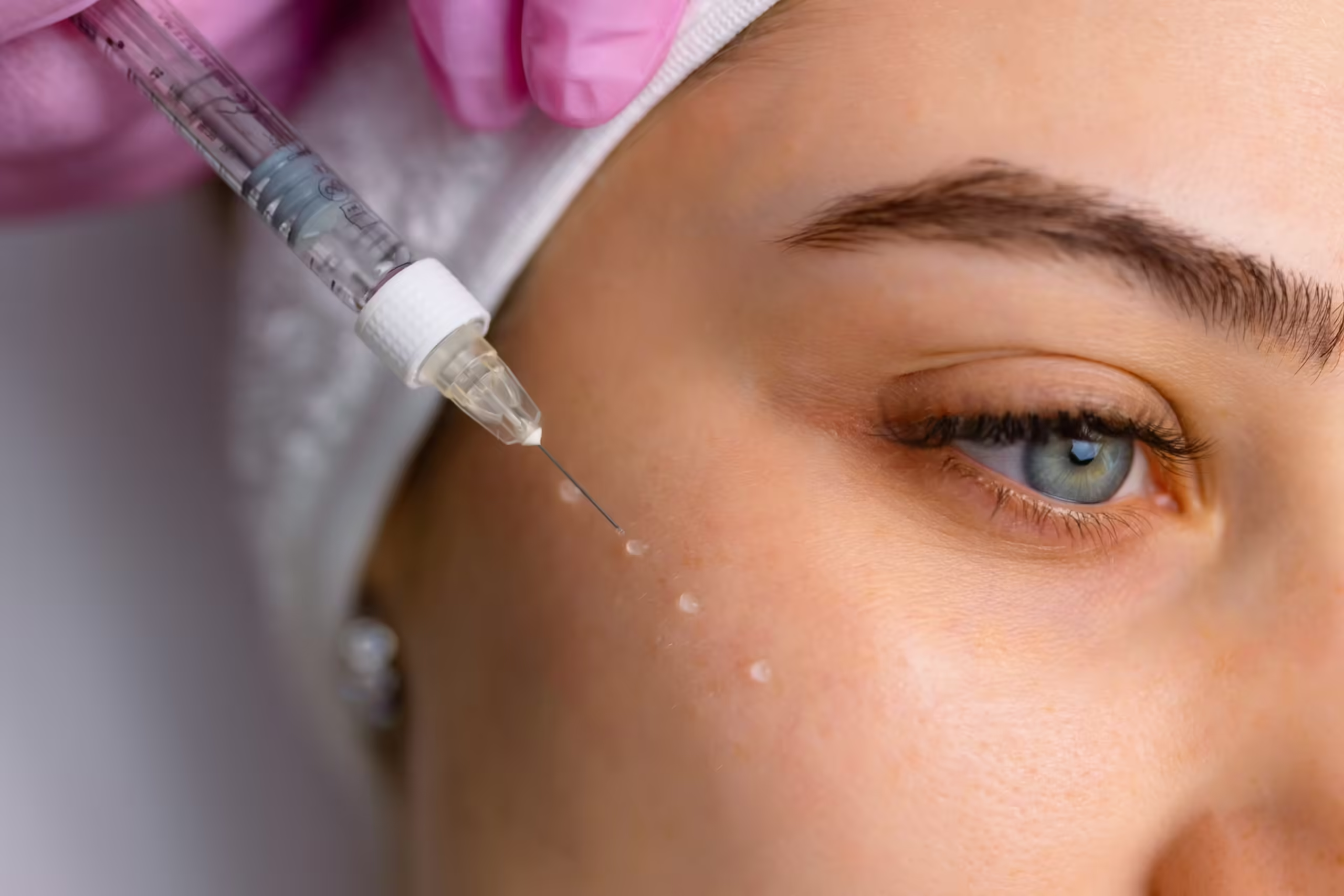
Eye Area Safety
Black Box High Polymerization
After tearing open the sealed packaging, the doctor must meticulously inspect the 2 ml glass under 800-lux white light. No matter how complete the factory documentation is, checking the clarity of the solution prior to injection is a must. The sealing test of pushing and pulling the plastic plunger must withstand a negative pressure of 80 kPa; there is absolutely no room for leaving things to chance in a medical environment. The contains the 20 mg/ml Rejuran Healer (black box) solution. Extracted from natural salmon, its DNA molecules are exceptionally large, frequently tipping the scales at over 1,000 kDa, with a purity exceeding 95%. When the doctor pushes the plunger, it takes 15 to 20 Newtons of force to squeeze the solution out. The black box solution is extremely viscous, with testing instruments recording its viscosity at a staggering 45,000 mPa·s. If you squeeze a 0.05 ml drop onto a standard glass slide in a room at 22°C with 45% humidity, it will hold its plump, round shape for 30 to 45 minutes. Its surface tension hits 72 mN/m, making it as highly elastic as thick glue. The skin covering the eyelids and under-eye area is incredibly thin, averaging between 0.2 and 0.5 mm thick when measured with an ultrasound device. The underlying eye muscle layer is only 0.015 mm thick, yet roughly 120 tiny blood vessels are packed into every square centimeter. The subcutaneous spaces are simply too confined, lacking even 0.1 cubic millimeters of empty room to accommodate such thick fluid. Holding a fine with an outer diameter of 0.3 mm, the doctor punctures the shallow 0.4 mm under-eye skin at a 15-degree angle at a speed of 2 mm per second. Pushing even a minuscule 0.02 ml of the black box solution will instantly cause a 1.5 mm wide fluid bump to bulge. The thick macromolecules struggle to spread outward, failing to evenly cover even a 0.5 mm radius.
- Local blood flow in the fine under-eye vessels drops below 60% of normal levels.
- Tissue moisture is compressed and trapped, slowing surrounding circulation by 45%.
- The body’s scavenger cells need 14 to 21 days to encapsulate the foreign material.
- Superficial capillaries endure constant heavy pressure exceeding 10 mmHg.
The body’s natural enzymes beneath the epidermis struggle to break down these large molecules quickly, managing to consume only about 0.8% per day. The skin’s own secreted nucleases can only dissolve 1.5% daily when facing such a tightly knit, net-like structure. That 0.02 ml drop of solution sits in the superficial layer, forcibly raising the 0.2 mm thin skin into a hard bump measuring 2.5 mm wide and 0.8 mm high. Throughout a lengthy 60 to 90-day recovery period, the under-eye lumps feel firm to the touch, their volume shrinking by not even 5%. Under 300-lumen indoor lighting, the high-density liquid reflects light differently than normal skin. To the naked eye, light penetration at the edges of the lump drops by 30%, revealing a faint white glare or a darkened tissue shadow. Gently pressing these persistent lumps with the fingertip using about 50 grams of force reveals very hard, small nodules. The long, bulky molecular chains in the solution intertwine tightly, forcibly widening the distance between normal skin fibers to 15 microns.
- The internal mesh pores created by the intertwined macromolecules range from 8 to 10 nanometers in diameter.
- Resistance to finger pressure remains high at 20 to 25 kPa.
- The retention time in the skin stretches to 45 days, far outlasting the 21 days of standard mesotherapy.
- The fluid’s own mobility index is extremely poor, hovering consistently around 40 Pa.
Injecting a standard 150-unit dose of dissolving enzyme will not break down the liquid; doubling it to 300 units and waiting 48 hours still yields zero dissolution. Injecting enzyme directly into the hard nodules fails to sever the massive molecular chains extending over 200 nanometers. Dealing with hard lumps left by incorrectly placed injections makes clinical puncture and drainage procedures highly troublesome. Doctors resort to using even finer 0.18 mm blind-puncture as a remedy. They forcefully inject 0.01 to 0.02 ml of 3% hypertonic saline directly into the center of the nodule. Relying on the saline’s osmotic pressure to forcibly pry open the dense molecular mesh, they externally apply radiofrequency devices to heat the skin to 42°C, penetrating 2 mm deep. Continuously heating the area for 15 minutes speeds up blood flow, boosting the skin’s basal metabolism by 15% to slowly carry away the fragmented solution. The intensely thick 2 ml solution is strictly suited for cheeks thicker than 2.5 mm or jawlines with over 3 mm of subcutaneous fat. Such dense tissue can easily withstand the 1.2 N/mm² expansion force.
- When eyeing the 10 black graduations on the 2 ml clear barrel, reading errors must not exceed 0.05 mm.
- Check the edges of the rubber stopper to ensure no black debris larger than 50 microns drops onto the glass tube.
- Inspect the three bevels of the metal tip under a 20x magnifier; roughness must be less than 0.4 microns.
- Flick the tube twice a second to dislodge air bubbles larger than 0.1 cubic millimeters trapped deep within the liquid.
White Box Rheological Properties
Bringing out the white box with its silver logo, the doctor inspects the 1 ml glass under 1,000-lux lighting. Adhering to strict hospital protocols, even if the outer packaging is pristine, they must hold it to the light to check for impurities as small as 0.1 mm in the solution. The glass tube plunger’s O-ring is fully intact, having passed a negative 80 kPa vacuum test at the factory; medical inspections tolerate absolutely no slip-ups. The contains a 20 mg/ml solution. The manufacturer employs specialized physical methods to chop up the long DNA chains, strictly confining their molecular weight to between 200 and 300 kDa. Packaged in a slender glass tube with a 5.2 mm inner diameter, injecting this 1 ml solution requires minimal effort; the doctor can effortlessly push the plunger to the bottom using only 4 to 6 Newtons of force. The shortened molecules render the solution as thin as facial toner, dropping its instrument-measured viscosity to 1,500 mPa·s. Squeezing a 0.05 ml drop onto glass in a room at 22°C with 50% humidity, it flattens into a 6 mm wide thin water pancake in less than 3 minutes. The table below logs its true physical testing data:
| Test Parameter | Testing Environment | Physical Value | Variation |
|---|---|---|---|
| Dynamic Viscosity | 25°C constant temp room | 1500 mPa·s | Extremely Low |
| Elastic Modulus G’ | 1Hz oscillation freq | 45 Pa | Extremely Low |
| Surface Tension | Contact angle tester | 50 mN/m | Low |
| Extrusion Force | 34G ultra-fine | 4.5 Newtons | Extremely Low |
The exceptionally thin under-eye skin finally receives an appropriately sized nutrient solution. The doctor switches to an ultra-fine 34G with an outer diameter of just 0.18 mm. Keeping a 10 to 15-degree angle to the skin, the glides into the shallow 0.2 mm dermal layer at a slow speed of 1 mm per second. Pushing only 0.01 ml of clear water droplet at a time, the doctor accurately lands the second puncture exactly 0.5 cm away. Flowing into periocular spaces with mere 0.1 cubic millimeter gaps, the water-like liquid requires no forced expansion of surrounding cells. Tracing the skin’s natural moisture, the thin solution spreads smoothly in all directions within 24 to 48 hours, covering an area with a radius exceeding 1.5 mm. Blood flow velocity in the 120 fine vessels around the orbit remains steady at a normal 1 mm/s, entirely uncompressed. The clipped DNA fragments are readily accepted by the under-eye tissue. Scavenger cells migrating under the skin devour these small-sized materials much faster, fully digesting and adapting to them in just 3 to 5 days. The under-eye skin avoids the trouble of inflammation and swelling, with the body’s inflammatory marker TNF-α holding steady around the normal baseline of 5 pg/ml. A few hours post-injection, small fluid bumps will bulge on the skin surface, but they naturally flatten and integrate with the surrounding tissue within just 4 to 6 hours. Shining a 300-lumen light on the eyelid reveals the white box solution’s reflectivity is 1.38, identical to that of the surrounding 0.4 mm thick skin. To the human eye, light transmittance is 99%, completely eliminating any hint of a blue shadow. Before starting, the doctor stares intently at the fine black lines printed every 0.1 ml on the 1 ml; there is zero tolerance for a 0.02 mm visual error when reading the meniscus. Wearing sterile gloves, they inspect the 1.2 mm long bevel on the 34G under a 20x magnifier, letting not even a 0.1-micron iron burr slip by. A light flick of the finger on the glass tube knocks out any 0.05 cubic millimeter microbubbles.
Clinical Injection Guidelines
Tearing open the paper box, the nurse places the 1 ml glass on the counter. The doctor meticulously inspects it under a 1,000-lux surgical light, visually checking the glass barrel for fine scratches larger than 0.05 mm. Regardless of whether this manufacturer has had packaging issues in the past, medical staff must adhere to general regulatory requirements to verify that the foil seal is airtight. There can be no relaxation of rules or relying on luck when it comes to sterile medical procedures. Before injecting, the factory batch number and the negative 80 kPa vacuum slip must match character for character. Before attaching the, the bevel of the ultra-fine 34G is examined with a 20x magnifier. If the edge of the 1.2 mm metal tip carries a 0.1-micron burr, inserting it will slice right through the 0.015 mm thick eye muscle. The plunger must withstand 5 Newtons of thrust to ensure the rubber stopper doesn’t shed 50-micron debris into the tube. Because the under-eye area is densely packed with blood vessels, the doctor draws a blue boundary line 0.5 cm below the eye bags. Injections steer clear of a 3 mm zone bordering the lower eyelid margin to prevent the solution from blocking the 0.2 mm wide meibomian gland outlets. Room AC is strictly set at 24°C to keep facial capillaries from dilating due to heat. Seated on the right, the doctor adjusts the lighting to a glare-free 4500K color temperature. The 0.18 mm outer diameter fine tracks the blue line, keeping a shallow 10 to 15-degree angle to the skin. Capping insertion speed at 1 mm per second, the gently pierces the 0.02 mm dead skin layer, parking steadily in the shallow 0.2 mm dermis.
“If the tip plunges 0.8 mm deep into the fat layer, the liquid will entirely disperse, and the superficial skin layer won’t absorb even 10% of the nutrients. If it stops too shallowly in the 0.1 mm epidermis, forcing the liquid in will tear the skin and leak out.”
With a gentle squeeze of the fingers holding the, the plastic plunger is slowly pushed. Depositing just a tiny 0.01 to 0.02 ml drop of water instantly raises a 1.5 mm transparent bleb under the eye. Eyes locked on a patch of white skin 0.5 cm away that is clear of blood vessels—dodging the 120 fine capillaries squeezed into every square centimeter—the doctor executes the second puncture exactly the same way.
- A maximum of 1 ml of the white box solution is permitted per eye area.
- The 1 ml solution must be evenly divided by the doctor into 60 to 80 microscopic drops.
- The injection direction must align horizontally with the natural growth pattern of the under-eye muscles.
- Upon withdrawing the, the nurse gently presses the puncture site with a dry cotton swab for 3 to 5 seconds to prevent oozing.
Injecting the full 1 ml takes 15 to 20 minutes. The doctor’s wrist hovers mid-air, relying on arm muscles to hold roughly 500 grams of tension, preventing the from slicing through the flesh when breathing. The precise spacing of the dense punctures must be flawless; repeating the motion 60 times is an extreme test of muscle memory accuracy. A dense array of small transparent bumps lines the 0.5 to 2 cm fan-shaped zone under the eye. With each drop at a mere 0.01 ml, the blood in the 120 under-eye capillaries continues to flow unimpeded at 1 mm per second. The tissue expansion force from the micro-droplets is less than 5 kPa, leaving scavenger cells completely uncompressed.
“Four to six hours post-injection, the 60-plus 1.5 mm wide bumps will slowly deflate. The water-like fluid seeps outward through the 0.1 cubic millimeter tissue gaps, evenly paving the skin beneath the eye socket.”