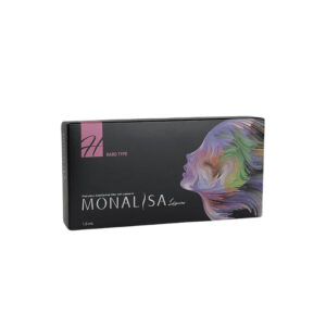
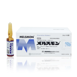
The key difference between Rejuran I and Rejuran S lies in their viscosity and injection depth.
Rejuran I has a thin, fluid texture and is injected superficially into the superficial dermis at a depth of about 1 mm.
It is specifically designed for the delicate under-eye area, targeting fine lines and dark circles while effectively minimizing the risk of subcutaneous nodules.
Rejuran S, by contrast, has a much higher viscosity and must be injected more deeply into the mid-to-deep dermis, directly at the base of acne scars. Its strong structural support allows it to physically fill depressed scars while promoting tissue repair.
Under-Eye Repair
The skin around the eyes is only about 0.5 mm thick and contains very few sebaceous glands. Rejuran I was specifically designed for this anatomical area. Its polynucleotide (PN) concentration is maintained at 20 mg/ml, and its molecular characteristics have been specially optimized to create an ultra-low-viscosity, water-like solution. It is administered with an ultra-fine 34G micro-, which has an outer diameter of just 0.18 mm.
Dermal Thickening & Concealment
The tissue beneath the lower eyelid is only 0.4 to 0.6 mm thick. The cheek is roughly three times thicker. Beneath that thin layer sits a dense network of purple-toned veins. These local microvessels are about 0.1 to 0.2 mm in diameter. When the skin is too thin to mask the color of those vessels, incoming light is refracted in a way that makes the under-eye area appear blue or purple to the naked eye. To thicken this thin layer, doctors use an ultra-fine 34G micro-. Its outer diameter is just 0.18 mm, even finer than a human hair. The contains polynucleotides (PN) at a concentration of 20 mg/ml. This material is derived from wild salmon sperm DNA, and its pH is kept between 7.2 and 7.4, which closely matches the body’s natural environment. The injection angle is extremely precise, held strictly between 10 and 15 degrees. The tip is placed only 1.0 to 1.5 mm below the epidermis in the superficial layer. The doctor’s hand must remain exceptionally steady, and the amount delivered with each injection is very small, usually controlled at 0.01 to 0.02 ml.
- Injection depth: 1.0-1.5 mm
- angle: 10°-15°
- Volume per injection: 0.01-0.02 ml
- Spacing between entry points: 3-4 mm
Once the solution is placed into the superficial dermis, it begins interacting with fibroblasts over the first 72 hours. Fibroblasts act like the skin’s production factories. Once stimulated, their proliferation rate increases by 20%. These cells then begin producing new collagen and elastin to fill the previously depleted spaces under the skin. Laboratory measurements show the changes most clearly. Secretion of Type I collagen, which provides structural support, rises by 30%. The proportion of Type III collagen, which supports flexibility, also increases to 15%. Binding sites for hyaluronic acid multiply, and average local tissue hydration rises by 12%. After three full treatment sessions spaced 21 days apart, ultrasound measurements of the under-eye area show a real increase in thickness. Skin that originally measured about 0.5 mm thick increases to around 0.61 mm. Overall under-eye thickness rises by an average of 15% to 22%. That additional 0.11 mm of tissue acts like a natural concealer built by the body itself. When light hits the under-eye area again, the thicker tissue blocks more of its penetration. Much of the light is refracted earlier by the dense network of newly formed collagen fibers, so fewer photons reach the underlying vessel network. As a result, the purple tone of the veins becomes much less visible. It is not only the vessels being concealed. Local blood flow around the eyes also improves. Components in the formula promote mild dilation of the microvasculature, widening the vessel diameter and increasing blood flow. High-resolution infrared thermography shows that by Week 8, average skin temperature around the eyes rises by 0.3 to 0.5°C. As temperature rises, circulation speeds up as well.
- Melanin index: minimal change, less than 2%
- Erythema intensity: reduced by 18%
- Water loss rate: reduced by 10%
- Roughness (Ra) value: improved by 25%
Dark, deoxygenated blood that tends to stagnate in the capillary corners is cleared more efficiently once circulation improves. By Week 12, the erythema index used to measure vascular color has fallen by a full 18%. This treatment does not do much for pigment-based dark circles, however. Melanin reduction stays below 2%. Its main role is to thicken the skin and improve the appearance of visible vascular discoloration. Under the microscope, the collagen structure in the reticular dermis also changes. Fibers that were once fragmented and disorganized become re-aligned into a compact, wave-like pattern. The undulating contour at the dermal-epidermal junction becomes more pronounced. The epidermis and dermis adhere more closely, and the contact surface between them increases. Nutrient exchange becomes more efficient, and the under-eye skin gains more resilience. Even when laughing or making exaggerated facial expressions, the under-eye area is less likely to fold into fixed, static dry lines.
Matrix Remodeling & Fine Line Smoothing
The skin under the eyes is only about 0.5 mm thick, yet the eyes blink tens of thousands of times every day. Within that delicate support network, water and natural hyaluronic acid are quietly lost at a rate of about 1% per year. What was once a full, cushioned structure gradually deflates, like a ball losing air. As a result, dry lines with depths ranging from 0.1 to 0.5 mm begin to form on the surface. [Image of extracellular matrix skin structure] The injected material is polynucleotide (PN), a long-chain molecule derived from salmon DNA. Its molecular weight ranges from 1000 to 2000 kDa, and its structure contains a large number of hydrophilic groups. It behaves like a super-absorbent sponge, with 1 gram of material able to bind more than 10 grams of water. Once a solution at 20 mg/ml is injected into the thin under-eye skin, it immediately starts absorbing surrounding tissue fluid and swelling. At a depth of about 1.5 mm below the epidermis, that swelling physically separates the collapsed tissue planes and re-creates three-dimensional space.
- Injection depth: 1.5 mm below the epidermis
- Product concentration: 20 mg/ml
- Water-binding ratio: more than 1:10
- Initial visible response: within the first 48 hours
The change is easy to see on high-frequency ultrasound. In less than two days, absolute hydration within the invisible subdermal structure increases by 14% to 18%. Static dry lines that were originally as deep as 0.2 mm are pushed upward by the water-loaded macromolecules beneath them, making the surface look smoother. But hydration alone only explains the first few days of plumpness. Long-term smoothing of fine lines depends on the “factories” beneath the skin. These large molecular chains specifically target fibroblasts, almost like setting off an alarm that forces dormant cells back into action. A cell division cycle that normally takes 28 days is shortened to 15 to 20 days. Once those cellular factories begin operating at full speed, new structural material starts to accumulate under the skin:
- Type I collagen, which provides structural firmness, rises by 30% by Week 4
- Type III collagen, which supports elasticity, climbs to more than 14%
- Natural hyaluronic acid binding sites increase dramatically
- By Week 12, the subdermal support structure becomes 0.11 mm thicker
| VISIA Tracking Metric | Baseline | Week 8 | Week 16 |
|---|---|---|---|
| Average crow’s feet depth (mm) | 0.35 | 0.22 | 0.18 |
| Local skin hydration (%) | 28.5 | 42.1 | 39.4 |
| Surface roughness (Ra) | 12.4 | 9.8 | 8.5 |
| Rebound time after finger pressure (s) | 2.1 | 1.4 | 1.2 |
On 3D skin texture analysis, the roughness parameter that reflects surface unevenness drops by 20.9% at Week 8. Pressing the under-eye skin with a fingertip also shows a visible difference. A loose lower eyelid that once took 2.1 seconds to rebound now returns in just 1.4 seconds. The deep grooves formed at the outer corners of the eyes are essentially structural voids. Wrinkles that were originally 0.35 mm deep are gradually filled by newly formed collagen. By Week 16, that depth is reduced to 0.18 mm. When light is shone from the side, the deep shadow that once appeared under the fold becomes only a faint line. The injected material itself does not remain in the body for very long. Within two to three weeks, it is broken down by enzymes. But nearby cells do not discard those fragments as waste. Instead, they use them as nutrients. These breakdown products become raw material for ATP synthesis, continuing to support metabolism in the under-eye area. Even after the material is gone, the collagen network it stimulated remains beneath the skin. That newly formed mesh can continue supporting the dermis for 6 to 9 months. Once hydration starts to decline again, a small maintenance treatment using a 2.5 mm ultra-fine blunt along the muscle plane can quickly restore moisture to above the 40% safety threshold. This water-like solution also behaves in a very controlled way once injected:
- Injection force is kept at a very low 0.5 N
- Only 0.02 ml is dispersed smoothly every 0.5 seconds
- The risk of nodules or lumps is below 0.01%
- No surrounding erythema develops even within the nearby 10 mm vascular network
Because the formula contains no crosslinking agent, its fluidity remains extremely high, allowing it to spread naturally through tissue planes. The highly sensitive immune cells in the under-eye area show excellent tolerance to this salmon DNA-derived material and do not mount an inflammatory response. Without visible redness or swelling, the epidermis quietly becomes better supported from below. The rebuilt foundation anchors the loose epidermis more firmly to the dermis underneath. Mechanical testing shows that shear resistance between the two layers nearly doubles compared with baseline. Even when makeup remover pads are rubbed across the eye area, that side-to-side pulling is far less likely to distort the previously fragile lower eyelid skin. New elastic fibers are also formed like tiny springs embedded within the collagen mesh. Under the microscope, the number of these spring-like fibers rises from fewer than 150 per square millimeter to about 280. This dense array of microscopic “springs” helps distribute the pressure generated by the muscles around the eyes. When smiling, the force of contraction is no longer concentrated enough to compress the epidermis into permanent static folds.
Formula Design & Edema Control
The drainage channels under the eyes—the lymphatic capillaries—are extremely delicate. Their internal diameter is usually only 10 to 50 µm. Even a small amount of pressure can impair their function. If a thick, paste-like filler is placed into skin that is less than 0.6 mm thick, it can easily compress these tiny drainage pathways and block tissue fluid outflow. Once that drainage is blocked, the under-eye area slowly swells like a reservoir filling with water. Edema caused by this kind of physical obstruction can persist for 3 to 6 months and may still not fully resolve. To avoid burdening such a vulnerable area, the laboratory uses an extreme level of refinement when preparing the polynucleotide (PN).
Special enzymes are used like microscopic scissors to cut the long molecular chains into much shorter fragments. The finished solution flows almost like purified water.
On rheological testing, its viscosity stays tightly within an ultra-low range of 20 to 30 cP. For comparison, conventional materials used to build nasal height often have viscosities of 100,000 cP or higher, behaving more like semi-solid glue. When this water-like solution is placed about 0.5 mm beneath the skin, it does not collect into a dense lump the way a gel would. Instead, it spreads rapidly through the spaces between cells, much like water soaking into sand.
- entry diameter: 0.18 mm
- Injection spacing: 3-5 mm
- Volume per injection: 0.01-0.05 ml
- Spread radius beneath the skin: about 2.5 mm
Using a 34G ultra-fine with an outer diameter of only 0.18 mm, the doctor delivers the product in numerous tiny deposits. This highly distributed technique, combined with the solution’s exceptional fluidity, minimizes tissue pressure in the thin under-eye area. Immediately after treatment, the skin may show small white bumps about 2 to 3 mm wide. These bumps are not firm nodules. They are simply tiny collections of very dilute fluid. Over the next few hours, osmotic exchange begins, and tissue fluid rapidly mixes with and absorbs the solution. Follow-up records show that in more than 85% of cases, these small white wheals are fully absorbed by the surrounding tissue within 24 to 48 hours. The hair-thin lymphatic channels under the eyes are not subjected to prolonged external pressure, so after a few hours of reduced function, drainage returns to normal.
The formula is completely free of chemical crosslinking agents. Its optical refractive index is almost identical to that of the body’s own tissue fluid.
Even when this very dilute solution is placed at an extremely superficial depth of just 0.2 mm, incoming blue light in the 400 to 500 nm wavelength range is not distorted into an unnatural bluish cast. Doctors can even place the tip less than 1 mm from the lash line and inject very slowly. This area near the lashes has almost no fat pad for cushioning. It consists only of a very thin orbicularis oculi muscle layer and a dense capillary network. If a conventional thick material were injected there, the next morning the eye could swell so much it would barely open. By contrast, this ultra-low-viscosity PN solution slips around capillary walls that are only about 5 µm thick.
- Safe distance from the lash line: less than 1.0 mm
- Chemical residue after degradation: 0%
- Optical refraction deviation from normal: less than 0.05
- Risk of delayed swelling months later: below 0.1%
The specific gravity of this water-based solution is precisely adjusted to 1.005 to 1.015, which closely matches the density of plasma in human tissue fluid. Because the densities match so closely, the solution does not sink or migrate downward under gravity after injection. Heavier, gel-like fillers often have a higher density. Since the muscles around the eyes move constantly with blinking, materials that are too heavy can shift downward along the fascial plane and create a false tear trough below the eye bag. By contrast, the ultra-light PN solution remains confined within its original 2.5 mm diffusion zone from the moment it is injected. The long molecular chains in the solution are gradually broken down by tissue nucleases over 14 to 21 days. As each portion degrades, the volume it occupied is replaced seamlessly by the body’s own newly formed granulation tissue. The entire process occurs without generating any acidic byproducts that might irritate the tissue. Throughout this period, the pH of the under-eye microenvironment stays within a healthy range of 7.35 to 7.45. Because there is no chemical residue left behind, the treatment avoids the recurring swelling that can appear months later with other materials. Some patients who receive conventional products may find that after staying up late or eating salty food, the under-eye area swells dramatically even six months later. That kind of delayed puffiness usually happens because residual crosslinking agents continue to irritate the surrounding lymphatic network. A water-based PN formula avoids that problem from the start. By Day 30, measured degradation exceeds 98%. A patient treated on a Friday afternoon may go home with tiny mosquito-bite-like bumps around the eyes, but by Sunday night, the 20 mg/ml reparative solution has already dissolved quietly into the thin 0.5 mm under-eye skin. 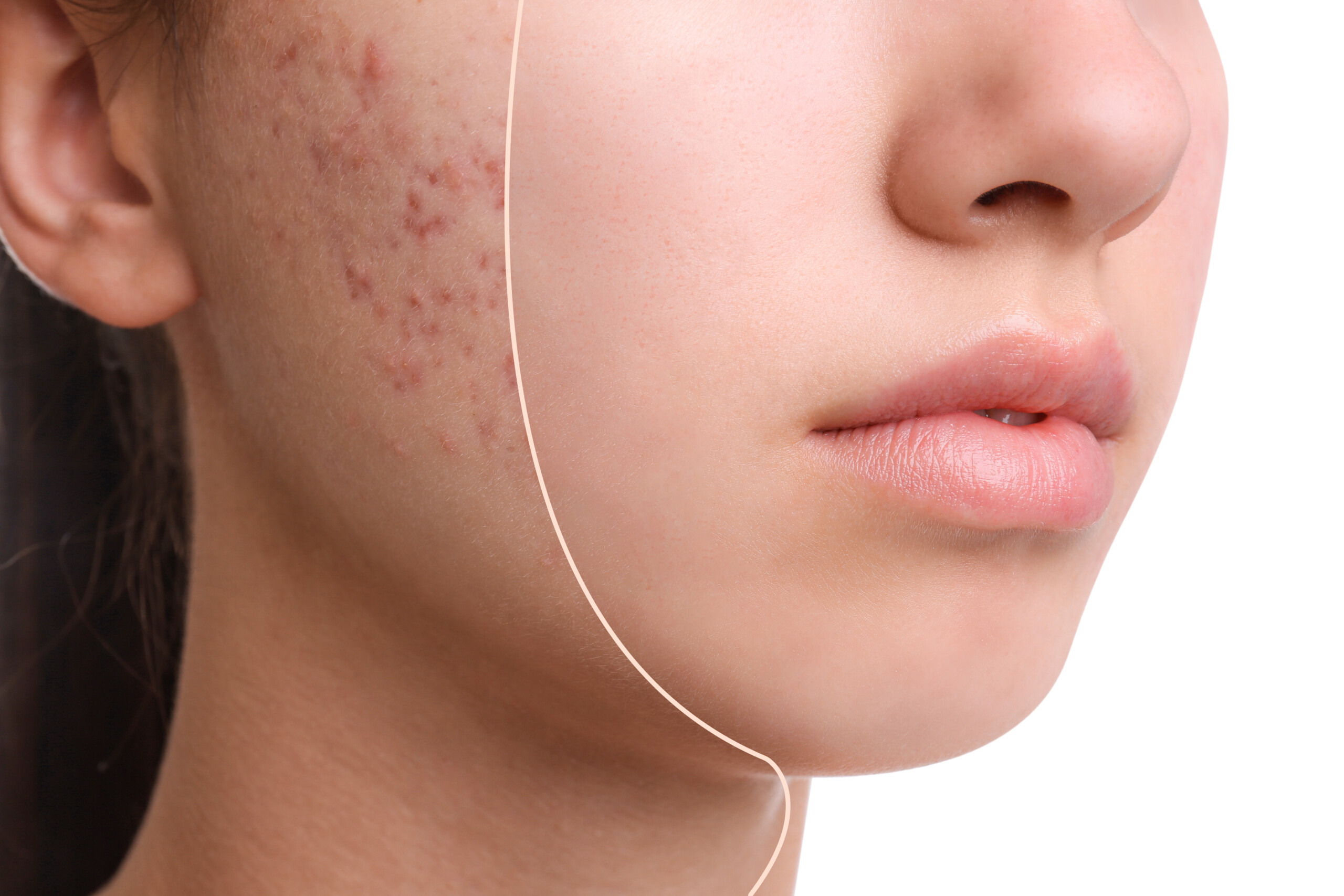
Acne Scars
Atrophic acne scars are three-dimensional tissue depressions caused by collagen loss within the dermis. Rejuran S contains 20 mg/mL of polynucleotides (PN) and has a viscosity of 1,000,000 mPa·s (cP), far higher than the standard versions. It is injected into the mid-to-lower dermis at a depth of 1.5 to 2.0 mm to create structural volume support. The PN molecules have an in vivo half-life of about 21 days. During this period, they attract fibroblasts and increase local Type I collagen synthesis by 20% to 30%. A single treatment typically requires 1 to 2 mL, and 3 to 4 sessions are usually needed at 4-week intervals before visible smoothing can be seen.
Viscosity & Concentration Differences
Both Rejuran I (white box) and Rejuran S (black box) contain polynucleotides (PN) extracted from wild salmon. The real difference lies in their concentration and viscosity. The white-box formula is as light as a watery toner, while the black-box formula is closer to a dense malt syrup. The PN concentration in the white-box formula is set at 10 mg/mL. In European and American clinics, when treating the delicate under-eye area, doctors usually inject only 0.05 mL per point. Because the solution is extremely fluid, it can be absorbed quickly by eyelid skin that is only 0.5 mm thick, making nodules much less likely. The black-box formula increases the concentration to 20 mg/mL. A single 1 mL contains twice the number of long-chain DNA molecules. This high-density composition is specifically designed for depressed acne scars with severe dermal damage. Doubling the concentration dramatically changes the product’s physical behavior. In medicine, this is measured by dynamic viscosity. The white-box formula has a viscosity of less than 100,000 mPa·s (cP). The black-box formula reaches 1,000,000 mPa·s. When injected 1.5 mm deep into the lower dermis, it stays firmly in place like a supportive silicone pad, physically lifting the base of the depressed scar.
- The white-box formula works with an ultra-fine 34G with an outer diameter of just 0.18 mm, and injects very smoothly
- The black-box formula requires a thicker 30G or 27G , with an outer diameter of around 0.3 mm
- Injecting the black-box formula requires more than 15 N of pressure
The two products behave very differently once placed beneath the skin. When the white-box formula is injected into the superficial dermis, it spreads outward within 24 hours, with a diffusion radius of up to 1.5 cm. Its role is broad, even distribution for fine-line hydration. The black-box formula shows almost no movement once injected. Instrument monitoring shows that 14 days after injection, the gel has shifted by less than 0.2 cm. It remains anchored directly beneath boxcar-type scars, maintaining an upward lifting force. The length of the molecular chains also affects degradation speed. In the body, macrophages take about 14 days to clear the thinner white-box formula. Under-eye maintenance treatments are therefore often scheduled every two weeks. The black-box formula contains much longer molecular chains and is much more resistant to breakdown. Enzymatic degradation takes 21 to 28 days. That gives fibroblasts nearly a full month to remain within the scar base and gradually synthesize Type I collagen.
- The white-box formula has a shorter half-life, so follow-up treatments are usually scheduled every 14 days
- The black-box formula resists enzymatic breakdown more effectively, extending its residence time to 28 days
- In anti-aging clinics across Europe and the US, rolling scar treatments are typically scheduled at 4-week intervals
Differences in viscosity also determine the injection depth. The white-box formula can only be placed superficially, around 0.5 mm below the skin. Doctors keep the bevel facing upward, gently lifting the skin to create tiny 2 mm papules. When dealing with ice pick scars that extend 2 mm or more into the tissue, the black-box formula must be injected into the mid-to-deep dermis at 1.5 to 2.5 mm. Doctors often use a retrograde injection technique, withdrawing the while slowly filling the subdermal cavity with the viscous material. This strong resistance during injection produces a very different pain profile. White-box injections feel more like mosquito bites, and patients can usually tolerate them easily after 20 minutes of topical numbing cream. Injecting the extremely viscous black-box formula through a 27G creates a much stronger tearing sensation beneath the skin. Overseas doctors often apply a 10% compounded lidocaine gel and leave it on for more than 40 minutes before treatment. The production process also explains the dramatic difference in viscosity. To make the fluid white-box formula, the factory processes wild salmon DNA at 40°C, cutting the long strands into shorter chains. To produce the 1,000,000 mPa·s black-box formula, the process uses 80°C heat together with 2.5 atmospheres of pressure. This high-heat, high-pressure method preserves a stronger three-dimensional network.
- The white-box formula undergoes 40°C low-temperature cleavage, resulting in a liquid texture with excellent flow
- The black-box formula undergoes 80°C high-temperature, high-pressure compression, resulting in a solid-like network structure
- These two different manufacturing routes give each product a clearly defined role in tissue repair
There is also a marked difference in hydrophilicity. PN molecules naturally attract water. The white-box formula expands by 15% within 12 hours after injection, creating a hydrated, luminous effect in the under-eye area. The denser network structure of the black-box formula absorbs water much more slowly. After 48 hours, its volume expansion is only 5%. This slow water uptake helps prevent acute swelling in scar tissue and leaves more room for dermal regeneration. The under-eye area contains a dense capillary network, which accelerates clearance of the white-box formula. With its lower concentration of 10 mg/mL, it is metabolized quickly through the vascular network and usually fades within one to two weeks. By contrast, fibrotic tissue at the base of acne scars has poor blood supply. The 20 mg/mL black-box formula remains in this relatively ischemic environment, degrading 3 to 5 days more slowly than it would in normal skin, which contributes to its longer-lasting filling effect.
Scar Type & Treatment Response
When a dermatologist in New York shines a strong side light across acne-scarred skin, the true shape of each depression becomes immediately obvious. The 20 mg/mL gel in the black-box formula performs very differently depending on the type of scar it is placed into. The Mayo Clinic has examined biopsy samples taken from skin after treatment. After three consecutive sessions, Type I collagen in the dermis increased by an average of 22%. That 22% increase in new tissue can translate into very different visible filling results depending on scar shape, ranging anywhere from 10% to 55%. When selecting candidates for treatment, doctors pay very close attention to the slope of the scar edge. Large, shallow scars are classified as rolling scars. Their width usually exceeds 4 mm, and their edges slope gradually. In these cases, the tissue is not dramatically lost; instead, the scar is held down by fibrous tethering bands beneath the surface. To treat this broad, sloping pattern, doctors use an 18G . First, they cut the fibrous bands beneath the scar to create a cavity about 5 mm across. Then they immediately inject 0.1 mL of the black-box gel into that space. With a viscosity of 1,000,000 mPa·s, the gel is dense enough to act like a miniature jack, physically lifting the collapsed tissue upward. After four sessions spaced 28 days apart, these wide scars can improve by about 45% to 55%. Once the downward pulling force is released, the high-concentration PN molecules remain in place throughout their 28-day degradation cycle, gradually activating the surrounding fibroblasts. A Beverly Hills clinic in Los Angeles reported that after four sessions, skin with an orange-peel texture could regain roughly 50% of its smoothness. The second major category is the boxcar scar. These scars usually measure 1.5 to 4 mm wide and have sharply defined edges, almost like vertical walls cut at a 90-degree angle. Even if a doctor manages to place 0.02 mL of gel directly at the base through a fine 30G, the material tends to spread across the flat base rather than climb the steep edges. Dermatologists in Manhattan therefore often use a multi-point microdroplet technique, placing several injections along the scar borders. The interval between sessions is kept at four weeks or longer to give fibroblasts enough time to proliferate.
- The edges are sharply vertical, and the width ranges from 1.5 to 4 mm
- Only 0.02 to 0.05 mL can be injected per point
- Even after four sessions, the chance of meaningful tissue filling is only about 25% to 35%
Follow-up records confirm that the numbers remain modest. These sharply defined square scars usually show improvement only in the 25% to 35% range. The most difficult type is the ice pick scar. The opening is usually less than 2 mm wide, but the depth extends more than 2 mm, often reaching into the fat layer. The entire base is essentially a narrow void. A with an outer diameter of 0.3 mm cannot enter such a narrow opening properly. Even if 0.03 mL of gel is forced in, there are too few viable cells at the base to respond to the regenerative signal. When Rejuran S is used alone on deep ice pick scars, the filling effect is often less than 10%. For these deep, V-shaped pits, doctors in Europe and the US usually begin with 100% trichloroacetic acid (TCA) applied using a wooden stick. The acid creates a controlled injury at the base of the scar, stimulating roughly 1 mm of upward tissue regrowth. Once the acid-induced scab falls off, the previously deep crater becomes a slightly shallower scar. While the newly formed capillary network is still active, doctors then place 0.05 mL of the black-box formula at the scar base every 28 days. Working along those new vessels, the product gradually fills upward from below. In clinic scheduling, different scar types are assigned very specific procedure times and treatment expectations.
| Scar Type | Physical Size | Injection Volume per Session | Combined Approach | Expected Improvement After 4 Sessions |
|---|---|---|---|---|
| Rolling | Width > 4 mm, sloping edges | 0.05-0.10 mL | Subcision with an 18G | 45%-55% |
| Boxcar | Width 1.5-4 mm, near-vertical edges | 0.02-0.05 mL | Multiple injections along the edges | 25%-35% |
| Ice Pick | Width < 2 mm, depth > 2 mm | Not effective as monotherapy | 100% TCA first | With combination treatment, only about 20% |
When treating an entire face with acne scars, an experienced doctor often needs to use 2 to 3 , each containing 1 mL. Because the product is so viscous, injection becomes increasingly difficult. After about half a, the often needs to be replaced to prevent the dulled tip from tearing the skin. When 20 mg/mL PN is injected into the dermis at a depth of 1.5 mm, the first three days usually involve red, swollen, firm lumps measuring 3 to 5 mm in diameter. Macrophages clear them very slowly. Complete breakdown of the injected material takes a full 28 days. Records from Boston medical centers clearly state that the next session should not be scheduled until the lumps have fully resolved and the connective tissue beneath has stabilized. Patients undergoing scar treatment are therefore usually scheduled for follow-up visits at intervals of about 30 days. No clinic uses exactly the same dose for every scar across the whole face.
Combination Treatment in Clinical Practice
Overseas dermatologists rarely rely on injections alone to treat long-standing acne scars. In Beverly Hills clinics, doctors often use the 20 mg/mL black-box formula almost like a structural putty, combining it with devices and other procedures. “First release the tether holding the skin down, then immediately place a dense material under it to prop it up.” That single line in a Manhattan aesthetics training manual summarizes the approach clearly. Instead of a standard, the doctor uses an 18G Nokor . The is inserted 1.5 mm deep beneath the skin and swept back and forth to cut the fibrous bands that tether the scar downward. Once the fibrous attachments are released, a small cavity roughly 5 mm wide is created. If that space is not filled immediately, blood and fluid will collect inside it, and within 48 hours a new scar may form, reattaching the skin. This is exactly where the 1,000,000 mPa·s black-box formula comes in. Before the fresh wound closes, the doctor injects 0.05 to 0.1 mL of the dense gel into the base of each scar through the same microtunnel.
- An 18G cuts the fibrous tethering bands at a depth of 1.5 mm
- A 5 mm cavity is created and immediately filled with about 0.05 mL of gel
- The dense formula acts as a physical spacer to prevent the wound from re-adhering
The gel locks into the scar base and physically lifts the depressed skin by about 1 mm. It takes macrophages a full 28 days to break down these large molecular structures. That one-month window gives the newly formed granulation tissue enough time to mature, preventing the surface from collapsing downward again. A completely different approach is CO₂ laser resurfacing, which is far more aggressive. The device uses a 10,600 nm wavelength, acting like a microscopic drilling tool to vaporize hundreds or even thousands of tiny thermal channels across the cheeks. The entire epidermis is thermally ablated, and tissue as deep as 2 mm contracts rapidly under heat. In the past, patients in New York often had to rely only on thick layers of petrolatum after laser treatment, enduring 7 to 10 days of oozing and crusting while the skin healed. However, physicians at Mount Sinai in Los Angeles reported that when a superficial 1 mL layer of the black-box formula was applied immediately after laser treatment, redness and crusting resolved more than 3 days faster. The dense microchannels created by the laser also provide a pathway for the black-box formula to penetrate deeper. The 20 mg/mL PN solution flows down these 1.5 mm-deep thermal channels and bathes the heat-injured cells.
- The 10,600 nm laser creates channels across the skin
- The 20 mg/mL polynucleotide solution travels through those channels to a depth of 1.5 mm
- Once the wound environment is saturated with the formula, collagen production increases by 15%
Overseas clinics have also measured TEWL after treatment. On skin treated with laser alone, water loss during the first three days reached 25 g/m²/h. On the side treated with the black-box gel, TEWL was kept below the warning line of 15 g/m²/h. For patients who are not comfortable with the smell and recovery of ablative laser, doctors in Texas often switch to radiofrequency microneedling. The tip carries 24 or 36 gold-plated , which penetrate up to 2.5 mm into the tissue and then deliver heat. Once the release energy, the surrounding tissue temperature quickly rises to 65°C, causing aged collagen to coagulate like cooked egg white. While the nearby capillaries are still dilated, the doctor then uses a 30G to layer the black-box formula into the area. At a clinic in Boston, RF microneedling and the black-box formula are routinely bundled together as a combined treatment. Microneedling alone depends on the skin’s own slow regenerative pace. With the added PN formula, the rate of cell division is effectively doubled. The thermally injured dermis urgently needs material for repair, and the DNA chain fragments in the black-box formula provide exactly that support. Instead of needing six RF microneedling sessions before improvement becomes visible, the same smoothing effect can often be seen after just three to four sessions, with scar filling reaching 40%. The timing of these combination protocols is extremely strict. The redness from laser or RF microneedling typically settles after about 5 days, but the injected black-box gel must remain in the tissue for a full 4 weeks before it is fully metabolized. No responsible doctor would schedule another high-heat procedure at Day 20. Scar repair becomes a slow, month-by-month process. For example, when treating a 3 mm-wide boxcar scar, the first session may involve releasing the fibrous tether and placing 0.05 mL of gel. Four weeks later, RF microneedling may be performed, followed by another 0.05 mL layer of the formula. After repeating this cycle over three months, only then does strong side lighting begin to reveal a slight flattening of the scar. 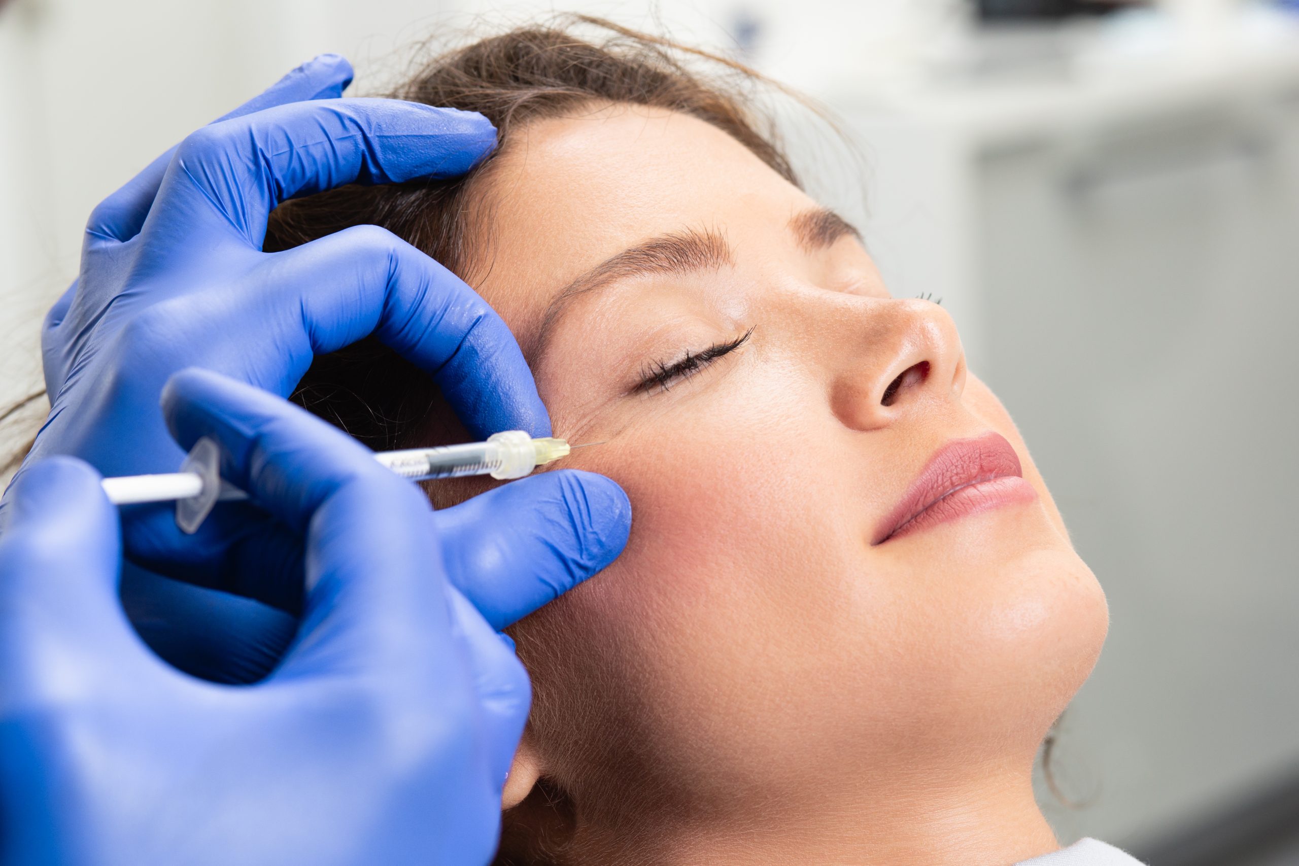
Injection Depth
The injection depth of Rejuran I and Rejuran S is primarily determined by the anatomical thickness of the target area and the concentration of polynucleotides (PN). Eyelid skin measures between 0.2 mm and 0.6 mm in thickness. Rejuran I is specifically placed into the superficial dermis, with the injection depth controlled at 0.1 mm to 0.15 mm. Used with a 34G , the low-viscosity gel spreads evenly through the superficial reticular fibers, helping minimize the risk of subcutaneous nodules. In contrast, the tissue over depressed cheek scars exceeds 1.5 mm in thickness. Rejuran S must be injected into the mid-to-deep dermis, extending to the junction with the subcutaneous layer, with an injection depth of 1.5 mm to 2.0 mm.
Thickness Measurement & Layer Positioning
Before any injection, the first step is to determine how thick the skin actually is. In specialist clinics across Europe and the United States, doctors routinely scan the face using 20 MHz to 50 MHz high-frequency ultrasound probes or OCT optical imaging devices. The skin beneath the eyelid is extremely thin, usually reading between 0.35 mm and 0.6 mm. On the cheeks, where atrophic acne scars are present, the displayed tissue thickness rises to 1.5 mm to 2.5 mm. During measurement, the probe must be held perfectly perpendicular to the skin. Even a 5-degree tilt can create a false reading of about 0.1 mm on the screen. In the eye area, where the skin may be only 0.4 mm thick, doctors use an ultra-fine 34G with an outer diameter of just 0.18 mm. The bevel faces upward, and the is inserted almost flat at an angle of 10° to 12°, gently lifting the stratum corneum as it enters. The tip is positioned precisely within the papillary dermis at a depth of 0.1 mm to 0.15 mm. Because the tissue here is loose, a very light thumb pressure of only 2 to 3 newtons is enough to inject the product. Each point receives only 0.02 cc to 0.05 cc of solution. Under-eye injection parameters:
- Insertion angle: 10°-12°, nearly parallel
- depth: 0.1-0.15 mm
- Volume per point: 0.02-0.05 cc
- Thumb pressure: 2-3 N
The dermis in the cheek area is about 2 mm thick and structurally much denser. A thick, highly viscous crosslinked gel cannot be placed that superficially. If it is injected within 0.5 mm of the surface, its strong structural support can physically push the epidermis upward into a hard lump. That can leave visible papules that persist for three to five months, sometimes with a bluish refractive effect under light. For deep facial scars, doctors switch to a thicker 30G with an outer diameter of 0.31 mm. The insertion angle is increased to 30° to 45° so the can pass through the dense reticular dermis. The depth must fall within the 1.5 mm to 2.0 mm range. The fibrous septa at this level are firm, so injection requires more than 8 newtons of mechanical pressure. The dose per point is also increased to 0.05 cc to 0.10 cc. At that depth, the product forms a rounded deposit that lifts the depressed epidermis from underneath. Cheek injection parameters:
- Insertion angle: 30°-45°, oblique
- depth: 1.5-2.0 mm
- Volume per point: 0.05-0.10 cc
- Thumb pressure: greater than 8 N
If the injection goes too deep—beyond 2.5 mm into the subcutaneous fat layer—the result becomes problematic. Blood flow is much faster there, and the product is rapidly carried away by circulation. Material expected to provide support for months may be cleared by active macrophages in under three weeks. During treatment, doctors often continue checking thickness with a high-frequency probe. After one injection, they may wait five minutes. As the tissue absorbs water and expands physically, the 0.4 mm under-eye skin may temporarily swell to 0.7 mm. When placing the next injection nearby, the original 10-degree angle may need to be adjusted to 15 degrees, otherwise the 0.25 mm bevel can easily slip back out through the edematous tissue. On the cheeks, after 0.1 cc has been placed, the local tissue may be physically elevated by around 0.8 mm. If an ultrasound probe is pressed down to remeasure the area, the pressure can temporarily displace subcutaneous fluid, making the reading appear 0.05 mm to 0.08 mm thinner than it would in a relaxed state. When reviewing OCT images, experienced doctors mentally compensate for these tiny discrepancies. Before treatment, physicians in Europe and the US often use Doppler ultrasound to map out blood vessels. At a depth of 1.5 mm in the cheek, numerous small branches of the facial artery are present, with vessel diameters of only 0.2 mm to 0.5 mm. A 30G inserted at 45 degrees can easily pierce a 0.3 mm facial venule. If that happens, an expensive injection can simply be lost into the bloodstream, while the scar beneath remains unfilled. When the eyelid is extremely thin—less than 0.3 mm—the bevel of a 34G can take up almost the full thickness of the dermis. Even a tiny tremor of less than 0.1 mm can cause the product to leak out through the eyelid. In these cases, doctors switch to a retrograde injection technique. The enters at 15 degrees to a depth of 0.3 mm, then the is slowly withdrawn while the plunger is gently pressed, allowing the low-viscosity solution to spread evenly through tiny spaces at 0.1 mm to 0.2 mm. In acne-scarred skin, the fibrotic adhesions beneath the surface appear on ultrasound as hypoechoic dark bands measuring 0.5 mm to 1.2 mm thick. As the probe passes across them, the thickness reading can fluctuate rapidly. The tip must stop with extreme precision, just 0.1 mm below this dark fibrotic band. At the moment the thumb applies pressure, the hyperechoic gel mass on the screen visibly displaces the surrounding soft tissue, lifting the 1.5 mm deep scar band upward to the 1.0 mm level. A very dry stratum corneum can also interfere with the process. When the epidermis is dehydrated, it reflects too much of the OCT light source, causing the displayed dermal reading to differ from the true tissue status by nearly 15%. To correct this, doctors apply 0.05 cc of medical ultrasound coupling gel to the skin, filling the tiny 0.02 mm surface grooves in the stratum corneum. Once applied, image clarity improves by about 40%, making it much easier to identify the correct injection depth.
Different Physical Forms
When physicians in Western clinics handle these two PN injectables, the resistance they feel on the plunger is completely different. Their viscosity is measured using a rheometer, a precision instrument that stirs the sample at a frequency of 1 radian per second. For the eye-specific I formulation, the reading stays within a narrow range of about 80 to 100 pascals (Pa). Visually, this product looks almost identical to normal saline, with fine ripples when the is shaken. The S formulation, designed for deep scars, sends the rheometer reading instantly above 400 Pa, sometimes even beyond 450 Pa. Each milliliter contains 20 mg of highly concentrated polynucleotides arranged in a dense crosslinked structure. When slowly extruded from the, the S formulation comes out as a thick, semi-solid gel. That difference of several hundred pascals fundamentally changes how the handles the. A very low viscosity creates a light, effortless injection feel. For the eyelid area, doctors use an ultra-fine 34G with an inner diameter of only 0.08 mm. The thumb applies just 2 to 2.5 newtons of force. The fluid enters smoothly into the tissue at a depth of 0.15 mm with almost no resistance. Behavior of the fluid formulation:
- Diffusion pattern: freely spreads within a 5 mm radius of the injection point
- Tissue elevation: only a faint 0.1 mm micro-elevation, barely visible
- Hydration response: absorbs 20% additional water from surrounding tissue within 15 minutes
- Resolution time: excess fluid clears through the lymphatic system within 24 to 48 hours
A semi-solid gel with a viscosity above 400 Pa cannot be pushed through such a fine. For the cheeks, the switches to a 30G with an inner diameter of 0.15 mm. The thumb must press firmly with 8 to 10 newtons of force to push the viscous gel into the deeper tissue. If that high-viscosity material is forced into the superficial skin at only 0.2 mm, the tightly packed collagen network has no room to accommodate it. A Beverly Hills dermatology clinic in Los Angeles included the following rheological comparison in its internal training manual:
| Measurement | Thin Formula (I Type) | Dense Formula (S Type) | Clinical Sensation |
|---|---|---|---|
| Elastic modulus (G’) | 80-100 Pa | 400-450 Pa | Water-like / gel-like injection feel |
| Matched lumen | 34G (0.08 mm) | 30G (0.15 mm) | Extremely smooth / clearly resistant |
| Mechanical injection resistance | 2.0-2.5 N | 8.0-10.0 N | Light pressure / forceful الضغط |
| Diffusion radius | Greater than 5.0 mm | Less than 1.0 mm | Broad hydration / remains localized |
The more viscous the product, the greater its capacity to hold physical space in the tissue. A 0.05 cc deposit of the S formulation placed 1.8 mm deep into the lower dermis stays exactly where it is and does not flow outward. It creates a small mound about 0.6 mm high, physically lifting the depressed skin above it. In the laboratory, DNA extracted from salmon milt is cut into chains of different lengths. The I formulation retains short-chain oligonucleotides and has very low surface tension at 25°C. A single drop placed on a smooth glass plate spreads into a water-like disc measuring 12 mm in diameter within 3 seconds. The S formulation retains long-chain polymers and has much higher surface tension at the same temperature. The same volume dropped onto glass still holds a height of about 3 mm even after 10 minutes. That highly deformation-resistant hemispherical shape makes it ideal for filling atrophic scars that are 2 mm deep. Once placed within dense subdermal tissue, the high-elasticity gel is able to resist the repeated mechanical strain of facial movement hundreds or thousands of times per day. At 37°C, the two formulas absorb water at very different rates. The short-chain I formulation is strongly hydrophilic and absorbs water equal to 20% of its own weight within 15 minutes of injection. That is why a faint whitish micro-swelling may appear beneath the lower eyelid at a depth of 0.35 mm. The long-chain, high-density S formulation absorbs water much more slowly. In the reticular cheek layer at 1.5 mm, there is little visible swelling during the first three days. Its initial water uptake stays below 5%, relying almost entirely on its inherent hardness of over 400 Pa for support. By Day 7, as tissue fluid gradually enters the structure, a secondary expansion of about 8% to 10% occurs. That small increase is enough to lift the overlying depressed epidermis by several dozen microns. Using confocal microscopy, dermatologists in New York have observed how long these subdermal gels persist. By Day 14, the water-like I formulation placed at 0.15 mm has already blended completely into the surrounding reticular fibers. No reflective boundary remains under the microscope—it has broken down into micron-scale fragments. On the same face, however, the S formulation placed at 2.0 mm remains round and full even on Day 14. Its hardness of 450 Pa makes it difficult for incoming macrophages to break down. It is not until around Day 45 that a pale 0.05 mm degradation halo begins to appear around the outer edge. Clinic refrigerators are typically maintained at 4°C. When the S formulation is taken directly from cold storage, its viscosity increases temporarily by about 15%. Nurses therefore leave the at room temperature for 10 minutes before use. If injected while still cold, even strong finger pressure may not be enough to push the gel through a 30G. By contrast, the water-like I formulation shows less than 0.1 newton of change in injection resistance even after being refrigerated for a full week.
Degradation Half-Life & Tissue Environment
Whether the injection is placed superficially or deeply makes a major difference, because the tissue environment is completely different. On high-frequency ultrasound, the area just 0.15 mm beneath the surface is filled with dense red and blue microvascular signals. At 1.5 mm, those signals almost disappear, replaced by pale gray blocks of dense fibrous tissue. The survival time of the injected material differs greatly between these two environments. The water-like I formulation, delivered with a fine 34G into the superficial layer at 0.15 mm, enters a region packed with tiny vessels measuring only about 0.01 mm in diameter. Blood flow is rapid, and as soon as the fluid spreads, macrophages begin moving in. A California medical laboratory observed this under the microscope: within 24 hours of superficial injection, more than 50,000 macrophages had accumulated within a 2 mm radius of the puncture site. In this highly vascular, highly active superficial layer, the I formulation does not last long. Its delicate components are broken down quickly and consumed by surrounding cells. Within 14 to 21 days, the thin 0.1 mm fluid band is no longer visible on ultrasound. Measured characteristics of the superficial injection environment:
- position: 0.15-0.30 mm depth
- Dense vascularity: more than 150 vessels per mm²
- Immune response: surrounded by 50,000 cells within 24 hours
- Survival time: fully cleared within 14-21 days
Now compare that with the S formulation, delivered through a 30G into the base of a depressed acne scar at 1.5 mm to 2.0 mm. This area is more like a sealed basement. Thick fibrous collagen bundles divide the tissue into poorly perfused compartments, and some vessels are thinner than 0.005 mm. A semi-solid gel with a hardness of 400 Pa placed into such a hypoxic, poorly perfused space is extremely difficult for macrophages to break down. The enzymatic activity that is highly active at 0.15 mm is reduced to only about 20% of that level at this depth. Follow-up records from New York clinics show that when 0.05 cc of the S formulation is placed at 1.8 mm, more than 75% of its original volume can still remain after 60 days. Fibrotic tissue at the base of old scars effectively shields the gel from degradative enzymes. The dense, high-concentration gel only erodes slowly at the surface while its core remains firm. Because enzymes cannot easily access it, the lifespan of the S formulation can be extended to 90 or even 120 days. That is exactly why doctors use it as a structural support that remains in place for three to four months. Facial muscles generate thousands of movements every day, constantly pulling on the cheeks. The gel must withstand 10 to 15 newtons of traction while the surrounding tissue slowly forms new blood vessels and repairs itself. If the water-like I formulation is accidentally injected into the fat layer at 2.5 mm, the treatment becomes essentially wasted. That fat layer contains large veins measuring around 0.5 mm in diameter. Once such a fluid product enters that space, it is rapidly carried away by circulation and can be fully cleared within three days. Injecting the S formulation too superficially—at around 0.5 mm—creates an even bigger problem. At that level, the superficial macrophages cannot digest a 400 Pa gel mass efficiently, so the body begins reacting against it. Within three days, the injection site may develop a hard, red nodule about 0.2 mm high. That lump can remain visible and palpable for as long as six months. Measured characteristics of the deep scar environment:
- position: 1.5-2.0 mm depth
- Sparse vascularity: fewer than 20 vessels per mm²
- Muscular traction: constant force of 10-15 N
- Survival time: acts as a structural support for 90-120 days
In addition to blood supply, local temperature and pH also affect how long the material lasts. The skin surface usually remains around 32°C, but at 2.0 mm beneath the surface the temperature approaches 37°C. For every 1°C increase, enzymatic degradation accelerates by about 5%. Old scar tissue is also slightly acidic, often with a pH around 6.8. In this warmer, more acidic environment, the dense S formulation ages and breaks down somewhat faster than laboratory data alone would suggest. That is why clinics typically schedule follow-up injections of the I formulation at around Week 3, while S formulation treatments are pushed back to Week 6 or even Week 8. Tissue hydration also differs sharply between superficial and deep layers. At 0.15 mm, water content is high, so the I formulation can absorb water equal to 20% of its own weight within 15 minutes. As it becomes more hydrated, it spreads more widely and is cleared more quickly. In contrast, within the dry scar base at 1.5 mm, the S formulation absorbs less than 5% water during the first three days, remaining relatively dry and stable. To overcome the stagnation of the deep scar environment, some Los Angeles physicians first use a long to release the fibrotic bands beneath the scar. This subcision step creates a fresh, blood-filled cavity about 2 mm high at a depth of 1.8 mm. The newly introduced blood brings nutrients and attracts a much larger number of macrophages. When 0.1 cc of the S formulation is placed into this newly created cavity, its degradation time shortens from about 120 days to 100 days because it is now surrounded by fresh blood flow. Although it loses about 20 days of persistence, the surrounding tissue regenerates nearly twice as fast. Without this release step, the gel may remain firm for 120 days, but the surrounding cells struggle to regenerate in such a poorly perfused space. Temperature and pH profile of the injection environment:
- Superficial skin temperature: around 32°C
- Deep tissue temperature: close to 37°C
- Scar-base pH: acidic, around 6.8
- Temperature penalty: every 1°C increase shortens lifespan by about 5%
European biochemical laboratories have simulated these conditions by placing both formulations in incubators set at 37°C and pH 6.8. Under those conditions, the water-like I formulation cannot remain intact beyond Day 10, and its molecular chains are fully broken down. The S formulation, by contrast, does not begin to soften and collapse until around Day 80.