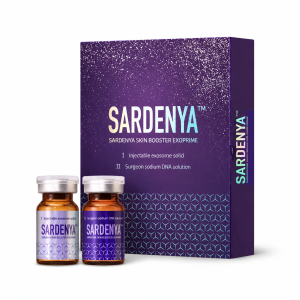
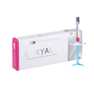
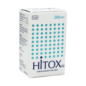
Kiara Reju skin booster combines salmon-derived PDRN and Niacinamide to provide you with professional skin repair and whitening solutions. Its core ingredient, PDRN, penetrates deep into the dermis to promote cell regeneration, while a scientifically concentrated dose of Niacinamide effectively blocks approximately 68% of melanin transfer to the epidermis. Clinical feedback shows that after 3 consecutive treatments, skin hydration is significantly improved, and dullness and spots are visibly faded.
Niacinamide Benefits
Niacinamide (the free form of Vitamin B3) works by blocking melanin and repairing the stratum corneum. A concentration of 2%-5% Niacinamide acting on the skin for 4 weeks can intercept approximately 35% to 40% of melanosome transfer to the epidermis, reducing skin yellowness (b* value) and the area of spots. In Kiara Reju, it is combined with 5mg/ml PDRN to penetrate the skin, stimulating the self-production of ceramides, with the transepidermal water loss rate (TEWL) decreasing by an average of 24%.
Pigmentation Interception and Brightening
Anatomical data shows that one melanocyte is typically connected to 36 keratinocytes, forming an epidermal melanin unit. The free Vitamin B3 derivative intervenes in this physiological process by interfering with the expression of protease-activated receptor-2 (PAR-2). In vitro culture experiments have determined that specific concentrations of Niacinamide can reduce the transfer rate of melanosomes by 35% to 68%. The composite preparation reaches the junction of the dermis and epidermis at a depth of 0.5 mm to 1.5 mm below the skin via minimally invasive methods. Traditional topical application is limited by the stratum corneum barrier, with a penetration rate of less than 5%. Mesotherapy delivers ingredients precisely to the target area, increasing the absorption rate to nearly 100%.
- The activity of protease-activated receptor-2 (PAR-2) is inhibited.
- The physical phagocytosis rate of melanosomes between cells decreases by about 40%.
- The proportion of untransferred pigment degraded by lysosomes within melanocytes increases by 20%.
- Intracellular superoxide dismutase (SOD) levels rise.
Clinical skin tone assessment widely uses the CIE L*a*b* color space system. Instrumental measurements show that in photoaged skin after 8 weeks of continuous intervention, the L* value, representing brightness/darkness, increases by an average of 1.2 to 1.5 units. The visible whiteness of the skin is positively correlated with the increase in the L* value. A dull, yellowish facial appearance is often related to protein glycation and lipid peroxidation. A 5% concentration Niacinamide solution acting on human skin for 4 weeks will reduce the b* value, which reflects yellowness, by approximately 0.5 units. The fading of yellow tones makes the overall skin texture appear clearer under natural light. For post-inflammatory hyperpigmentation (PIH) caused by acne, high-concentration local action can accelerate the fading of marks. Skin microcirculation is improved under anti-inflammatory properties. Compared to natural metabolism, the fading period of reddish-brown acne marks is shortened by an average of 2 to 3 weeks.
- Day 14: Multispectral imaging shows that the edges of superficial light-colored spots begin to blur.
- Day 28: The area of potential UV spots beneath the dermis shrinks by approximately 15%.
- Day 56: The color difference between aging sunspots on the cheeks and surrounding tissue is reduced by 25%.
- Day 84: Surface light reflectance (Radiance) increases by about 20%.
People with Fitzpatrick skin types III and IV are more prone to pigmentation. In a double-blind controlled trial for melasma, a 4% concentration of the ingredient used continuously for 8 weeks resulted in an average decrease in the Melanin Index of 5% to 10%. The micro-trauma caused by physical microneedling triggers transient mechanical erythema. The formula system exerts a calming effect by reducing capillary dilation. By the 7th day post-treatment, the a* value representing skin erythema generally decreases by 10%, significantly reducing the risk of post-inflammatory hyperpigmentation (rebound darkening). Melanosomes that fail to transfer to the epidermis do not accumulate indefinitely in the basal layer. The intracellular autophagy mechanism is activated to process the excess pigment granules. Histological staining observation showed that the total number of pigment granules within a single melanocyte decreased by about 30% after 12 weeks.
- The total dosage for a single facial procedure is controlled between 2ml and 3ml.
- The diffusion radius of the solution within the subcutaneous tissue is approximately 1 cm around the injection point.
- The peak concentration in the basal layer occurs 24 to 48 hours after the procedure.
- The basic course of treatment is set for 3 to 4 sessions, with an interval of 21 to 28 days each.
Ultraviolet radiation is the strongest external factor triggering melanin synthesis. A defense mechanism is established within the tissue to block the transmission of alpha-melanocyte-stimulating hormone (α-MSH) signals induced by UV rays. Within 48 hours of exposure to long-wave ultraviolet (UVA), the intensity of the pigment response is significantly inhibited. With age, the metabolic renewal cycle of epidermal cells gradually extends from 28 days to over 45 days. Old keratinocytes carry a large amount of pigment and remain on the skin surface. Biochemical intervention accelerates the differentiation rate of keratinocytes, assisting in the exfoliation of old cells containing pigment.
Lipid Barrier and Water Retention
The stratum corneum consists of 15 to 20 layers of flat, enucleated dead cells. The spaces between cells are filled with a lipid matrix, forming a physical structure similar to bricks and mortar. The lipid matrix contains approximately 50% ceramides, 25% cholesterol, and 10% to 20% free fatty acids. After the free Vitamin B3 derivative enters the skin tissue, it upregulates the biochemical reaction rate of serine palmitoyltransferase. This enzyme is the rate-limiting step in the sphingolipid biosynthetic pathway. Exposure to a 2% concentration solution for 4 weeks increased ceramide synthesis in epidermal cells by 34%. The synthesis rate of free fatty acids simultaneously increased by approximately 67%. High concentrations of lipids are secreted into the extracellular space, rearranging into a dense lamellar structure. On a microscopic level, micro-cracks in the stratum corneum caused by damage are filled by newly generated lipids. Transepidermal water loss (TEWL) is a quantitative indicator of barrier integrity. Measured using a Tewameter, the TEWL value in specific facial areas decreased from 25 g/m²h to 18 g/m²h after 4 weeks of intervention, a decrease of over 24%.
| Biochemical Assessment Item | Initial State Value | Value After 28 Days | Overall Change |
|---|---|---|---|
| Total Ceramide Concentration | 1.2 mg/cm² | 1.6 mg/cm² | +34.0% |
| Free Fatty Acid Content | 0.8 mg/cm² | 1.3 mg/cm² | +67.0% |
| Stratum Corneum Water Content | 14.5% | 22.3% | +53.8% |
| Transepidermal Water Loss | 25.1 g/m²h | 18.5 g/m²h | -26.2% |
The synthesis rates of Filaggrin and Involucrin increased by approximately 20% after biochemical intervention. Structural proteins cross-link within keratinocytes, forming a sturdy shell resistant to mechanical forces. Mesotherapy delivers ingredients to the reticular dermis at a depth of 1.0 mm to 2.0 mm. The 20mg/ml non-crosslinked hyaluronic acid molecules in the formula absorb 1000 times their own weight in water molecules within the dermis. The thickening of the epidermal lipid barrier blocks the evaporation of water from the dermis at the surface. Hyaluronic acid acts as a reservoir beneath the skin, while the tough stratum corneum above acts as a physical occlusive film. Their synergy stabilizes the water content of the epidermal stratum corneum at a healthy threshold of 20% to 25%. In dry environments with relative humidity below 30%, the rate of water loss from fragile skin doubles. Skin subjected to a full cycle of intervention showed a TEWL increase of only one-third compared to the untreated state under the same low-humidity exposure. A healthy hydrolipidic film is slightly acidic. The increase in lipid synthesis assists in restoring the pH value of the epidermal surface.
- Skin surface pH drops from a slightly damaged state of pH 6.2.
- Probe data stabilizes between pH 5.4 and 5.6.
- The diameter of local capillary dilation decreased by approximately 15% under histological observation.
Barrier damage is accompanied by low-grade chronic micro-inflammation. The release of inflammatory mediators such as interleukin-1alpha (IL-1α) and prostaglandin E2 (PGE2) in the epidermis decreases. The increase in barrier thickness improves physical tolerance to surfactants. Patch tests using 1% sodium dodecyl sulfate (SDS) showed that the intervention group’s erythema reaction was delayed by 48 hours compared to the control group. The normal metabolic renewal cycle of keratinocytes is approximately 28 days. When the lipid matrix is sufficient, desmosome enzyme activity is at its optimal state. Old keratinocytes shed regularly at a rate of about 1 to 2 layers per day. High-resolution ultrasound imaging shows that after 8 weeks of continuous treatment, the average thickness of the epidermis increased by 2 to 3 microns. The increase in physical thickness resists the penetration of particulate matter with a diameter less than 2.5 microns into the hair follicles. Aquaporin-3 (AQP3) on the cell membrane is responsible for transporting water and glycerol to various layers of the epidermis. In vitro biochemical assays found that AQP3 expression levels were upregulated by approximately 25%. [Image of aquaporin-3 function in skin] Using 34G ultra-fine for facial grid-like application, the distance between each point is about 1 cm. The injection dose per point is controlled between 0.02ml and 0.05ml. The micro-liquid forms a papule about 3 mm in diameter under the skin, which then diffuses evenly to surrounding tissues within 24 hours. Microcirculatory blood flow in the dermis increased by about 18% within 48 hours after injection. Blood delivers oxygen and nutrients to the basal layer of the epidermis, providing energy substrates for the synthesis of lipids in the upper cells. Water fullness changes the mechanical properties of the epidermis. The plasticity of the stratum corneum increases, and the elastic modulus decreases. 3D skin topography analysis shows that the number of superficial fine lines with a depth of less than 0.1 mm in the eye and cheek areas decreased by about 32% after 4 cycles. For every 1 degree Celsius drop in ambient temperature, sebum secretion decreases by 10%. Exogenously supplemented lipid production compensates for the physiological gap caused by low temperatures. 
Sebum Regulation and Pore Contraction
Sebaceous glands are widely distributed in the dermal tissue, with a distribution density in the forehead and nose areas reaching 400 to 900 per square centimeter. Glandular cells mainly synthesize triglycerides, squalene, and wax esters. When a 2% concentration of the free Vitamin B3 derivative enters the tissue, it interferes with the biochemical activity of diacylglycerol acyltransferase (DGAT). This enzyme controls the rate of lipid generation in the triglyceride biosynthetic pathway. Sebaceous gland cells in vitro culture showed an average reduction of approximately 25% in the volume of lipid droplets in the cytoplasm on the 6th day of exposure to a 2% concentration solution. The overall secretion of the sebaceous glands is inhibited, resulting in a quantitative decrease in free oil levels on the skin surface. Clinical studies use Sebutape microporous polymer patches to collect excess facial oil. After 14 days of continuous intervention, the oil adsorption area on the patches decreased by 18%. Data from 28 days showed that the sebum excretion rate (SER) decreased from 3.5 micrograms to 2.7 micrograms per square centimeter.
- Triglyceride secretion decreased by about 20%.
- Free fatty acid concentration decreased by 15%.
- Squalene oxide generation rate slowed by 30%.
- The sebum film surface pH is maintained at around 5.4 to 5.6.
- The formation cycle of micro-comedones is extended to over 45 days.
Excessive sebum secretion physically stretches the follicular infundibulum. In follicles and sebaceous gland ducts under long-term high-load oil discharge, surrounding keratinocytes undergo abnormal hyperplasia. The decrease in oil discharge eliminates the continuous physical expansion pressure on the inner walls of the ducts. Data from 3D skin topography analysis (Visia-CR) shows that decreased sebum secretion is accompanied by a contraction of pore diameter. After 4 weeks of applying high-concentration ingredients, the number of visible enlarged pores on the face decreased by about 22%. The cross-sectional area of individual pores shrank by an average of 15% to 20%. Enlarged pores are caused not only by oil stretching but also by micro-inflammation of surrounding tissues triggered by sebum oxidation. Squalene in sebum is easily oxidized to squalene peroxide under UV radiation. The antioxidant properties of the free Vitamin B3 derivative reduce the generation of peroxides. Histological staining observation found that the infiltration density of inflammatory cells around hair follicles decreased by about 35% after 8 weeks of intervention. The reduction in inflammatory mediators slows the degradation rate of collagen fibers around the follicles. The dermal matrix supporting the pores restores its original mechanical firmness.
- Abnormal keratinization of the follicular infundibulum is inhibited.
- The recurrence rate of blackheads is reduced by 40%.
- The rate of collagen loss around pores decreased by 25%.
- The skin surface texture smoothness index increased by 18%.
Mesotherapy delivers the liquid 1.0 mm subcutaneous via 34G ultra-fine. Active ingredients bypass the epidermal barrier and accumulate in high concentrations around the sebaceous glands. The drug concentration in the tissue fluid peaks 24 hours after injection, with approximately 80% of the ingredients absorbed by the glandular cells. Polydeoxyribonucleotide (PDRN) in the formula promotes the secretion of Type I and Type III collagen by fibroblasts. New collagen fibers form a dense network around the hair follicles. Enhanced mechanical support counters pore sagging into water-drop shapes caused by gravity. Measured under a confocal laser microscope, the dermal thickness in the cheek area increased by approximately 0.2 mm after completing 3 minimally invasive interventions. The thickened dermal tissue squeezes the follicular sebaceous glands from the outside in. Physical squeezing combined with reduced internal oil discharge acts bi-directionally on pore diameter. Sebum secretion is influenced by fluctuations in internal hormone levels, especially the stimulation of dihydrotestosterone (DHT). In vitro biochemical assays show that specific doses of free Vitamin B3 can fine-tune the activity of 5-alpha reductase, reducing the synthesis rate of testosterone to DHT by approximately 10%. Subjects stayed in a high-humidity environment with temperatures as high as 30 degrees Celsius for 2 hours. The nasal sebum secretion of the untreated group rose by 45% from baseline. Tissues that received 4 continuous cycles of biochemical intervention showed a sebum increase of only 15% under the same conditions. The 20mg/ml non-crosslinked hyaluronic acid introduced via micro-invasion locks in a large amount of water molecules within the dermis. Improved tissue hydration enhances the optical transparency of the epidermis. Shadows produced by pore depressions become blurred under light diffuse reflection, and the measured surface roughness (Ra value) decreased by 12%.
- The visual area of pore shadows on the cheeks shrank by 20%.
- The density of reticular fibers in the dermis increased by 30%.
- The stratum corneum desquamation rate restored to the normal 28-day value.
- Epidermal light reflectance increased by 15%.
Whitening Effect
The whitening mechanism of Kiara Reju is built on the direct delivery of 2% Niacinamide and PDRN to the dermis. Clinical observations show that Niacinamide in the dermis can block 35%-68% of melanosome transfer to keratinocytes. PDRN increases the proliferation rate of fibroblasts, shortening the pigment metabolism cycle of the basal layer of the epidermis from 28 days to approximately 21 days. After completing 3 injections (at 3-4 week intervals), the skin’s ITA° value (a measure of skin brightness) rises significantly, the melanin density in spotted areas decreases, and the overall skin tone presents a uniform brightness.
Reducing Pigmentation
When ultraviolet B (UVB) with wavelengths between 290-320nm penetrates the skin surface, melanocytes in the basal layer receive oxidative stress signals. Tyrosinase within the cells, catalyzed by copper ions, oxidizes tyrosine into dopa, synthesizing melanin granules. Kiara Reju uses 30G or 32G ultra-fine to deliver the liquid via micro-droplet injection, targeting the superficial dermis 1.0-1.5 mm below the skin. The formula contains standardized Niacinamide to block signal transduction in the protease-activated receptor-2 (PAR-2) pathway. Pigment granules encapsulated in melanosomes would normally be transported to surrounding keratinocytes through the dendritic structures of melanocytes. Niacinamide molecules injected into the dermis can stop 35%-68% of melanosomes from transferring to the peripheral stratum corneum. Lacking the replenishment of new pigment granules within epidermal cells, existing dark areas gradually fade as approximately 30,000 to 40,000 dead skin cells are naturally shed daily. Another pathological mechanism causing pigmentation is local inflammation, known medically as post-inflammatory hyperpigmentation (PIH). Acne ruptures or micro-trauma from laser treatments lead to macrophages releasing interleukin-6 (IL-6) and tumor necrosis factor-alpha (TNF-α). Inflammatory mediators stimulate vasodilation and force nearby melanocytes to over-secrete pigment as a defense mechanism. Kiara Reju’s polydeoxyribonucleotide (PDRN), extracted from salmon milt, has a molecular weight controlled within the 50-1500 kDa range, highly matching human DNA fragments. PDRN molecules rapidly bind to A2A purinergic receptors on the cell surface after entering the dermal matrix. Once the receptor binding reaction is triggered, the secretion of local inflammatory cytokines decreases by 30%-40% within 48 hours. Abnormal capillary dilation is inhibited, and the fading cycle of reddish-brown acne marks is shortened from 3-6 months to 4-6 weeks. PDRN also promotes dermal fibroblast proliferation, accelerating the reconstruction of the collagen synthesis network. The division speed of basal epidermal cells increases accordingly, compressing the keratinocyte metabolism cycle from the standard 28 days to 21-24 days. Stubborn pigment deposited in the middle and lower layers of the epidermis reaches the surface of the stratum corneum and sheds earlier due to the accelerated upward movement of cells. To quantitatively evaluate the depigmentation process, dermatological clinical practice usually monitors changes in the following physiological indicators:
- Day 7: Dermal vascular bed micro-inflammation subsides; skin red area (a* value) decreases by 12-15%.
- Day 14: Melanin content in keratinocytes decreases; dermal water content increases by over 20%.
- Day 21: New cells in the epidermal basal layer exceed 60%; superficial pigment spot area shrinks.
- Day 30: ITA° value (Individual Typology Angle, higher values mean brighter skin) increases steadily by 3-5 units.
Beyond biochemical pigment degradation, the 20mg/ml non-crosslinked hyaluronic acid (HA) added to the formula changes the visibility of spots at an optical-physical level. Non-crosslinked hyaluronic acid is highly hydrophilic, capable of binding 1000 times its own weight in water molecules within the extracellular matrix. When the dermal interstitial water content is highly saturated, the refractive index of skin tissue changes. When light hits well-hydrated skin, the proportion of diffuse reflection is significantly reduced, and high-contrast pigment spots visually fade by 15%-20%. The half-life of effective ingredients delivered in a single injection within the subcutaneous tissue is approximately 7-14 days. According to medical aesthetic operation standards, a melanin-fading course requires a scientifically planned intervention frequency. Patients usually receive 3-4 cycles of procedures, with the specific timeline linearly related to pigment distribution:
- Initial Phase: Inject 2-3ml dose every 3 weeks, distributed across 80-100 micro-droplet points face-wide.
- Metabolic Phase: After two operations overlap, the dermis accumulates 1.5-2.0mg/ml PDRN concentration to phagocytize old pigment.
- Stabilization Phase: After the 3rd intervention, the interval extends to 4-6 weeks to reconstruct a low-pigment extracellular microenvironment.
- Maintenance Phase: Perform 1 supplement every 3-6 months to resist cumulative damage from daily 2-3 hour UV exposure.
External care intervention during the treatment period seriously affects the final outcome of pigment metabolism. UVA waves in sunlight (320-400nm) have strong penetrative power and can reach the dermis to reactivate melanocytes. Patients must apply broad-spectrum sunscreen with SPF 50+ and PA++++ during the day, at a dosage of 2 mg per square centimeter of skin. Within 14 days post-procedure, the stratum corneum is in a micro-trauma recovery stage, barrier function is weakened, and transepidermal water loss (TEWL) rises. Use of alpha hydroxy acids (AHA) exceeding 5%, salicylic acid (BHA), and any concentration of retinol (Vitamin A derivatives) must be stopped. Exfoliating acid components can cause compensatory inflammation in the dermis, inducing new pigment deposition in the reticular dermis. Macrophages migrate through the lymphatic network after engulfing decomposed pigment fragments. Facial lymphatic drainage speed is approximately 0.5-1 mm per minute, and pigment-carrying particles eventually enter the cervical lymph nodes. Consuming 2000-2500 ml of pure water daily helps maintain blood osmotic pressure and accelerates the excretion rate of pigment metabolites.
Improving Skin Refractive Index
When ambient light hits the human face, about 5% of it undergoes regular reflection on the surface of the stratum corneum. The remaining 95% of light beams penetrate the epidermis and enter the dermal matrix at a depth of about 1.5 mm. In a healthy state, the water content of the stratum corneum is maintained between 15% and 20%, and the cells are arranged flat and tightly. Once the water content drops below 10%, the edges of keratinocytes curl slightly, forming tiny scale structures. Optical laws in physics indicate that light hitting a rough scaly surface produces diffuse reflection, drastically reducing the luminous flux entering the human eye. The skin thus presents a visual characteristic lacking luster, and the Gloss Value on measuring instruments is typically below 10. Kiara Reju is delivered to the subcutaneous tissue using 32G ultra-fine, and the 20mg/ml non-crosslinked hyaluronic acid (HA) it carries rapidly spreads within the extracellular matrix. Hyaluronic acid molecules with a molecular weight of 1 to 1.5 million Daltons possess a strong spatial network structure. Every 1 gram of hyaluronic acid can firmly lock in 1000 ml of water molecules in the dermis.
The surge in dermal water content fills the gaps between withered collagen fibers with high-density hydrated gel. When light passes through this plump hydrated medium, the scattering path is shortened, and photons can bounce back more densely along the original path.
Tiny wrinkles on the surface of the stratum corneum are physically flattened by internal hydraulic pressure, and skin surface roughness (Ra value) decreases by about 25%. A smooth physical surface promotes a shift in light reflection mode from diffuse reflection to Specular Reflection. The directional light beams reaching the retina increase, forming high-light reflection areas of about 2-3 square centimeters on the cheekbones and forehead. Salmon-derived 50-1500 kDa polydeoxyribonucleotide (PDRN) is responsible for basal repair in improving the refractive index. It binds to the A2A receptors of dermal fibroblasts, accelerating the synthesis of Laminin-5. Laminin-5 is the structural protein connecting the epidermal basal layer and the dermal papillary layer. The densification of this region reduces light attenuation by 12%-18% when passing through the dermo-epidermal junction (DEJ). Within a continuous 28-day epidermal metabolic cycle, ceramide synthesis in the stratum corneum increased by about 34%. Ceramides, free fatty acids, and cholesterol are arranged in a 3:1:1 molar ratio to fill the gaps between keratinocytes. An intact lipid barrier keeps transepidermal water loss (TEWL) below 15 g per hour per square meter. To quantify changes in skin refractive index in 3D space, laboratories often use Visia detectors or Skin-Glossmeters for optical measurement:
- Day 5-7: Superficial dermal hydration peaks; instrument-measured surface reflected light intensity (Gloss value) increases by 15-20 units.
- Day 14-21: Epidermal microstructure completes initial reconstruction; diffuse reflection luminous flux in facial highlight areas reduces by about 30%.
- Day 30-45: Dermal matrix density increases; ultra-fine textures below 0.5 mm deep are physically flattened by over 40%.
A smooth stratum corneum is like a polished glass surface, while the highly hydrated dermis acts as a high-quality reflective backing. The physical superposition of the two determines the final refractive brightness captured by the naked eye.
Within 72 to 96 hours post-injection, free HA molecules begin enzymatic cleavage. The endogenous collagen and hyaluronic acid secreted by fibroblasts stimulated by PDRN take over the supporting role of exogenous components. The half-life of self-synthesized high-molecular-weight substances in dermal tissue fluid is as long as 15 to 20 days. Environmental humidity management within 48 hours post-procedure has a huge impact on the establishment of the initial refractive index. In an air-conditioned room with relative humidity (RH) below 40%, the rate of water evaporation from the epidermis to the air doubles. A humidifier is needed indoors to maintain ambient humidity in the 55% to 65% range. Apply an occlusive cream containing 5% panthenol (Vitamin B5) and biomimetic lipids daily, with a thickness of 0.1 mm. The occlusive oil film forms an artificial barrier on the skin surface, physically blocking the diffusion path of water molecules from the stratum corneum. Stability of epidermal osmotic pressure ensures that the biochemical binding of PDRN with cell receptors in the dermis is not disturbed for up to 14 days. When cleaning the face, the water temperature must be strictly controlled at a lukewarm state of 32℃ to 35℃. Hot water exceeding 38℃ will wash away a large amount of the natural lipid film, causing the surface refractive index to drop significantly within 2 hours. 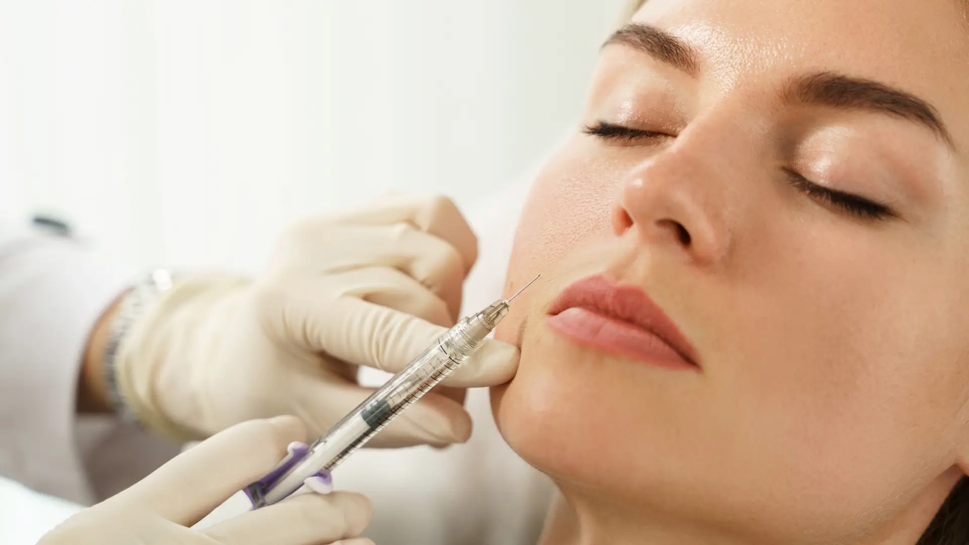
Treatment Schedule and Expected Changes
The standard Kiara Reju facial delivery protocol is set for 3 to 4 consecutive operation cycles. The doctor will use multi-negative pressure equipment or a single 32G microneedle to distribute a 3ml liquid at micro-droplet doses of 0.02-0.05ml across about 100-120 injection sites on the face. The compound containing 20mg/ml non-crosslinked hyaluronic acid and 50-1500 kDa molecular weight PDRN forms tiny reservoirs in the reticular dermis 1.0-1.5 mm subcutaneous. Within the initial 24 to 48 hours, mechanical micro-trauma will trigger transient erythema and mild tissue edema. Macrophages in the tissue fluid clear cell debris from the tiny wounds within 72 hours. Free hyaluronic acid molecules reach their maximum water absorption threshold on days 3 to 5, and the transepidermal water loss (TEWL) drops from a post-op 25g/m²·h back to the physiological norm of 10-12g/m²·h. Patients will observe changes in the physical texture of the stratum corneum between days 7 and 14. Free water molecules flatten dry lines with a depth of 0.1-0.2 mm, optical diffuse reflection on both sides of the cheeks is reduced, and instrument-measured gloss values increase by about 15%. For the superficial physical changes in the first 14 days, skin clinical practice usually monitors 3 objective parameters:
- Dermal Water Content: Ultrasound testing shows the deep tissue echo band widening by 1.2 mm.
- Erythema Index (EI): Microvascular reaction subsides; EI value decreases by 8-10 units.
- Smoothness (Ra): Stratum corneum scales close; surface roughness decreases by 20%.
On days 21 to 28 after the first operation, the epidermis completes a full 28-day cell replacement cycle. Niacinamide molecules in the dermis block approximately 40% of melanosome transfer to keratinocytes, and overall facial dullness shows visual fading under daylight. A second 3ml subcutaneous delivery is performed at week 4. The binding rate of A2A purinergic receptors in the dermis to PDRN reaches its peak, and the proliferation rate of fibroblasts is about 30% higher than in the natural state. The density of the collagen fiber network increases by 15-20% between days 45 and 60. The upward propulsion of basal layer cells is enhanced, and stubborn melanin granules deposited in the granular layer accelerate their exfoliation accordingly; UV spot area on the Visia detector shrinks by 10-12%. Entering the third operation at week 8, the skin’s underlying microenvironment shifts from dehydration and micro-inflammation to high hydration. In follow-up data at 90 days, over 70% of patients’ facial ITA° values (Individual Typology Angle) improved by 3-5 units compared to baseline. Epidermal thickness increased from an average of 0.08 mm to over 0.09 mm. To accurately compare histological and visual changes at each stage, dermatological clinics use a unified assessment system to record physiological parameter fluctuations over 12 weeks:
| Procedure Node | Days from First Visit | Histological Parameter Change Measurement | Clinical Visible Brightening Performance |
|---|---|---|---|
| 1st 3ml Delivery | 14-21 days | Dermal hydration ↑25%, TEWL back to 12g/m²·h | Cheek dryness disappears; skin tone appears full and lustrous during morning washing. |
| 2nd 3ml Delivery | 45-60 days | Melanosome transfer blocked ↑45%, epidermal turnover days ↓3 days | Superficial acne marks fade; edges of pigment deposition areas on the forehead and around the mouth blur. |
| 3rd 3ml Delivery | 75-90 days | Collagen density ↑18%, ITA° value rises 4-5 units | Overall skin tone uniformity improves; skin appears translucent and naturally glowing even without makeup. |
After completing 3 standard injections, the non-crosslinked hyaluronic acid in the dermal interstitium will be degraded by endogenous hyaluronidase within 30-45 days.
To extend the timeline where cells remain in a high-metabolic and low-pigment state, the 4th and subsequent maintenance interventions are stretched to every 8-12 weeks. Each supplement of 2-3ml dose combats oxidative stress generated under daily UV radiation of 2-3 mW/cm².
Washing the face with hot water over 40℃ daily or entering a sauna with relative humidity over 80% will accelerate blood circulation in the dermal capillaries. Local blood flow speed rising from 0.5 mm to 1.5 mm per second will cause free HA and Niacinamide molecules to be metabolized out of the body prematurely via micro-veins and the lymphatic network. The expected 21-day brightening maintenance cycle will shrink to 10-14 days. Apply a cream containing 3% ceramide and 2% squalane morning and night to the surface of the stratum corneum, keeping the thickness at 0.05 mm. The artificial lipid film can reduce the evaporation rate of water from the epidermis by 40%, providing a stable environment for dermal osmotic pressure. In clinical statistics, the compliance rate for pigment metabolism cycles is as high as 85% for patients who strictly apply SPF 50+ sunscreen and control facial temperature. Individuals who do not practice strict photoprotection show a 5-8% rebound in UV spot area when tested on day 45.