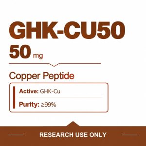
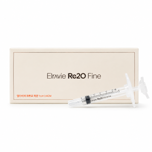
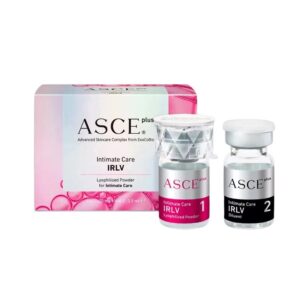
Rejuran S is a professional Polynucleotide (PN) injectable specifically designed for the repair of depressed acne scars. With its high viscosity characteristics, specialized doctors precisely inject it into the deep dermis (approximately 1 to 2 mm deep), allowing it to act like a “scaffold” to directly support atrophic scar tissue while improving local rough texture. Its core principle is to achieve true skin tissue remodeling by continuously stimulating the regeneration of underlying collagen. Clinically, it is usually recommended that patients undergo 3 to 4 sessions (with intervals of approximately 4 weeks), which can significantly reduce scar depth and greatly enhance skin smoothness after the full course.
Scar Texture
Rejuran S contains 20mg/ml of Polynucleotides (PN). Its high-molecular network structure’s viscosity far exceeds that of ordinary aqueous solvents, specializing in the textural repair of atrophic acne scars. Scar textures are classified by morphology into rolling scars (width > 4mm, depth 1-2mm), boxcar scars (width 1-4mm, U-shaped cross-section), and ice pick scars (depth > 2mm, V-shaped opening < 2mm). After injection into the deep dermis, its high viscosity properties establish an immediate physical support scaffold under the skin. Within 4 to 6 weeks, local fibroblast proliferation occurs, and Type I collagen secretion increases by approximately 20%, smoothing out epidermal irregularities by filling the volume defects in the dermal matrix.
Rolling Depression Repair
Rolling scars typically have a diameter exceeding 4 mm and present as smooth, wave-like undulations on the surface. Observed under magnification, abnormal vertical fibrous bands exist beneath the epidermis. One end of the tissue connects to the bottom of the dermal layer, while the other end pulls on the subcutaneous fascia layer. The physical pulling force forces the surface skin to depress downward, forming a drop of 1 to 2 mm. In dermatology clinics in New York and Los Angeles, treating this usually begins with subcision of the subcutaneous tissue. Doctors use an 18G or 22G Nokor to penetrate the skin surface, entering above the sub-dermal vascular plexus. Moving the in a horizontal fan-shaped motion, they cut the fibrous tissue pulling downwards. As the fibrous bands break, the depressed epidermis loses its downward pull and rebounds upwards by approximately 30% to 50%. If the tiny wounds left by subcision are left untreated, the exudation of tissue fluid can easily lead to re-adhesion within 7 to 14 days. The epidermis would collapse again; Rejuran S intervenes at this moment as a physical space-filling material. It contains a high concentration of 20mg/ml Polynucleotide (PN) with a molecular weight of over 1000kDa. Its structural viscosity is several times higher than that of ordinary mesotherapy preparations. Doctors switch to a fine 27G or 30G at the same injection site to inject the gel. Each subcised cavity is injected with a dose of 0.05ml to 0.1ml. The high-viscosity gel forms a physical isolation barrier with a thickness of about 0.5 to 1 mm between the dermis and the subcutaneous fat layer.
- Occupies the subcised wound space
- Blocks incorrect cross-linking of collagen fibers
- Supports the epidermis to maintain horizontal height
- Resists post-operative local tissue contraction forces
Changes within 48 hours after injection can be observed under an ultrasonic skin detector. The dermal base is completely lifted by the high-density gel, forming a three-dimensional mesh scaffold. It is capable of withstanding the compression caused by local facial muscle movement. The macromolecular scaffold degrades slowly in the body over a period of 14 to 28 days. As the physical material gradually degrades over two to four weeks, PN fragments bind to Adenosine A2A receptors in the surrounding tissue. Once the receptors are activated, the division and proliferation rate of local fibroblasts increases by approximately 20%. New cells begin to synthesize large amounts of Type I and Type III collagen, as well as elastin. Clinical data shows that by the 4th week after the first injection, the extracellular matrix density of the local dermal layer increases by 10% to 15%. Original dead spaces are filled with new, healthy dermal tissue. Significant changes in data appear after 3 consecutive treatments at 4-week intervals. 3D skin imaging shows an increase in dermal thickness of approximately 0.2 to 0.4 mm. Precisely delivering PN material to the designated depth requires high dexterity from the doctor. For rolling depressions at a depth of 1.5 mm, the insertion angle is usually maintained between 10 to 15 degrees. Doctors use a retrograde linear injection method, uniformly injecting the solution while slowly withdrawing the.
- insertion at 10-15 degrees
- Positioning at the base of the dermal layer
- Retrograde linear injection
- Releasing gel while withdrawing the
During the process, the epidermis will show slight white ridges, which then flatten under light pressure. Injections that are too superficial will cause the high-viscosity gel to form visible nodules under the epidermis, which may take weeks to subside. If the injection is too deep and enters the subcutaneous fat layer, the polynucleotides cannot reach the dense fibroblasts. Consequently, the regeneration rate of new collagen will decrease. A single full-face rolling depression repair session typically consumes 1 to 2 (1ml per ) of material. Slight post-operative facial swelling and marks occur, with a recovery period of 3 to 5 days. In some cases, because subcision cuts micro-vessels, bruising with a diameter of 2 to 5 mm appears in the injection area. Bruising requires 7 to 10 days for natural metabolic absorption. In European clinical skin practice, the proportion of large-area depression repair using single injection is not high. Clinics overlay non-ablative 1565nm Erbium-glass laser in the 2nd week after subcision and material injection. The microscopic thermal zone (MTZ) depth generated by the laser is around 800 microns. High-density thermal stimulation penetrates the epidermis, accelerating the biological response of the PN components residing in the deep dermis. Polynucleotides provide abundant DNA synthesis raw materials for damaged tissue, shortening the post-laser erythema period by about 40%. Three months after receiving combined treatment, doctors measure the skin surface using the Roughness Index (Ra). The amplitude of undulations drops from an average of 1.2 microns before treatment to 0.6 microns. Macrophages aggregate at the injection site within 24 hours post-operation, initiating a mild inflammatory response. They are responsible for clearing cell debris and minimal hematoma generated by subcision. The anti-inflammatory properties of the macromolecular material inhibit the excessive release of pro-inflammatory cytokines such as Interleukin-6 (IL-6). The severity of local swelling is therefore reduced by about 30%. On the 7th day post-operation, the inflammatory phase transitions smoothly to the proliferative phase. New capillary networks slowly extend inward under the guidance of polynucleotides. The establishment of microcirculation supplies fibroblasts with sufficient oxygen and amino acids.
- Macrophages clear tissue debris
- Interleukin-6 secretion is inhibited
- Micro-vessels grow inward at 0.1 mm per day
- Blood perfusion increases by 25% after 6 months
Vascular networks grow inward along the edges of the high-viscosity gel at a rate of about 0.1 mm per day. Sufficient blood supply is the prerequisite for the formation of a long-lasting collagen fiber network. Six months after completing three sessions, ultrasound scans show that blood perfusion in the new tissue is 25% higher than in the original area. By this time, the polynucleotides have been metabolized into water and carbon dioxide and expelled from the body. The smoothness of the epidermis is maintained long-term, and the half-life of the collagen fiber matrix synthesized by the patient’s own fibroblasts lasts as long as 10 to 15 years.
Boxcar Edge Smoothing
The diameter of boxcar scars is typically between 1.5 and 4.0 mm. Vertical edges form an approximately 90-degree right angle with the depressed base. The depth of superficial forms is between 0.1 and 0.5 mm, while the depth of deep forms exceeds 0.5 mm. Light shining on the U-shaped edges creates harsh shadows, making the skin surface appear extremely uneven. Injecting high-viscosity Rejuran S (20mg/ml) alone into the base of the depression can lift the collapsed tissue. The physical volume of the gel pushes the flat base upward by 0.2 to 0.3 mm. The vertical 90-degree edges still maintain their original physical form after the base is lifted. In North American dermatological practice codes, it is routine to combine mechanical edge ablation with biological base filling.
- Assess the smoothness of the depression base
- Measure the vertical drop of the edge
- Determine the spacing of injection points
- Select ablation laser wavelength
Doctors use 10,600nm Carbon Dioxide fractional laser or 2940nm Erbium laser to vaporize sharp scar boundaries. The laser beam outputs energy at 15 to 25 mJ per micro-beam. The 90-degree vertical edges are precisely ground into gentle slopes of 20 to 30 degrees. The ground slopes naturally blend the scar boundary with the surrounding healthy epidermis, eliminating shadow discrepancies. Within 15 minutes of laser vaporization, the doctor switches to an ultra-fine 30G or 32G. The tip precisely penetrates the exact center of the U-shaped depression. The injection depth is strictly controlled at 1.0 to 1.5 mm, reaching the mid-to-deep dermis. Doctors use a micro-droplet fractional injection technique to control the output volume of the liquid. Each injection point delivers 0.01 to 0.02 ml of polynucleotide gel. The physical spacing between injection points is strictly maintained at 2 mm. For a boxcar depression with a 4 mm diameter, 3 to 4 micro-droplet points are usually deployed. The high-viscosity gel forms a tight matrix network within the dermal layer, supporting the ground area from below. Tissue fluid begins to infiltrate the tiny channels generated by the laser within 24 hours. The polynucleotide fragments in the Rejuran S formula act as a molecular scaffold for fibroblast migration. The cell division and proliferation rate in the targeted base area increases by approximately 18%.
| Edge Morphology | Laser Energy Setting | Single Point Injection Dose | Expected Tissue Lifting Rate |
|---|---|---|---|
| Superficial U-type (< 0.5mm) | 10-15 mJ/MTZ | 0.01 ml | 40% – 50% |
| Deep U-type (> 0.5mm) | 20-25 mJ/MTZ | 0.02 ml | 25% – 35% |
| Fused Wide Edge | 15-20 mJ/MTZ | 0.015 ml | 30% – 40% |
Type III collagen is rapidly synthesized within the first 14 days after treatment. The extracellular matrix thickness at the base of the scar increases by 12% to 15%. In the subsequent 30 to 45 days, Type III collagen is slowly remodeled into higher-density Type I collagen fibers. Combined treatment triggers a strong wound-healing response at the dermo-epidermal junction. Due to the dual physical destruction of laser thermal effects and puncture, erythema will persist for 5 to 7 days. The 2% concentration of polynucleotides actively down-regulates pro-inflammatory cytokines such as Tumor Necrosis Factor-alpha. Compared to laser grinding alone, the duration of local inflammation is shortened by up to 3 days. Transepidermal water loss peaks at 48 hours and returns to normal baseline by day 7. The structural integrity of the new collagen fiber network depends on sufficient hydration of the extracellular matrix. Three treatments constitute a full tissue remodeling cycle, with each interval strictly controlled at 4 weeks. Clinical measurement data from the third month shows that 90-degree shadow edges are completely replaced by gentle fused tissue. Overall visual severity decreased by 2 to 3 grades on the Goodman and Baron quantitative scale. Severe boxcar tissue defects deeper than 1.0 mm are often combined with punch elevation prior to injection. Doctors use a 1.5 or 2.0 mm biopsy punch to cut the physical connection between the scar base and underlying tissue.
- Punch cuts deep physical traction
- Lifts micro-tissue plug to the surface level
- Suture or medical adhesive fixes the edges
- Inject polynucleotide gel into the bottom cavity
- Apply antibiotic ointment to seal the wound
Injecting 0.05 ml of Rejuran S into this gap prevents the tissue plug from sinking again due to gravity. Sidewall capillaries begin to grow into the fibrin clot and gel matrix within 24 hours. Angiogenesis advances at a rate of 0.15 mm per day, completing blood flow reconstruction of the lifted tissue within 5 days. With the support of the high-viscosity physical scaffold at the bottom, the survival rate of the mechanically lifted base reaches 95%. For tiny edge lesions with a diameter of 1 to 2 mm, European clinics favor punch excision combined with 6-0 Prolene sutures. Tiny linear surgical wounds are immediately injected with 0.02 ml of repair material after suturing. The tensile strength of the linear wound reaches 20% of normal skin by the 3rd week. Polynucleotides accelerate the cross-linking reaction of collagen fibers around the suture, reducing abnormal tension pull. The final flat linear mark width is kept below 0.1 mm, visually blending into normal skin texture. Skin elasticity recovery in the treatment area is quantified using a Cutometer skin elasticity tester. The R0 value, representing skin firmness, increased by 18% after the second injection cycle. Mechanical stress produced by facial expression muscle contraction no longer pulls inward on the already softened scar edges. The degradation half-life of long-chain polynucleotides in the human dermal layer is 14 to 21 days. Macrophages gradually engulf the gel material, leaving behind a high-density autologous collagen matrix. Tissue biopsy sections at month 6 confirm that the local elastic fiber density has increased by 22%.
Ice Pick Deep Limitation
Ice pick lesions have an opening diameter strictly less than 2.0 mm and present in a sharp V-shape. Their physical penetration depth usually exceeds 2.0 mm, with some lesions ending directly in the subcutaneous fat layer. Epidermal cells grow downward along the V-shaped funnel, forming a complete epithelialized sinus tract. Polynucleotides (PN) at a 20mg/ml concentration have extremely high hydrodynamic viscosity. Even with an ultra-fine 30G injection with an outer diameter of only 0.3 mm, doctors cannot accurately push the gel into the narrow V-shaped bottom.
In clinical pathology section observations at the University of California, forcibly injecting high-viscosity gel into a 0.5 mm narrow base causes mechanical tearing of the surrounding normal dermal tissue.
Gel will overflow toward the epidermis along the path of least resistance under physical pressure. The true tissue defect dead space at the bottom cannot be effectively filled by the 3D physical scaffold.
- Narrow opening prevents standard entry
- High-viscosity material cannot reach extremely deep bottoms
- Blind pressurized injection leads to tissue tearing
- Gel material easily refluxes to the epidermal layer
Destroying the tough epithelialized sinus wall at the base is a prerequisite for initiating deep dermal tissue regeneration. In standardized clinical pathways in North America, Trichloroacetic Acid (TCA) at 70% to 100% concentration is used to trigger local chemical peeling (TCA CROSS). Doctors dip a special wooden applicator with a tip diameter of only 0.1 mm into the high-concentration TCA solution. The applicator carries about 0.01 ml of acid solution, precisely dotting it at the extremely deep bottom of the V-shaped opening. Within 10 to 15 seconds of contact with epithelial cells, the solution triggers intense protein denaturation and coagulation. The lesion area instantly exhibits a clear-bordered white frosting phenomenon.
Measurement data from a joint dermatology clinic in Florida shows that chemical necrosis produced by 100% TCA can reach a depth of 1.5 to 2.0 mm, completely destroying abnormal epithelial arrangement.
Over the next 48 to 72 hours, the necrotic tissue loses moisture and forms tiny, hard black scabs. Local fibroblasts initiate an intense inflammatory repair mechanism under strong acid stimulation. Within 5 minutes after TCA application, macromolecular nucleotides are introduced as accelerators for tissue regeneration. Doctors inject it into the healthy superficial dermis 2 to 4 mm away from the chemical burn margin. A multi-point micro-droplet injection method is used around each lesion, with a total injection of 0.05 ml of gel. Macromolecular nucleotide fragments slowly diffuse toward the central inflammatory area through interstitial fluid infiltration of the extracellular matrix.
- Blocks excessive penetration of TCA into surrounding healthy tissue
- Provides nucleotide raw materials needed for large-scale DNA synthesis
- Down-regulates the expression of pro-inflammatory factors such as Interleukin-1 beta
- Induces polarization of macrophages toward the M2 anti-inflammatory phenotype
High concentrations of polynucleotide networks construct a water-rich microscopic environment within the dermis. Transepidermal water loss (TEWL) after chemical burn is reduced by about 25% within 24 hours after injection. Sufficient hydration provides an ideal physical medium for the extension of new capillaries. The microvascular network advances rapidly toward the center of the TCA-induced necrosis at a physical speed of 0.2 mm per day. Increased blood perfusion brings large amounts of oxygen and amino acids to the damaged area. Under adenosine receptor activation, the mitotic rate of fibroblasts peaks on day 5, a 20% increase compared to using TCA alone.
A clinical report from the Anti-Aging Medicine World Congress (AMWC) noted that when combined with PN material, the shedding time of tiny scabs produced by TCA was shortened from an average of 14 days to 9 days.
After scabs shed naturally, the V-shaped pit base is gradually filled from the bottom up by new red granulation tissue. The steep physical defect originally as deep as 2.0 mm sees a depth reduction of 0.8 to 1.2 mm after a single treatment. Initial new tissue consists primarily of Type III collagen, and the epidermis shows obvious telangiectatic erythema. Macromolecular nucleotides inhibit excessive proliferation of abnormal vessels, shortening the post-operative erythema period by about 35%. In the 4th week after the initial treatment, the clinic performs a second cycle of combined intervention. For residual depressions that have already become shallower, the TCA concentration is usually down-regulated to 70%.
- Measure the thickness of new granulation tissue at the bottom of the pit
- Assess the percentage of fading of peripheral capillary erythema
- Lower the acid concentration for the second chemical peel
- Maintain the 0.05 ml peripheral matrix injection dose
At this stage, polynucleotides primarily promote the transformation of Type III collagen into tougher Type I collagen fibers. Collagen fiber cross-linking density increases by 30% by week 8, providing sufficient tensile strength for the new tissue at the base. After three standard treatment cycles (total of 12 weeks), high-frequency ultrasound probes show that the V-shaped sinus tract has been completely closed. The physical drop originally greater than 2.0 mm is leveled to a superficial epidermal pit within 0.5 mm. Optical Coherence Tomography (OCT) images confirm that the epidermal layer at the base has restored a normal thickness of about 0.1 mm. Visually, deep holes as coarse as those pierced by an ice pick are transformed into slightly enlarged normal pore morphologies. Compared to the clinical control group using TCA CROSS therapy alone, the probability of post-inflammatory hyperpigmentation (PIH) in the combined treatment group dropped from 28% to 12%. Indirect regulation of melanocyte dendrite growth by polynucleotides provides protection. An independent clinical laboratory in Los Angeles analyzed follow-up data after 6 months using 3D surface imaging. For these deep localized atrophic textures, 85% of patients achieved an improvement of at least 1.5 grades in physical morphology on objective scales. 
Injection Depth
As patients, we need the doctor to precisely inject the into the middle and lower layers of the facial dermis, at a depth of 1.5 to 2.0 mm. Rejuran S has a Polynucleotide (PN) concentration of 20mg/ml and exhibits high viscosity. If the injection depth is less than 1.0 mm, obvious papules will appear on the epidermis lasting for 2 to 4 weeks; If the depth exceeds 3.0 mm and enters the subcutaneous fat layer, the high-concentration PN will be rapidly metabolized, failing to form physical support beneath the collapsed scar. Each micro-droplet injection of 0.01 to 0.05 ml must be precisely controlled within the dermal layer.
Middle and Lower Dermal Layers
The thickness of the epidermal layer in different areas of the human face is usually around 0.1 mm. Below that is the dermal layer, which reaches a thickness of 1.0 to 4.0 mm. The dermis is divided into the upper papillary layer and the lower reticular layer. The target area is in the middle and lower part of the dermal reticular layer, 1.5 to 2.0 mm from the skin surface. Within this anatomical interval, there are approximately one million fibroblasts per square millimeter. Collagen and elastin are synthesized by fibroblasts. The physical manifestation of atrophic scars is a reduction in volume caused by the loss of collagen in the local dermal reticular layer. Physiological reactions can only be generated by precisely delivering the drug to the anatomical depth where fibroblasts are densely concentrated. The molecular weight of the Polynucleotide (PN) in Rejuran S is as high as 1000 kDa. The high molecular weight gives the solution a high viscosity of 20mg/ml. Doctors use a 30G fine with an outer diameter of 0.3 mm or an ultra-fine 32G with an outer diameter of 0.23 mm for the operation. The physical length of the is standardized at 4 mm or 13 mm. To reach the accurate depth of 1.5 to 2.0 mm, the insertion angle must be maintained between 30 to 45 degrees. Doctors rely on hand feedback to perceive the slight physical resistance when the tip passes through the epidermis into the dense dermis. The dosage of a single injection must be strictly controlled between 0.01 to 0.05 ml.
- 90-degree Vertical: Applied to thick skin areas of the cheeks, tip inserted 2.0 mm deep
- 45-degree Slant: Applied to thin skin areas of the forehead, insertion length controlled within 1.2 mm
- 15-degree Parallel: Applied within the very shallow epidermal layer; not used for Rejuran S
- Retrograde Injection Method: After the is pushed in 2.0 mm, 0.05 ml of solution is released while withdrawing
At a depth of 1.5 to 2.0 mm, the high-viscosity solution acts like a micro-physical scaffold to push open the atrophic tissue. The connective tissue of the dermal reticular layer is extremely dense, and temporary blanching occurs locally after injection. Epidermal blanching will subside within 10 to 15 minutes along with blood return. The surrounding dense capillary network provides oxygen and nutrients to the stimulated fibroblasts. Polynucleotide macromolecules will remain in the dermal reticular layer for 2 to 4 weeks. During this period, surrounding cells gradually proliferate and synthesize Type I collagen to fill the physical three-dimensional space occupied by the solution. The dermal papillary layer (0.5-1.0 mm depth) below the epidermis is very loose. Injecting high-viscosity Rejuran S at a very shallow position will form obvious papules with a diameter of 2 to 4 mm.
- Days 1 to 3: Papules appear as high hemispherical protrusions, reaching 3.0 mm in diameter
- Day 7: Volume reduces by about 30% after water absorption, but the skin surface remains uneven
- Day 14: Most patients observe a granular sensation of 1.0 to 2.0 mm with the naked eye
- Day 28: Patients with slow metabolism must wait four weeks to recover complete smoothness
Shallow injection causes aesthetic concerns lasting for weeks and uses up the dosage meant to function in the deep layers. The shallow layer lacks enough fibroblasts to respond to the pharmacological stimulation of polynucleotides. Depths exceeding 3.0 mm usually enter the subcutaneous fat layer. The fat layer mainly consists of adipocytes and loose connective tissue. Injecting expensive polynucleotides into the fat layer causes a great waste of dosage. The fat layer has an extremely rich microvascular and lymphatic circulation metabolic system. The injected 20mg/ml high-concentration PN gel will be metabolized and excreted from the body in just a few days.
- Loss of Mechanical Support: The texture of the fat layer is extremely soft and cannot provide upward counter-thrust
- Zero Collagen Production: Lack of fibroblasts as target cells to receive PN signals
- Surge in Metabolic Speed: Drug half-life is shortened from four weeks in the dermis to within three days
- Zero Appearance Improvement: No protrusion on the surface, and the scar volume does not change at all
Differences in dermal thickness in various facial anatomical regions require doctors to make millimeter-level depth adjustments. The absolute thickness of the dermal layer on the nasal bridge and forehead is often less than 1.5 mm. When treating boxcar scars on the forehead, the 30G tip can only be advanced to 1.0 to 1.2 mm below the skin. Moving to the cheek and jawline areas, the physical thickness of the dermal layer reaches 2.5 mm or even 3.0 mm. Severe rolling scars require the insertion depth to be precisely lowered to 2.0 to 2.5 mm. Operating at a 1.2 mm thin skin depth in a 3.0 mm thick skin area is equivalent to an injection that is too shallow. For wide boxcar scars with a diameter exceeding 5.0 mm, the drug released from a single injection point cannot cover the entire base. Doctors will perform grid-like multi-point micro-droplet injections in the dermal reticular layer at the base of the scar. The physical spacing between two adjacent injection points is maintained at a constant distance of 1.0 to 2.0 mm. Deploy 5 to 8 micro-droplets of 0.01 ml each densely at a depth of 1.5 mm. Grid-like arrangement can uniformly lift the entire depressed area from the middle and lower dermis. Dispersed micro-droplets avoid excessive local tension and drug leakage into surrounding normal tissues caused by a single-point large injection of 0.05 ml.
Matching Different Scars
Boxcar scars appear as U-shaped depressions on the skin surface, with the edge cross-section being 90-degree vertical to the epidermal layer. The diameter of the defect base is usually between 1.5 mm and 4.0 mm. The physical depth usually stops at the dermal papillary layer at 0.5 mm. The number of fibroblasts in the damaged area is about 40% less than in the surrounding healthy skin. Doctors choose a 30G and insert it at a 30-degree slant. The tip must penetrate the epidermis and stay in the dermal reticular layer 1.0 to 1.5 mm directly below the base of the depression. The operator pushes 0.02 to 0.05 ml of Rejuran S gel at each injection point. The high-viscosity solution forms a liquid scaffold with a volume of about 0.05 cubic centimeters at a depth of 1.5 mm. The physical support force pushes up the atrophic tissue with a thickness of 0.5 mm. For wide boxcar bases with a diameter exceeding 3.0 mm, single-point injection will cause the liquid to overflow toward areas with less edge resistance. Doctors use a grid-like micro-droplet technique to distribute the solution evenly.
- The physical distance between adjacent entry points is set to 1.0 mm
- Each site releases only 0.01 ml of gel
- 5 to 6 micro-droplets are required to cover a 4.0 mm diameter area
- Injection depth is uniformly maintained at 1.5 mm below the dermal layer
Rolling scars typically have a diameter greater than 4.0 mm and present with gentle wave-like physical undulations. The epidermis and upper dermis themselves do not have serious volume loss. Physical depression stems from abnormal hardened fiber bundles existing between the lower dermis and the subcutaneous fat layer. Abnormal fiber bundles pull down the entire piece of skin, which is 2.0 to 3.0 mm thick, like an anchor. Injecting Rejuran S alone cannot generate enough physical thrust to counter the downward pull of the fiber bundles. Intervention levels must descend to the subcutaneous junction at 2.0 to 3.0 mm. Doctors use an 18G Nokor with an outer diameter of 1.2 mm or a 22G blunt with an outer diameter of 0.7 mm for physical subcision. The performs fan-shaped horizontal reciprocating movements at a depth of 2.5 mm under the skin. The operator cuts through hard collagen fiber bands with a diameter of about 0.5 to 1.0 mm based on tactile feedback. Subcision creates a physical cavity approximately 5.0 mm in both length and width at a depth of 2.5 mm. The interior of the cavity is filled with trace amounts of blood and tissue fluid. At this time, 0.05 to 0.1 ml of high-concentration PN is immediately injected into the area.
- Injection depth reaches 2.5 to 3.0 mm subcutaneously
- The volume of the solution accounts for more than 60% of the total cavity volume
- The gel physically blocks broken fibers from re-adhering within 48 hours
- Platelets in the blood work synergistically with PN to stimulate Type I collagen production within 30 days
Ice pick scars have very small openings on the epidermis, with a diameter usually less than 2.0 mm. The physical form is V-shaped, narrowing sharply downward like a funnel. The depth of the fibrotic duct often exceeds 2.5 mm, penetrating the entire dermal layer to the subcutaneous tissue. It is extremely difficult for the 20mg/ml high-viscosity PN gel to be pushed to the bottom along the narrow and hard fibrotic duct. Forcibly releasing 0.05 ml of solution in a very narrow space will lead to local physical tension overload. Excessive tension will force the solution to overflow backward to the skin surface along horizontal dermal fissures. Doctors move the entry point to a healthy skin area 1.0 to 1.5 mm away from the edge of the ice pick scar. A 32G ultra-fine is inserted at a 45-degree angle to a depth of 1.5 mm in the dermis. Circular multi-point micro-droplet operations are performed around the periphery of the V-shaped defect.
- 3 to 4 entry points are arranged around the ice pick opening
- Injection depth is controlled at 1.0 to 1.5 mm
- Each point is injected with a very tiny amount of 0.01 ml of solution
- The physical radius from the center point of the scar is 1.5 mm
Healthy fibroblasts in the dermal reticular layer receive the stimulation from the circularly distributed high-concentration polynucleotides. Cells within a 2.0 mm range accelerate division in the following 21 to 28 days. New collagen protein squeezes and contracts from the periphery toward the center, physically reducing the opening diameter of the ice pick defect.
| Geometric Morphology | Physical Diameter | Tissue Defect Depth | Tip Dwell Depth | Single Point Dosage |
|---|---|---|---|---|
| U-shaped Vertical Edge | 1.5-4.0mm | 0.5-1.0mm | 1.0-1.5mm | 0.02-0.05ml |
| Gentle Wave-like Undulation | > 4.0mm | 2.0-3.0mm (Pulling) | 2.5-3.0mm | 0.05-0.10ml |
| V-shaped Narrow Funnel | < 2.0mm | 2.5-3.0mm (Penetrating) | 1.0-1.5mm (Peripheral) | 0.01ml Micro-droplet |
In the dermal papillary layer at 1.0 mm, microvessel diameters are only 10 to 15 microns. Less blood circulation causes the injected gel to stay in the shallow layer for more than 14 days, forming visible papules. Reaching the deep dermal reticular layer at 2.0 mm, the internal diameter of vessels increases to 30 to 50 microns. The rich blood supply decomposes the macromolecules into small-molecule nucleotides that cells can utilize within 7 to 10 days. The metabolic activity of fibroblasts at this depth is 3 times that of the shallow layer. Operators adapt to the physical pressure at different depths by adjusting the speed at which the plunger is pushed. In dense tissue at a depth of 1.5 mm, slowly inject at a flow rate of 0.01 ml per second. If the flow rate exceeds 0.03 ml per second, the physical impact force will tear surrounding normal collagen fibers. For different types of depressions coexisting on a single patient’s face, the doctor needs to frequently switch strategies during a 45-minute session. A boxcar depression 2.0 mm deep on the forehead and a rolling depression 3.0 mm deep on the cheek require completely different hand muscle memories.
Combined Subcision
The physical basis of rolling acne scars is the abnormal fiber bundles formed between the lower dermis and the SMAS fascia layer. Dense collagen fibers with a thickness between 0.5 and 1.5 mm pull the surface skin vertically downward like cables. When treating wide depressions with a diameter exceeding 5.0 mm, surface injection cannot generate a reverse thrust greater than the approximately 0.3 Newton downward pull of the fiber bundles.
Physical subcision is a necessary operation to cut the abnormal stress structure. The 18G Nokor has a flat lateral blade similar to a micro-scalpel, which can easily cut through hardened connective tissue bands with a diameter of 1.0 mm or more.
The cutting operation occurs in the shallow subcutaneous fat layer 2.0 to 4.0 mm below the facial skin surface. The operator inserts a 40 mm long 18G at a tiny horizontal angle of 10 to 15 degrees. The tip performs fan-shaped reciprocating movements subcutaneously, cutting through a fiber network covering an area of 1.0 to 2.0 square centimeters. The separation will rupture capillaries with a diameter of around 50 microns.
- 18G Nokor for severe adhesions, 22G blunt for moderate pulling
- Entry area maintained at 2.5-3.5 mm subcutaneously, avoiding deep trunk vessels
- Subcision area usually exceeds the visible scar edge by 1.0-2.0 mm
- Physical bleeding at each subcision point must be strictly controlled within 0.1-0.2 ml
A trace amount of blood solidifies in the subcutaneous cavity at a depth of 2.5 mm, forming a micro-hematoma with a volume of about 0.1 cubic centimeters. Released platelets release microvesicles containing Platelet-Derived Growth Factor (PDGF) over the next 24 to 48 hours. If no physical barrier is introduced, about 35% of the broken fibers will re-adhere and grow downward along the hematoma scaffold within 14 days.
Rejuran S high-viscosity gel intervenes at this point as a physical spacer. The 20mg/ml concentration of polynucleotides has a strong space-occupying ability, which can block the physical contact between the two layers of fibroblasts.
At the same second the subcision is withdrawn, the operator immediately replaces it with a 30G containing Rejuran S. The tip precisely explores the physical cavity just created at a depth of 3.0 mm subcutaneously. Each subcision area of about 1.0 square centimeter requires 0.05 to 0.1 ml of solution. The injection flow rate is maintained at 0.02 ml per second to prevent high pressure from destroying the fragile initial hematoma structure. After the solution enters and mixes with the 0.1 ml hematoma, it physically props up the height of the subcutaneous cavity by 1.5 to 2.0 mm. The three-dimensional mesh structure of high-molecular polynucleotides effectively reduces local fibrinogen concentration, causing the re-adhesion rate to plunge from 35% to less than 5%. Free nucleotide fragments begin to attach to A2A receptors on the cell surface on day 3.
- Days 1-3: 0.1ml mixture maintains an upward physical height of 1.5mm in the cavity
- Day 7: 20% tissue water metabolism, liquid volume reduces to 0.08 cubic centimeters
- Day 14: Number of fibroblasts increases by about 300% under polynucleotide stimulation
- Day 28: New Type I collagen thickness at the base reaches 0.5-0.8 mm
Six months after subcision alone, the average physical volume recovery rate of depressed scars is 30%. After introducing 0.05 ml of Rejuran S, the 30-day short-term volume recovery rate reaches 60%. While providing mechanical support, polynucleotides stimulate the growth of local capillary networks into the scar base lacking blood supply within 14 days.
Local blood circulation reconstruction speed increases by 40%. Microvascular diameters expand from less than 20 microns to 35 microns, providing the amino acids and oxygen needed for collagen synthesis.
In anatomical areas with a thicker fat layer on the outer cheek, the subcision depth needs to descend to 4.0 mm subcutaneously. The density of nerve endings here is about 60% lower than in the 2.0 mm shallow layer, so the physical pain is weaker. Injecting 0.1 ml of solution at this depth relies on the compression of the overlying 3.0 mm thick tissue to prevent the gel from physically shifting to the upper healthy dermis. For the temporal region, skin thickness is usually only 1.0 to 1.5 mm, closely overlying the superficial temporal fascia. Subcision operations are strictly limited to a depth of 1.5 to 2.0 mm subcutaneously. The drug dose injected each time in this area is reduced to 0.02 to 0.03 ml to prevent large volumes from compressing superficial temporal vein branches, which can reach 2.0 mm in diameter. When the tip stays at a depth of 3.0 mm, the plunger must be pulled back to generate 0.5 ml of negative pressure and held for 5 seconds. Confirm that no red blood return is seen in the lumen before releasing the 20mg/ml polynucleotide gel, avoiding high concentrations of macromolecules entering the circulatory system. Histological section follow-up for 90 days shows that collagen fibers within the combined operation area present a parallel three-dimensional structure. In pure subcision areas without drug intervention, new collagen fibers are distributed in nodular clumps, with thickness usually not exceeding 0.3 mm. The thickness of the orderly collagen layer induced by high-concentration PN reaches 0.8 to 1.2 mm, with physical properties close to the surrounding healthy dermis. 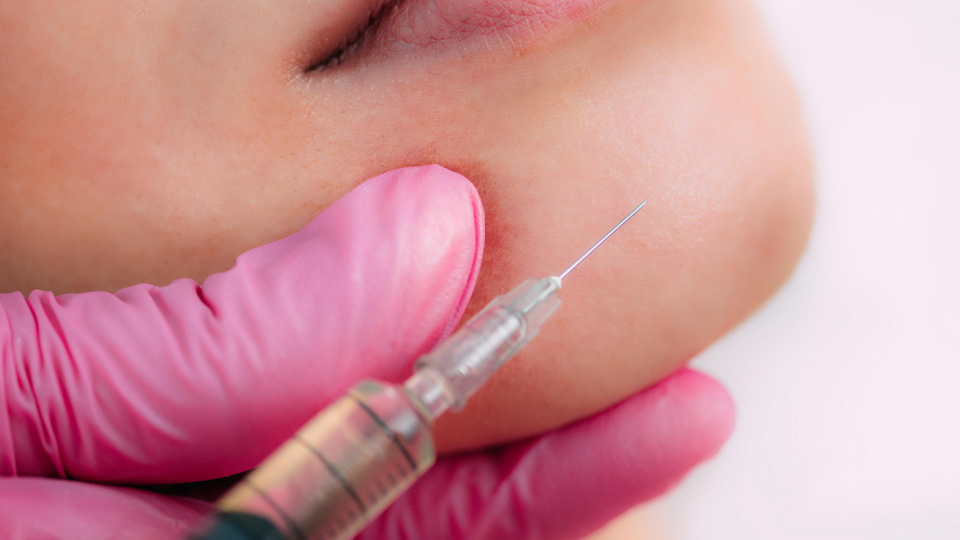
Expected Results
After completing 3-4 standard sessions (at 4-week intervals), the dermal thickness increases by an average of 20%-30%. The peak synthesis of Type I and Type III collagen appears at weeks 8-12 after the first injection. The overall depth of atrophic scars can be reduced by 30%-50%. High-viscosity polynucleotide (PN) remains in the subcutaneous tissue for approximately 14-28 days, generating real tissue volume through fibroblast proliferation and directly producing autologous connective tissue.
Improvement by Type
The surface morphology of atrophic scars determines the residency behavior of the polynucleotide (PN) solution in the subcutaneous tissue. Rolling scars with a diameter greater than 4 mm and a depth of 0.5 to 1.5 mm have extensive fibrous adhesions at the base. When high-viscosity Rejuran S is injected into the deep dermis, it can spread laterally to cover 2 to 3 square centimeters of the wound base. The physical space-occupying effect peaks within 48 hours after injection, with the average thickness of the local dermal reticular layer increasing by approximately 0.8 mm. Fibroblasts begin directional migration along the monomer scaffold formed by PN degradation on day 14. For Caucasian patients with Fitzpatrick skin types I to III, the volume flattening rate reaches 15% four weeks after a single injection. Dermatologists in Europe and America often use combined intervention protocols to treat wide-base depressions:
- 18G or 21G blunt for subcision to cut the pulling fibrous bands
- Immediate injection of 0.05ml of solution into the same tunnel after subcision
- Combination with Microneedling RF thermal stimulation
- External application of 0.1% Tretinoin cream during intervals to accelerate keratin metabolism
- Local cold compress for 15 minutes post-procedure to reduce capillary oozing
Clinical tracking data from three consecutive combined treatments shows that the overall depth reduction value of rolling scars stabilizes at around 45% after 6 months. Ultrasound imaging shows that the anechoic area at 1.2 mm subcutaneously is replaced by a new low-echoic collagen fiber network. The local skin light reflection angle decreases from greater than 30 degrees to less than 10 degrees. Boxcar scars, with edges sinking vertically at 70 to 90 degrees, show strong physical resistance to simple injection filling. The wound diameter is usually between 1.5 to 4 mm, lacking a smooth transition zone. A vertical injection of 0.2ml of PN solution directly underneath can only lift the base tissue by about 0.4 mm. Steep fibrotic edges restrict the uniform penetration of the solution into surrounding healthy tissue. In the EADV scale assessment, using biologics alone to treat boxcar depressions deeper than 2 mm usually results in a tissue filling rate staying within the 25% to 30% range. North American doctors often adjust intervention steps based on the wound diameter, rebuilding the absorption environment through physical destruction.
| Scar Diameter (mm) | Edge Morphology Features | Clinical Pre-intervention Operation | PN Injection Dose (ml/point) |
|---|---|---|---|
| 1.0 – 1.5 | Shallow, sharp angles | 2940nm Er:YAG laser ablation of edges | 0.02 – 0.03 |
| 1.5 – 3.0 | Middle layer, vertical walls | 10600nm CO2 gasification of scar tissue | 0.05 |
| > 3.0 | Penetrating deep to fat layer | Punch Excision | 0.05 injected 2 weeks after suturing |
After ablative lasers smooth the sharp edges, epidermal microchannels remain open for about 48 hours. Laying an injection of high-concentration polynucleotide in the shallow dermis (0.5 to 0.8 mm depth) can shorten the duration of post-laser erythema by 20%. The migration speed of new epithelial cells increases from 0.1 mm to 0.15 mm per day. Ice pick scars extending subcutaneously in a V-shape usually have an opening diameter of less than 2 mm. The complete loss of dermal structure leads to a lack of sufficient fibroblasts in the area to serve as target cells. Injected PN molecules cannot find an attachment scaffold and easily leak to other layers with tissue fluid. Three-dimensional volume measurements show that regardless of increasing dosage or frequency, the base elevation of ice pick scars is less than 10%. Clinical guidelines from Mount Sinai Hospital in New York state that high-viscosity biologics are not suitable for extremely narrow depressions that penetrate the full depth. Standard medical procedures require inducing a controlled chemical burn to close the narrow channel first:
- Use a thin wooden stick dipped in 70%-100% Trichloroacetic Acid (TCA)
- Precisely apply to the bottom of the ice pick opening to produce protein coagulation
- Wait 14 to 21 days for the local scab to fall off naturally
- Re-evaluate after the channel closes and converts to a shallow boxcar type
- Inject 0.01ml PN at the base on day 30 to induce regeneration
The absorption rate of polynucleotides in the base converted by TCA CROSS therapy can increase several-fold. Tiny coagulation necrosis zones are surrounded by new capillary networks, and local blood flow increases by 30% within 4 weeks after injection. Micro-droplet technology can precisely control the amount of drug delivered. Patients with severe acne usually present with a composite distribution of scars. A single cheek averages 15 to 20 depressions of various depths. A single full-face session usually requires 2 to 3 ml of high-concentration PN solution. There is an objective difference of 0.2 mm per second in the diffusion rate of the solution between tissues of different densities. Operators typically use 30G or 32G ultra-fine, inserting them at an angle of 10 to 15 degrees. The spacing between each injection point is strictly controlled between 0.5 to 1 cm to ensure uniform coverage of the dermal reticular layer. The mechanical expansion pressure from injection is approximately 15 to 20 mmHg. Trace tissue trauma itself triggers the wound healing cascade. Released Platelet-Derived Growth Factor (PDGF) reaches peak concentration within 6 hours after injection, creating a synergistic effect with PN macromolecules. Skin histology studies from UCLA confirm that under the dual physical and chemical stimulation of microneedling trauma and polynucleotides, the expression of local hyaluronic acid synthase is upregulated by 40%. Hydration of the extracellular matrix reaches its maximum in the 3rd week, with a visual skin light reflectance increase of 25%.
Tissue Regeneration Cycle
After high-concentration polynucleotide (PN) is injected into the dermal reticular layer, high-molecular-weight chains of 2.5 to 3.0 MDa begin to occupy physical space in the tissue gaps. In the first 48 hours after injection, local osmotic pressure briefly rises from the normal 280 mOsm/L to 310 mOsm/L. Water rapidly gathers in the injection zone, and the dermal water content increases by about 15% on day 3. Macrophages migrate around the injection site within 72 hours, releasing Matrix Metalloproteinase (MMP-1). The high-viscosity gel form degrades by less than 10% between days 7 to 14. Early visual scar flattening is mainly composed of the 0.05ml solution’s occupancy and 20% inflammatory edema. Local blood flow peaks on day 10, with Doppler ultrasound showing a capillary flow velocity increase of 0.4 cm/s. PN long chains are cut by endonucleases in the tissue, releasing free oligonucleotide monomers. [Image of the wound healing process: inflammation, proliferation, and remodeling phases] A2A purinergic receptors on the surface of fibroblasts are activated by a large number of monomers on day 14. The proportion of cells entering the S phase (DNA synthesis phase) jumps from a resting state of 2% to 18%. The number of proliferating fibroblasts increases by approximately 25% on day 21. In vitro culture data from medical laboratories in Europe and America show specific biochemical manifestations after receptor activation:
- Extracellular matrix secretion increases by 35%
- Hyaluronic Acid Synthase-2 (HAS2) gene expression increases by 2.5 times
- Endogenous hyaluronic acid concentration rises from 0.5 mg/ml to 0.8 mg/ml
- Fibronectin deposition speed increases by 15%
- Local antioxidant stress protein Superoxide Dismutase (SOD) levels rise by 20%
The assembly rate of procollagen peptide chains in the rough endoplasmic reticulum grows exponentially between days 21 to 30. Type III juvenile collagen fibers deposit along tissue tunnels at a rate of 0.02 mm per day. At this time, ultrasound detection will find decreased acoustic impedance in the dermis, forming a 3 mm wide low-echoic band. Although the Type III collagen grid is soft, its tensile strength is only 10% of Type I collagen. The improvement in scar smoothness recorded by high-frequency 3D imaging (Visia-CR) at week 4 is mainly supported by the accumulation of Type III collagen. Local erythema (PIE) persists due to the dilation of new microvessels. Extremely intense remodeling reactions occur within the tissue during weeks 8 to 12. Lysyl oxidase concentration rises by 40% on day 60, catalyzing the conversion of Type III collagen into Type I collagen. The level of hydroxyproline generated by cross-linking increases to 4 mg per 100 mg of tissue. At this point, high-frequency ultrasound examination of the dermis shows increased echogenicity, as dense connective tissue replaces the previous low-echoic hydration zone.
| Time Point | Dominant Collagen Type | Dermal Thickness Change | Collagen Fiber Tensile Strength |
|---|---|---|---|
| Day 30 | Type III (70%) | +0.4 mm | 0.8 MPa |
| Day 90 | Type I (50%) | +0.7 mm | 1.9 MPa |
| Day 180 | Type I (85%) | +0.6 mm | 2.8 MPa |
The diameter of thick Type I collagen fibers increases from 40 nm to 100 nm. The tensile strength at the scar base increases from the original 0.5 MPa to over 2.5 MPa. Tissue contraction at a depth of 1.5 mm subcutaneously reaches 12% on day 90. As connective tissue densifies, the microvascular network in the dermal papillary layer begins to regress. Blood flow drops to 110% of the normal physiological baseline by the 4th month. Post-inflammatory erythema (PIE) caused by the initial trauma fades at a rate of 15% per month. International multicenter clinical tracking data show various measurement indicators at the 6-month long-term stability period:
- Average volume reduction of atrophic scars is 45%
- Net increase in dermal reticular layer thickness is 0.6 mm
- Epidermal cell renewal cycle shortened to 24 days
- Transepidermal Water Loss (TEWL) reduced by 18%
- Hyperpigmentation index decreased by 1.5 points
The volume of new tissue generated by autologous fibroblasts no longer relies on the mechanical support of polynucleotide molecules. The injected high-molecular PN is completely metabolized into uric acid by day 180 and 100% excreted from the body. The volume increase deep in the dermis is composed entirely of the patient’s own collagen and elastin. Long-term matrix retention is subject to individual aging processes and the degree of photoaging. Skin aging research from the University of Sheffield confirms that exposure to an average daily UVB radiation of 200 mJ/cm² shortens the half-life of newly generated collagen fibers by 35%. To maintain the fullness of the scar base, some patients receive local supplementary injections of 0.05ml/point at month 12 or 18. The fibroblast proliferation response induced by supplementary injection can be observed in tissue sections by day 7, with the onset time being 50% faster than the first injection.
Long-term Efficacy Evaluation
By the 12th month after completing 4 standard sessions, Type I new collagen dominates the matrix repair of the damaged dermal reticular layer. Vectra H2 3D facial scanning results show that the best scar flattening volume achieved by patients at 6 months has a retention rate as high as 95% after 1 year. Tissue induced by high-concentration polynucleotide (PN) consists entirely of real connective tissue secreted by autologous fibroblasts. Cross-linked hyaluronic acid (HA) fillers are completely degraded by endogenous hyaluronidase within 9 to 12 months after injection. The half-life of mature Type I collagen fibers in the human dermis under normal physiological conditions is as long as 15 years. The volume increase induced by Rejuran S is not limited by foreign body metabolic cycles but strictly follows the biological clock of natural human aging.
The 2024 ESCAD long-term follow-up report points out: at the 24th month after injection, the atrophic scar regression rate in the HA filler group reached 82%, while the average volume regression rate in the polynucleotide (PN) group was only measured at 6.5%.
The 6.5% volume loss rate highly overlaps with the natural aging process of individual patients. For adults over 25, dermal collagen is physiologically consumed at an annual rate of 1.0% to 1.5%. The new tissue scaffold established by polynucleotide treatment also undergoes natural degradation over time. Long-term efficacy maintenance is severely interfered with by exogenous exposome factors. UVA rays with wavelengths between 315 to 400 nm can penetrate to the deep dermis 1 mm below the skin. Excessive UV radiation upregulates the expression of matrix metalloproteinases (MMP-1 and MMP-9), accelerating the cleavage of newly formed collagen peptide chains.
- Unprotected daily exposure to UVA for over 2 hours increases the breakage rate of new collagen by 22%
- Smoking more than 10 cigarettes daily decreases local microvascular oxygen carriage by 15%, causing an extra 8% volume atrophy
- High-sugar diets lead to the accumulation of Advanced Glycation End-products (AGEs), decreasing the elastic modulus by 30%
Mayo Clinic dermatology routinely prescribes L-ascorbic acid serum at concentrations of 10% to 15%. Daily local application can neutralize Reactive Oxygen Species (ROS) and delay the structural destruction of the new scar base by MMP enzymes. External retinoids are widely used in long-term maintenance protocols in North America. Apply 0.025% Tretinoin cream to the whole face three nights a week. Local medication for as long as 24 months can downregulate collagenase activity by 40%, extending the structural integrity of the new dermal matrix by 1.5 times.
The Stanford University School of Medicine facial remodeling guide suggests: receiving a single touch-up injection of 0.05ml/point between months 12 to 18 after completing the basic sessions to fill the 1.5% tissue consumption generated by annual natural metabolism.
Annual touch-up injections initiate the cell memory mechanism of fibroblasts. Local cell populations have already been pre-activated by purinergic receptors, significantly compressing the biological response time to booster doses. The start date of procollagen synthesis is advanced from day 14 of the first treatment to day 7.
- A single touch-up injection adds an extra 0.2 mm to 0.3 mm to the dermal reticular layer thickness
- The hydration index of the dermal papillary layer climbs rapidly by 28% within 15 days
- The density of the connective tissue at the scar base is maintained for over 36 months under 20 MHz high-frequency ultrasound
EADV conducted 3D quantitative measurements on 500 Caucasian patients with Fitzpatrick types II-III. At the 24-month detection point, wide-base rolling scars maintained a stable reduction rate of 41.5% in average total surface area. The synthesis cycle of elastin is much slower than that of collagen; its slow accumulation consolidates mechanical support over a long timeline. At month 18 after injection, the new elastic fibers increase the local skin elasticity value by 18%. The stiffness of the remaining edges of boxcar scars is visually weakened due to the increase in elasticity.