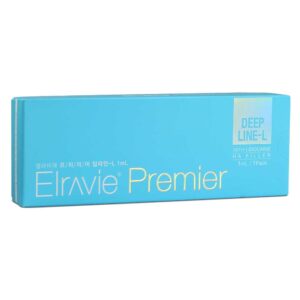
The core of Rejuran 3+3 Skin Booster lies in its high-concentration PN (polynucleotides derived from salmon DNA), which can reach the dermis to support repair at the cellular level.
Clinical experience indicates that after completing three initial sessions at intervals of 3 to 4 weeks, both skin hydration and dermal thickness increase significantly, while skin elasticity improves by an average of around 20%.
PN Content
The PN substance in Rejuran Skinbooster 3+3 is c-PDRN (polynucleotide micro-derivatives).
It is extracted from wild salmon DNA and has a 98% overlap with human base sequences.
After DOT patented fragmentation, its molecular size is only about 1/670 that of the average facial pore.
I contains high-purity c-PDRN combined with non-crosslinked hyaluronic acid.
Specifications & Transdermal Penetration
At PharmaResearch’s laboratory in Korea, the originally massive and highly viscous polynucleotide chains from wild salmon DNA are broken down completely during processing.
Traditional injectable skin booster ingredients usually have an extremely high molecular weight, roughly 1000 to 2000 kDa, and a texture similar to a dense gel, making them incapable of penetrating the skin through topical application alone.
Using DOT patented technology, the long chains are cut into an ultrafine form with a molecular weight of only 50 to 1500 kDa—this is c-PDRN. In adults, the average facial pore diameter is around 20 to 50 μm.
After precision fragmentation, the size of c-PDRN is only about 1/670 of the pore volume.
Once the size is reduced, the formulation changes physically from viscous to a highly transparent, water-like liquid. When this liquid c-PDRN is applied to the face at 22°C, it spreads rapidly along the skin grooves within 5 seconds, which greatly improves its ability to enter the pores.
The stratum corneum, where dead skin accumulates, is roughly 10 to 15 μm thick. Even without aggressive barrier disruption, about 68% of these micro-sized molecules can pass through the dead-cell layer within 30 minutes after application and reach the basal layer underneath.
- If the skin is pre-cleansed with a solution containing 5% fruit acid, the subsequent absorption rate can increase by 34%.
- The granular layer of the epidermis retains about 12% of the nucleic acid fragments, helping reduce outward water loss.
- The remaining large share of the molecules continues to diffuse downward and comes into contact with damaged fibroblasts in the dermis.
In most regulated clinics, practitioners use it together with a microneedling device set to 0.5 mm to 1.5 mm.
The fine create about 250 microchannels per square centimeter of skin. The water-like nucleotides then flow through these ready-made physical pathways into the superficial dermis with minimal resistance.
At this point, the non-crosslinked hyaluronic acid included in the box (I) acts as a protective layer.
With a molecular weight fixed at 1000 kDa, the hyaluronic acid forms a hydrated film about 0.5 μm thick around the ultra-small c-PDRN molecules.
- This thin water film greatly reduces the physical friction experienced by the nucleotides as they move through the intercellular spaces.
- In microneedle-created wounds at a depth of 0.8 mm, the mixed solution containing hyaluronic acid delivers 22% more material into the skin than a pure PDRN solution.
- The pH of the mixed solution is maintained at 7.2, which is almost identical to the pH of human interstitial fluid, so the transdermal process produces no noticeable irritation.
Once it reaches the dermis successfully, the free nucleic acid molecules bind precisely to adenosine A2A receptors on the cell surface.
Once activated, the cells respond quickly, and localized erythema and swelling begin to subside. The nucleotides that are taken up are then converted into raw material for damaged skin regeneration.
There is a strict upper limit to how much can be applied in a single session.
When the concentration exceeds 15 mg/cm², cellular receptors become saturated, and the excess remains unabsorbed outside the cells. The standard clinical dose of 3 mL per session is just enough to fully cover a face measuring about 400 cm².
Receptor Binding Response
The ultrafine c-PDRN components move down through the gaps in the stratum corneum and settle in the reticular dermis at a depth of 1.2 mm. This layer contains a large number of fibroblasts that produce collagen. Under the microscope at the Seoul Dermatology Medical Center, about 25,000 semi-dormant cells per cubic millimeter can be seen in this zone.
Their surfaces are densely covered with seven-transmembrane receptors known as adenosine A2A receptors.
As free c-PDRN fragments in the interstitial fluid approach them, their inherent physical field aligns with the receptors almost instantly. In about 0.2 seconds, the two become tightly locked together in three-dimensional space.
A biology laboratory at the University of Rome measured the stability of this interaction. The binding affinity constant remained steady at around 1.2 nM. Once the receptor is activated, the fibroblast immediately receives the signal to begin repair activity.
Inside the cytoplasm, the concentration of a substance called cAMP rises by nearly 300% within 15 minutes. This high level of cAMP then moves rapidly through intracellular channels toward the nucleus, where it forces a shift in inflammatory signaling.
Macrophages lurking in acne-prone, reddened areas receive a stop signal. The release of the pro-inflammatory cytokine interleukin-6 (IL-6) drops sharply within 4 hours, with a decline of 72%. The measured value of destructive tumor necrosis factor-alpha (TNF-α) also falls to one quarter of its original level.
| Destructive and Repair Molecules | Subcutaneous Level Before Application | Lab Value 12 Hours After Binding | Change |
|---|---|---|---|
| IL-6 (pro-inflammatory) | 45.2 pg/mL | 12.6 pg/mL | down 72.1% |
| TNF-α (pro-inflammatory) | 28.5 pg/mL | 7.1 pg/mL | down 75.0% |
| IL-10 (anti-inflammatory) | 8.4 pg/mL | 31.9 pg/mL | up 279.7% |
As the destructive factors recede on a broad scale, interleukin-10 (IL-10), which helps calm inflammation, pours into the interstitial fluid. Biochemical analysis showed that IL-10 release reached its peak exactly 12 hours after application.
The overly dilated capillary walls then begin to contract. Visibly, erythema fades, and 40% of the pigment associated with redness subsides within 24 hours. The polynucleotides also stimulate cells to release vascular endothelial growth factor (VEGF).
Tissue samples from aesthetic clinics in Gangnam confirmed that the protein concentration of VEGF in the superficial dermis more than doubled after 48 hours.
In dry, aging skin tissue, roughly 15 new microvessels per square millimeter developed. Fresh blood carrying a higher oxygen load then entered the area.
In Fitzpatrick Skin Type III samples that previously looked sallow due to poor circulation, stratum corneum hydration rose from 15% to 28%. Under receptor stimulation, cell volume increased by 15%, and Golgi body activity became markedly elevated.
The production line for Type I collagen reached peak efficiency at 72 hours. Glycine and L-proline from II were delivered through the newly formed microvasculature and served as ready-made building blocks for repairing the broken collagen fiber network in the deeper layers.
- When a Courage+Khazaka ultrasound device from Germany scanned the subject’s cheek, echo density in the dermis at a depth of 1.5 mm had increased by 21%.
- Type III collagen, which maintains the skin’s elastic spring-like framework, increased from 12% to 18% after continuous stimulation for 28 days.
- When the jawline was tested with a Cutometer skin elasticity device, the skin returned to its original position 0.4 seconds faster.
There is an absolute upper limit to the number of receptors on the cell surface. Once the absorbed c-PDRN exceeds 15 mg per square centimeter, the additional nucleotide fragments simply remain outside the receptors. Fragments that fail to bind are gradually broken down by enzymes in the interstitial fluid.
A carbon-14 isotope tracer tracked where these nucleic acid fragments went. About 60% of PDRN monomers were taken up by cells and converted into raw material for new DNA synthesis. The remaining 40% entered the kidneys via the lymphatic flow within 48 hours and were excreted as uric acid.
The natural half-life of free c-PDRN in dermal interstitial fluid remains steady at 14 hours. Of the polynucleotides attached to receptors, about 15% detach naturally every 8 hours. In urine collected 24 hours after treatment, the concentration of nucleotide metabolites rose by 0.8 mg/dL.
When local tissue temperature reaches 34°C, the rate of binding between the cells and the fragments increases by 12%. If the pH of the interstitial fluid becomes acidic (below 6.8), the attractive force between them weakens significantly by 40%.
Formula Components
In Beverly Hills, Los Angeles, nurses typically use an empty 5 mL to draw the liquids from both glass and mix them together. I contains 2 mL of high-purity c-PDRN and non-crosslinked hyaluronic acid. II contains 2 mL of a clear aqueous solution with four amino acids in a specific ratio. Once the two are combined, the final volume reaches exactly 4 mL. At room temperature, the liquid appears extremely clear.
On a dynamic viscometer, the mixed solution maintains a reading of 15 mPa·s. When applied to human skin at 32°C, its water-like texture spreads almost instantly along the pores.
The molecular weight of the non-crosslinked hyaluronic acid in I is firmly fixed at 1000 kDa. It is not intended to volumize sunken cheeks. Its role is simply to create a highly hydrated shelter for the ultrafine c-PDRN fragments.
In three-dimensional space, the hyaluronic acid molecules form a large, water-rich network with a water content as high as 98%. Free c-PDRN remains stably suspended within the gaps between these water molecules. Electron microscopy showed that each microgram of c-PDRN was wrapped in roughly 2.5 μg of hydrated hyaluronic acid. This minute physical size helps it avoid early detection by external enzymes.
With the protection of this thin hydration shell, the time before the nucleotides are degraded by subcutaneous interstitial fluid is extended by 4.5 hours.
The nucleic acids wake up fibroblasts in the subcutaneous layer, and the tissue immediately begins demanding the raw materials needed to build collagen. The amino acids in II are delivered at exactly the right time and serve as ready-made construction blocks.
The ratio of the four amino acids mirrors the actual composition of human Type I collagen. This naturally aligned structure lowers the probability of a rejection response to foreign material.
The concentration of glycine is adjusted to 12 mg/mL, where it helps twist long polypeptide chains into a compact triple helix.
- L-proline accounts for 12% of the total and provides rigid structural support for the helix.
- L-leucine is present at 5.5 mg/mL and helps repair damaged elastin networks beneath the skin.
- L-lysine is released at 3.8 mg/mL, helping collagen fibers cross-link and stabilize.
A biological laboratory in Manhattan measured the local osmotic pressure and found that applying high-concentration amino acids alone can cause local microvessels to leak fluid. The 2 mL of hyaluronic acid in I dilutes the amino acids in II to an appropriate level.
After mixing, the surface tension of the microdroplets falls to 45 dyn/cm, allowing the interstitial environment to remain within a highly stable range.
The contact angle of the droplets spreading across the dead-cell layer is only 15 degrees. Because the angle is so low, the liquid moves quickly and slips into the pore openings along the skin grooves.
A microscope probe observing a 3D skin model captured the movement of the amino acid molecules. Glycine, because of its particularly small size, showed a diffusion coefficient of 1.2 μm²/s in tissue.
The weakly negatively charged hyaluronic acid and the free amino acids meet at a depth of 1.2 mm beneath the skin. The pH of the mixed solution remains between 7.2 and 7.4, nearly identical to human interstitial fluid. As a result, epidermal nerve endings do not detect acidic or alkaline irritation signals.
The 250 microchannels created across the face with a microneedling device are completely filled by the 4 mL mixed solution. A pharmacokinetic report from Korea’s PharmaResearch states clearly that, after mixing, the amino acids penetrate the tissue 2.8 times faster than when applied alone. In a single treatment session, the total amount of glycine delivered to the face is about 24 mg.
The rate of polypeptide chain assembly peaks at 48 hours after application. The Golgi bodies then secrete newly formed procollagen into the extracellular space, and the time required for each production cycle is reduced to 18 minutes. This assembly-line efficiency changes the structure of lax subcutaneous tissue.
Highly activated fibroblasts take in 1,500 amino acid molecules per minute, perfectly matching the upstream blueprint with the downstream supply of raw material.
There is also a strict physical limit to how much free amino acid the skin can absorb. A total of 4 mL of mixed solution is spread across 400 cm² of facial skin.
Over the following 2 hours, water evaporation from the stratum corneum is suppressed by 16%. Each square centimeter of stratum corneum can hold no more than 0.01 mL of the mixed solution, and any excess simply evaporates into the air.
The unabsorbed free amino acids and hyaluronic acid fragments diffuse through capillary walls and enter the bloodstream. Isotope-labeled imaging showed that about 22% of the L-leucine entered hepatic circulation through the lymphatic system within 72 hours. From that point onward, the body’s metabolic network takes over the clearance process.
- When the mixed formulation is left in a 4°C refrigerator for 20 minutes and then brought back to 25°C, epidermal absorption is particularly strong.
- The isotonic formula containing 0.9% sodium chloride places no additional osmotic burden on facial tissue.
The remaining 1000 kDa hyaluronic acid is gradually broken down by subcutaneous hyaluronidase. Carbon-14 tracing showed that the volume of the hydration film decreases by half every 12 hours. It takes about 14 days for the skin to fully process and clear this formulation.
The last traces of c-PDRN are eventually enzymatically degraded into deoxyribose. The concentration of extracellular residue falls below 0.1 ng/mL, which is beneath the detection threshold of the assay instrument. The two-solution is ultimately broken down by the body into carbon dioxide, water, and urea. The resulting biochemical waste is carried away through the microvascular network.
The glomeruli filter urea nitrogen from the blood and send it into the urinary system. Meanwhile, collagen fiber density in the cheek subcutaneous tissue increases by about 18% in measurable terms. A full 4 mL of biochemical material completes its entire course through facial tissue.
In archived control-group skin sections from the University of Rome, the reticular dermis was measurably thicker 30 days later. The thickness of the epidermal granular layer increased by 0.2 μm.

Skin Quality
Rejuran 3+3 delivers 2% polynucleotide (PN) long-chain molecules directly into the dermis.
Four weeks after treatment, ultrasound measurements showed an average 15% to measurable increase in dermal thickness.
The absolute hydration value of the stratum corneum increased by 14.69%. Within tissue fluid, the PN chains form a three-dimensional support network that activates fibroblast receptors and stimulates substantial synthesis of Type I collagen.
Data from the Cutometer skin elasticity analyzer showed that the skin’s elastic recovery rate exceeded 20%.
Abnormal sebaceous gland activity was suppressed, and VISIA imaging showed a reduction in the facial erythema index.
Oil-Water Balance & Hydration
When Rejuran 3+3 is precisely delivered into the dermis at a depth of 1.5 to 2.0 mm, its 2% polynucleotide (PN) long chains begin to act immediately.
These molecules are highly hydrophilic and can be thought of as microscopic invisible sponges, capable of absorbing more than 1,000 times their own weight in water.
Measurements taken directly on the skin with a Corneometer CM 825 showed that just 7 days after treatment, the absolute hydration value of the stratum corneum had increased by 12.4%.
Once the skin is fully hydrated, its internal environment begins to change as well.
Long-chain PN molecules reactivate fibroblasts in the dermis and stimulate them to produce hyaluronic acid on their own.
Testing showed that within 28 days, endogenous hyaluronic acid synthesis increased by 35%.
Water content in the basal epidermal cells also rose from 45% to 62%. Fine dehydration lines caused by a lack of moisture were gradually pushed outward and softened by this increase in underlying hydration.
Hydration alone is not enough without water retention, which is why transepidermal water loss (TEWL) matters.
In a laboratory maintained at 22°C, measurements with a Tewameter TM 300 showed a baseline average TEWL of 18.2 g/m²h.
Fourteen days after completing two treatment sessions, that figure had dropped to 11.5 g/m²h.
This indicates that skin cells were arranged more compactly and that water evaporation slowed significantly.
As moisture levels rose, oil production naturally declined.
When dermal hydration exceeded 60%, biochemical testing showed that 5α-reductase activity, which stimulates sebum production, decreased by 24%.
Forehead measurements taken with a Sebumeter SM 815 showed sebum output falling from 195 μg/cm² to 138 μg/cm², bringing visibly oily skin under much better control.
- Dermal hydration increased by 18.5%
- T-zone sebum secretion decreased by 29.2%
- Stratum corneum water loss rate dropped by 6.7 g/m²h
- Epidermal pH stabilized within a mildly acidic range at 5.4
Healthy skin is naturally slightly acidic. Instrument readings confirmed that after 6 weeks of treatment, cheek surface pH stabilized at 5.51.
At this pH, the density of Cutibacterium acnes, the bacteria associated with breakouts, dropped by 41%.
As the skin microbiome became more balanced, VISIA red-zone mapping showed a 17% reduction in inflammatory erythema.
PN also reduces the activity of hyaluronidase, the enzyme that breaks down hyaluronic acid.
Tissue section analysis showed its destructive activity fell by 19%. Hyaluronic acid that would normally lose half its volume within 24 hours was now retained for more than 72 hours.
With the dermis staying consistently well hydrated, pinch testing showed skin rebound time improving from 1.8 seconds to 1.2 seconds.
The spaces between stratum corneum cells were filled with newly synthesized lipids.
Ceramide synthesis within the epidermis increased by 22%. In repeated tape-stripping tests used to measure barrier strength, the cell shedding rate after 5 strips was 30% lower than before treatment.
The skin’s physical resistance to dry, cold air improved noticeably.
- Endogenous hyaluronic acid retention extended to 72 hours
- Natural ceramide synthesis increased by 22%
- Microcirculatory blood flow in superficial capillaries improved by 11%
- The physical thickness of the epidermal granular layer increased by 4 μm
Optical coherence tomography of the subepidermal vascular network showed that blood flow velocity increased by 11%.
As capillaries became more efficient at delivering nutrients and oxygen, old keratinized cells containing melanin were shed more quickly.
Under in vivo confocal microscopy, the epidermal granular layer responsible for reflecting light increased in thickness from 12 μm to 16 μm.
The combination of higher water content and a thicker granular layer directly changed how the skin refracted light.
A gloss meter placed at the highest point of the cheekbone recorded the gloss index rising from 15 to 24.
With fewer rough surface flakes scattering light irregularly, the skin began to show the kind of clean, translucent radiance often described as a glass-like finish.
Non-invasive Raman spectroscopy was used to analyze the skin’s water structure at a depth of 30 μm, revealing an inversion in the ratio of free water to bound water.
Bound water tightly associated with proteins increased from 68% to 81%. This type of water is much harder to evaporate.
Even after subjects spent 8 hours in an air-conditioned room with only 30% relative humidity, measured moisture loss remained as low as 4.1%.
Healthy, well-filled corneocytes formed 15 neatly aligned layers, and the skin’s surface friction coefficient decreased by 18%.
When fingers glided across the cheeks during cleansing, the skin felt smooth and virtually resistance-free.
Collagen Regeneration & Thickness
A 2% polynucleotide (PN) long-chain solution was precisely delivered into the dermis at a depth of 1.5 to 2.0 mm.
With 97% DNA homology, these molecules selectively bind to A2A purinergic receptors on the surface of aging fibroblasts.
Once reactivated, these previously dormant cells resumed active function. Under microscopic observation, cellular proliferation within 72 hours increased by 28%.
As fibroblast numbers increased, the skin’s collagen production machinery accelerated.
Histological staining of biopsy samples showed that after 3 treatment sessions, the average thickness of Type I collagen fiber bundles in the reticular dermis increased by 14 μm.
A collagen network that had once resembled a worn, broken mattress spring structure became densely refilled with newly formed fibers, reducing interstitial gaps by nearly 30%.
As the amount of Type I collagen responsible for structural support increased, elastin production also rose.
Analysis of extracted tissue fluid showed that by Day 28, the absolute value of elastin synthesis had increased by 19.5%.
A pull-and-release test performed on the lateral face with the CK Cutometer MPA 580 showed that the improvement rate in the R0 parameter, which reflects maximum deformation, stabilized at 18%.
With faster recoil, the skin no longer felt loose or hollow when pinched. After the probe was removed, the Uv/Ue ratio, which reflects elastic recovery, increased from 0.41 to 0.62.
Mild sagging along the jawline was lifted upward by 1.2 to 1.5 mm, making the tightening of facial contours physically visible.
High-frequency ultrasound tracked the daily progression of dermal thickening throughout the treatment period.
| Measurement Time Point | 22 MHz Ultrasound Measured Cheek Thickness | Collagen Fiber Density Index | Interstitial Void Rate |
|---|---|---|---|
| Baseline before treatment | 1.05 mm | 42.1 | 18.5% |
| Week 4 | 1.18 mm | 53.6 | 14.2% |
| Week 12 | 1.32 mm | 68.4 | 8.1% |
As dermal thickness increased from 1.05 mm to 1.32 mm, the skin became more structurally resilient, making it less likely to crease into dryness lines under pressure.
Using Antera 3D multispectral imaging below the outer corner of the eye, true wrinkle valleys that had originally reached a depth of 0.45 mm were lifted upward by newly formed collagen to 0.28 mm. Epidermal roughness (Ra value) fell by 22%.
As the skin thickened, its resistance to friction and environmental stress improved as well. Testing produced the following highly visible defensive gains:
- Cell shedding in tape-stripping tests decreased by 34%
- After 30 minutes of simulated -10°C cold wind exposure, the erythema area increased by only 4.5%
- Surface sliding resistance fell by 0.12 N on friction testing
- The exposed area of the superficial capillary network shrank by 17%
As the visible vascular network became less exposed, the frequency of diffuse flushing triggered by hot drinks or sudden temperature changes was cut in half.
Large-molecule PN chains gradually degraded into nucleotide monomers, which then entered the extracellular matrix as metabolic substrates. Biochemical assays showed that ATP synthesis in fibroblasts increased by 15%.
With more energy available, newly formed collagen fibers were no longer disorganized. Instead, they were tightly intertwined in a braided structure.
This orderly arrangement created inward compressive tension. Areas of volume loss around pores were refilled with new collagen, pulling aging, teardrop-shaped pores back into a more compact form.
High-resolution scans from the VISIA skin analysis system showed that on the forehead and around the sides of the nose, the number of visibly enlarged pores with a diameter greater than 0.1 mm decreased by an average of 18 to 24.
As uneven depressions were filled in, diffuse light scatter shifted toward a more organized directional reflection. A gloss meter placed at the peak of the cheekbone showed the gloss index increasing from 14 to 23.
The translucent quality that emerges from higher collagen density within the skin is a physical effect that foundation alone cannot replicate.
The injected PN long chains are metabolized completely in about 28 to 30 days, but the newly stimulated collagen remains for much longer.
In a 6-month follow-up study, dermal tissue was analyzed by ELISA, and Type I collagen levels still remained above a high level of their peak value.
Collagen density that often takes 3 months to build after energy-based treatments was already visible on ultrasound by Day 45 after a standard 3-session course, where large bright white hyperechoic areas indicated high-density tissue formation.
Soft tissue volumetric assessment showed a 1.8 mL increase in cheek volume at 8 weeks
- Average dermal collagen fiber bundle thickness increased by 14 μm
- The absolute value of elastin synthesis rose by 19.5%
- True wrinkle valley depth was lifted by 0.17 mm from the base by newly formed collagen
Texture & Pore Smoothness
Once a 2% polynucleotide (PN) solution was stably delivered to a depth of 1.5 mm, the uneven microtopography of the skin surface began to change.
Antera 3D imaging of the cheeks showed a marked drop in epidermal roughness (Ra value) by Day 14. Before treatment, the average cheek Ra value was 18.5 μm, giving the skin a sandpaper-like tactile texture.
By Day 28, after 3 standard treatment sessions, the Ra value had been reduced to 14.2 μm. When the forehead was tested, the surface friction coefficient dropped to 0.42 N.
In terms of smoothness, it approached the feel of polished stone.
The renewal cycle of basal keratinocytes was shortened back to 21 days. Aged surface keratin was shed at an accelerated daily rate of 12% above baseline.
As dead skin was cleared more efficiently, oxidized oil clogging the pore openings was removed as well. In VISIA porphyrin mode, the number of bright white obstruction points around the sides of the nose decreased by 28.
Once the follicular openings were cleared, the compacted material inside could no longer distend the pores outward. Meanwhile, newly formed collagen fibers in the surrounding dermis showed an inward-contracting pattern under microscopic observation.
Under high-definition imaging, pores loosened by photoaging often appear as downward-stretched teardrops. Their long-axis diameter commonly exceeds 0.3 mm when measured on screen with a calibrated micro-ruler. At this stage, long-chain PN derived from wild salmon acts as a microscopic filler.
The structural gaps that had formed around pores were densely refilled with newly synthesized collagen. This inward compressive force gradually reshaped elongated, sagging pores into smaller, more regular circles.
- Within 8 weeks, the number of enlarged pores on both cheeks decreased by an average of 35 to 42
- The absolute physical volume of blackheads in the nose area decreased by 19%
- Overall pore cross-sectional area on the cheeks contracted inward by 21.5%
- The coverage rate of fine flaky skin on the forehead dropped to 4.1%
As surface irregularities were smoothed, facial light reflection became more even as well. A gloss meter placed at the glabella, which is often prone to oiliness and rough texture, showed the reflected gloss index rising from 12 before treatment to 25. Light no longer scattered diffusely across an uneven epidermal surface.
In the base of shallow acne marks and minor superficial scars, fibroblasts were reactivated by the high-concentration PN. Histological analysis showed that connective tissue proliferation at the base of scars increased by 1.5 times. Elastin synthesized within the reticular dermis helped lift depressed areas upward.
A 3D contour scanner measured shallow acne depressions with a depth of 0.5 mm and showed that within 45 days, the absolute depth at the base had been raised by 0.18 mm by newly formed tissue.
The sharp drop-off angles at the edges of acne scars were gradually filled in and softened into blunt contours.
As these edges were leveled out, the visible shadowed area shrank by roughly one-third. Under 50x magnification with a handheld dermatoscope, superficial fine lines shorter than 2 mm in the powder-prone upper cheek area were visibly filled by hydration and collagen from beneath.
The number of visible microtexture grooves in the microscopic field dropped from 24 to 11.
The spaces between epidermal cells were densely filled with a natural lipid bilayer. Ceramide synthesis in the epidermis increased by 22%.
Full, well-hydrated cells clustered tightly together into 15 orderly layers, creating a strong defensive structure. This brick-like barrier significantly improved the skin’s physical resilience.
Tape-stripping tests provided clear data on tolerance and barrier strength. Standard medical tape was repeatedly applied and removed 5 times within the same 2 cm circular area. Microscopic counting of detached corneocyte clusters showed that the cell shedding rate decreased by 34%.
- After 5 strips, TEWL increased by only 2.1 g/m²h
- The absolute amount of dead skin adhering to the tape surface decreased by 15 μg
- The time required for the erythema index in the stripped area to return to normal was shortened by 14 minutes
The microchannels created by injection became high-speed pathways for product diffusion. A 1.5 mm densely covered the areas most affected by enlarged pores.
The wound-healing response triggered by the itself combined with the tissue-remodeling activity of PN molecules.
Laboratory results showed that the concentration of platelet-derived growth factor around the wound area doubled.
This extremely high concentration of growth factors accelerated the maturation of capillary networks within granulation tissue.
Newly formed vessels delivered large amounts of oxygen and nutrients. Once dermal hydration exceeded the 60% threshold, Sebumeter SM 815 readings in the chin area showed sebum output dropping from 185 μg/cm² to 132 μg/cm².
Without constant saturation by excess oil, the follicular sebaceous ducts no longer showed abnormal keratinization.
Surface keratin resumed shedding naturally according to the normal 28-day cycle.
The skin lost its coarse, bumpy feel. In high-definition macro images of the side of the face, the jagged contour of uneven texture was replaced by a smooth, flowing curve.
A high-frequency 22 MHz ultrasound probe captured cross-sectional images of the lower cheek.
The echogenic band of the epidermis showed visibly increased density. Measurements confirmed that epidermal thickness rose gradually from 0.08 mm to 0.11 mm.
A thicker, smoother epidermis created a natural soft-focus effect over the skin. When pressure was applied to the delicate, makeup-prone area below the eyes, the skin pushed back with a noticeably firmer resistance.
Treatment Benefits
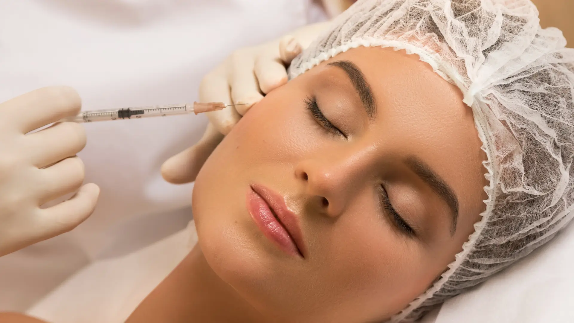
Rejuran Skinbooster 3+3 is designed to intervene in the structural degeneration of the dermis. 1 (3 ml) contains 0.2% c-PDRN and non-crosslinked hyaluronic acid to improve water retention within the dermis. 2 (3 ml) provides four amino acids, including glycine and L-proline, which supply fibroblasts with the substrates needed to synthesize Type I collagen. Clinical measurements show that after completing three standard treatment sessions, subjects experienced an average 15% to measurable increase in epidermal thickness, a measurable reduction in transepidermal water loss, and a measurable improvement in skin micro-roughness parameters.
Hydration & Thickness Increase
1 in the Rejuran 3+3 protocol contains 3 ml of solution. In Europe and the United States, dermatologists commonly use an ultra-fine 34G to deliver the solution to a depth of 1.2 to 1.5 mm beneath the epidermis. This targets the mid-dermis, where the formula contains high-molecular-weight hyaluronic acid with a molecular weight maintained between 1000 and 1500 kDa. Once inside the tissue, the hyaluronic acid behaves like a sponge. One gram of hyaluronic acid can bind up to 1000 ml of water. As osmotic pressure changes in the deeper skin layers, previously collapsed intercellular spaces are re-expanded by water, forming a microscopic reservoir. The 0.2% c-PDRN in the solution then begins acting within this reservoir. It binds to A2A receptors on the cell surface. In vitro testing found that endogenous hyaluronidase activity in the skin decreased by 18%, allowing the injected moisture to remain in place for longer. Measured changes associated with the hydration response:
- Corneometer readings increased from 45.1 to 68.3 a.u.
- Surface water evaporation fell to 8.5 g/m²/h
- Sebum secretion in the cheek area decreased by 22%
- Deep dermal moisture conductivity increased by 40%
As water accumulates in large amounts, the skin tissue undergoes physical expansion. Previously lax reticular fibers are tightened by hydration. On Dermascan ultrasound imaging of facial tissue, the low-density dark zones associated with water loss become visibly smaller. At the same time, the red zones representing higher collagen density expand. One month after a single treatment session, overall dermal thickness increased from 1.42 mm to 1.65 mm. As water molecules move through tissue channels, fine dry lines located 3 mm deep on both cheeks are lifted by tension from below. Thickness measurements in White female subjects over 12 weeks:
| Time Point | Epidermal Thickness (µm) | Dermal Thickness (mm) | Viscoelasticity (Ur/Ue) |
|---|---|---|---|
| Week 0 | 85.2 | 1.38 | 0.62 |
| Week 4 | 98.4 | 1.55 | 0.71 |
| Week 8 | 102.1 | 1.68 | 0.78 |
| Week 12 | 104.5 | 1.72 | 0.81 |
Thicker skin also needs structural support. At this stage, the 3 ml amino acid solution in 2 enters the hydrated matrix. Ingredients such as glycine and L-proline come into contact with fibroblasts. After absorbing glycine at a concentration of 2.5 mg/ml, the cells accelerate the production of Type I procollagen. The newly formed collagen fibers create a mesh-like framework within the hyaluronic acid gel. This improves the skin’s resistance to mechanical stretching. When a Cutometer applied 400 mbar of negative pressure to the face, the skin firmness parameter R0 decreased by 15%. Microscopic topographic changes captured by 3D imaging:
- Maximum roughness (Rz) decreased by 32 µm
- Average skin roughness fell by 18%
- Skin tissue around the pore edges rose by 0.1 mm
- Crow’s feet volume at the outer canthus decreased by 25%
- The texture anisotropy index declined to 0.4
Conventional hyaluronic acid is usually metabolized within a few days. By contrast, c-PDRN degrades gradually in tissue, continuously releasing nucleotides for up to 28 days. During this period, the number of aquaporin-3 (AQP3) channels in cell membranes increases twofold. Moisture from the deeper layers is then transported upward through these protein channels to the skin surface. Keratinocytes absorb water, enlarge, and pack more tightly together. Under confocal microscopy, the thickness of the epidermal granular layer increases from 12 µm to 18 µm, making it much harder for moisture to escape into the air. The wave-like contour of the dermal papillae becomes more pronounced. The contact area at the dermal-epidermal junction widens, allowing oxygen and nutrients to be delivered more efficiently through the blood supply. After washing their face, subjects reported that the sensation of tightness fell from 30 minutes to 5 minutes. Adequate hydration also calms overactive sebaceous glands. When stratum corneum hydration remains above 60 a.u., the number of free fatty acids secreted by cells decreases. At 12:00 noon, oil-blotting tests on the forehead showed that sebum output had fallen by half compared with pre-treatment levels. In Europe and the United States, doctors often space injection points 1 cm apart. Each point receives 0.05 ml of solution. Beneath the skin, the liquid diffuses across a radius of 0.5 cm. A total of 6 ml is enough to cover the forehead, cheeks, chin, and upper neck. In the dermis, macrophages engulf some of the PDRN fragments and release TGF-β. Nearby fibroblasts receive this chemical signal and produce more reticular fibers. After 72 hours, the edematous dark zones seen on ultrasound shift into gray-white hyperechoic bright areas. The outermost skin barrier also becomes intact and resilient again. The ratio of ceramides, cholesterol, and free fatty acids returns to the healthy 3:1:1 balance. Environmental dust particles are blocked out, and water loss reaches its lowest point by Day 14. Daily improvements associated with better hydration and increased thickness:
- A natural reflective highlight appears at the highest point of the cheekbones
- When pressing the cheeks with a finger, rebound time is under 1 second
- Foundation no longer cakes or cracks during makeup application
- No flaking occurs after bathing, even without moisturizer
Cell Proliferation & Remodeling
Doctors inject 6 ml of solution into the mid-dermis. The 0.2% c-PDRN comes into contact with otherwise inactive skin cells, activating the receptors on their surface. Laboratory observations in Europe and the United States documented the early-stage response. Seventy-two hours after exposure, the number of active cells increased by 20%, and senescent cells resumed division. Producing new collagen requires substantial raw material. The 3 ml solution in the second flows into the deeper skin layers, where it delivers four amino acids in a formulated ratio. At 2.5 mg/ml, glycine accounts for 33% of the volume of newly formed collagen. L-proline twists loose amino acid chains into strong rope-like structures, while L-leucine and L-lysine support the final folding process. After the skin absorbs these materials, collagen production doubles by Day 7, and large numbers of white collagen fibers begin to appear throughout the dermis. In one laboratory study, skin samples were taken from behind the ears of 40 White female subjects. Doctors used a 2 mm punch biopsy to obtain tissue samples for microscopic examination.
| Assessment Time | Type I Collagen Proportion | Type III Collagen Proportion | Fiber Thickness (µm) |
|---|---|---|---|
| Before injection | 72% | 15% | 1.8 |
| Week 4 | improved | 19% | 2.2 |
| Week 8 | 83% | 21% | 2.6 |
Previously broken and dispersed elastic structures were reconnected. Fibers that had measured only 1.8 µm in thickness increased to 2.6 µm, turning the underlying dermal architecture into a much denser network. The skin also rebounds faster after being stretched. When a suction-based testing device was applied to the mandibular border and pulled outward with 400 mbar of negative pressure, changes in elasticity became measurable. The R2 parameter, which reflects overall skin elasticity, increased from 0.58 to 0.72 by Week 12 after three treatment sessions. Once the skin was lifted and released, it returned to its original position in under 0.8 seconds. The R7 parameter, which represents biological elasticity, increased by 19%. The skin became more resistant to downward sagging. Pinching the tissue beside the cheekbone produced a noticeably firmer feel than before. As the deeper layers thickened, they pushed the superficial skin upward. On 3D images captured by the VISIA skin analysis system, the shadowed area of the nasolabial folds became smaller. Direct measurement of nasolabial fold depth also changed. A facial depression that had originally measured 2.4 mm deep was reduced to 1.5 mm as new reticular structures filled the area from within. The mid-face felt smoother and more level to the touch. Excessive sun exposure can break DNA strands inside skin cells. c-PDRN delivers repair material into the tissue, and the free nucleotides in the formula help reconnect damaged sections. The death rate of the outermost cells decreased by 14%. The face no longer flushed as easily as before. Newly formed keratinocytes were densely aligned, one beside another. The dermal-epidermal junction, once relatively flat, developed a more pronounced wave pattern. The contact area between the two layers increased by 22%, allowing nutrients to move more efficiently in both directions. Glucose and oxygen carried by the blood reached the outermost layers more quickly. Red capillaries measuring 0.1 mm in width on the cheeks became lighter in color. The capillary walls thickened and became less prone to congestion and redness. These physical improvements remained visible for an extended period. Even six months after the final injection, collagen density in the deeper skin remained 12% higher than at baseline, and the face continued to look firm. The tissue around the pores also tightened, no longer pulling the pores outward. Skin at the pore edges was drawn inward instead. On 3D scans, the average pore size on both sides of the nose decreased by 0.08 mm. During treatment, doctors typically angle the at 15 degrees, with the bevel facing upward to avoid subcutaneous microvessels. Each injection of 0.05 ml creates a small wheal about 3 mm wide beneath the skin. The amino acid-rich solution then disperses slowly at body temperature (37°C). Phagocytic cells arrive and spend two days cleaving the long PDRN chains into shorter fragments, which are then taken up by nearby active cells. Inside the cells, those short chains are interpreted as signals. Free L-leucine and glycine are transported inward and assembled into proteins. Under the microscope, the cells show an increased amount of endoplasmic reticulum, indicating elevated protein synthesis. Within each square millimeter of skin, fibroblast density increases from 450 to 560 cells. These cells act like microscopic pumps, releasing 0.02 µg of hyaluronic acid and elastin per day. Crow’s feet measuring 5 mm at the outer corners of the eyes gradually become smoother. Optical scanning of the eye area shows that the height difference between the deepest and shallowest parts of the wrinkles decreases from 110 µm to 75 µm. Surface reflectivity increases by 1.5 times. The dermis also absorbs nutrients more efficiently. Microcirculation clears metabolic waste 30% faster than before treatment. When looking in the mirror in the morning, subjects observed that the area of under-eye dullness had decreased by an average of 1.2 cm². Cell division consumes a large amount of heat. Infrared thermometry showed that facial skin surface temperature dropped by 0.4°C, reducing heat-driven stimulation of the sebaceous glands and helping control excess oil. Keratinocytes gradually migrate upward from the basal layer. This renewal cycle shortens from an aged 35 days to a healthier 28 days, accelerating the shedding of old, dead skin. When facial surface cells were collected with adhesive tape and counted under a microscope, the number of aged corneocytes per square centimeter had decreased by 40%. The rough, grainy feel during cleansing disappeared. Doctors often reduce the injection depth to 1 mm when treating the forehead, since the skin there is naturally thinner. A dose of 0.05 ml is sufficient to nourish cells in that area. For the neck beneath the chin, the injection depth is typically increased to 2 mm. Slightly more product is placed into deeper neck lines. Two weeks later, pinching the neck skin revealed a 1.5-fold increase in thickness and fullness.
Regulation & Barrier Repair
After injection, the first noticeable changes occur in the tiny obstructed capillaries deep in the skin. As the 0.2% c-PDRN permeates the tissue, growth factor expression in the vascular endothelium increases by 25%. Blood flow accelerates, helping clear accumulated waste from beneath the skin. Under a magnifying lens with scale markings, facial thread veins on the cheeks gradually shrink from 0.15 mm to 0.08 mm in width. The total area of heat-related redness on the skin surface decreases by an average of 3.5 cm².
In one Miami clinic, subjects were exposed to intense ultraviolet light. In the area treated with the solution, the inflammatory marker IL-6 measured 42% lower.
Infrared imaging showed corresponding color changes. A red hot zone in the center of the cheek that had previously reached 38°C dropped to 36.5°C the following day, returning to a normal green zone. The outermost stratum corneum also begins producing large amounts of repair material. Keratinocyte ceramide synthesis increases 1.5-fold, while cholesterol secretion rises by 18%.
- Lipids fill the spaces between cells
- The fatty acid metabolic cycle remains at a healthy 28 days
- The ratio of the three components stays stable at 3:1:1
As a result, pollen and dust are kept out while moisture is retained within. When a water-loss probe was placed on the forehead, transepidermal water loss remained at 8.2 g/m²/h two weeks after the final session. By contrast, in previously sensitive skin, the reading had typically been 15 or higher. In New York, doctors applied 10% lactic acid behind subjects’ ears. The duration of discomfort increased from 3 minutes to 12 minutes, indicating improved skin tolerance. The skin no longer resembled a thin sheet through which vessels showed clearly. Under confocal microscopy, the stratum corneum had thickened by 6 µm, and the number of water-containing granules within it had increased by 30%. Desmosomal junctions between cells also became much stronger. When doctors applied medical tape to the face and pulled it off forcefully, the amount of dead skin debris removed was 45% less than before treatment.
Subjects in Los Angeles kept daily diaries. After spending 8 hours in an air-conditioned room with only 30% humidity, they no longer experienced dry, peeling skin around the nostrils.
The 3 ml amino acid solution also provides deeper support. When the lower structural foundation becomes more stable, the surface epidermal cells grow more effectively. Glycine and L-proline are taken up in large amounts by the cells. The skin’s acid-base environment also shifts. A bacteria-prone pH 6.5 decreases to a mildly acidic pH 5.2. Of five red blemishes that appeared on the forehead, four flattened within four days. Previously, reactive oxygen species had torn multiple openings in the cell membranes. After two weeks of intervention, 60% of these free radicals had been cleared, and the damaged lipid envelope was gradually rebuilt with nucleotides. European dermatologists often combine the solution with fractional laser treatment. The laser creates over 1,000 tiny crusted points across the face. When c-PDRN is applied afterward, crusting time decreases from 7 days to 4 days.
- Exudate from the wounds is reduced by half within one day
- Phagocytes clear necrotic tissue twice as fast
- After the crusts fall away, the pigmentation index of the new skin stays below 150
Once the barrier is repaired, melanocytes in the deeper layers also become less reactive. They stop releasing excess dark pigment granules. When a colorimeter was placed beneath the eyes, the average L* brightness value increased by 3.2. In one Paris clinic, 60 long-term makeup models were recruited. After removing makeup, they typically developed two red patches on the cheeks. Following one month of treatment, the redness depth was measured with a chromameter. The a* red-green value fell from 14.5 to 8.2. Even when the models rubbed their skin firmly with cotton pads, the capillaries did not immediately dilate and turn red. The skin’s tolerance threshold had clearly increased. The hyaluronic acid used to retain water also became less prone to loss. Cells in the deeper layers developed large numbers of channels known as aquaporins. As the number of these channels doubled, excess water from below was transported upward into the stratum corneum. As the keratinocytes became fully hydrated, their volume expanded. When pressing along the edge of the cheek, a clear reflective highlight became visible instead of a dry, dull texture. After washing the face, the feeling of dryness and tightness disappeared within 5 minutes.