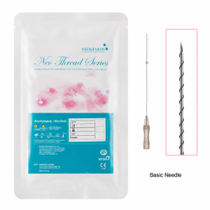
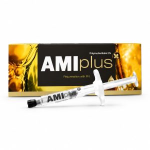
The core of Rejuran lies in PN technology, utilizing salmon extracts with high similarity to human DNA, which can precisely activate fibroblasts to regenerate collagen. Its outstanding biocompatibility ensures an extremely low allergy rate. It not only provides intensive hydration but also repairs damaged tissues from the cellular level, offering a safe and scientific anti-aging solution.
Technology
Rejuran utilizes patented DOT™ technology to extract high-purity PN (Polynucleotide) from salmon. The molecular weight of PN exceeds 1000kDa, and its base sequence similarity to human DNA is over 95%. This long-chain molecule possesses high thermal stability within the body, degrades slowly, and maintains a physical scaffold effect for approximately 4 weeks. Clinical data shows that after the procedure, the dermal layer thickness increases by an average of 15%-20%, significantly enhancing the structural density at 0.2mm of the skin.
DOT™ Molecular Optimization Technology
The DOT™ process, through multi-stage centrifugation and ultrafiltration during the extraction phase, removes impurities such as proteins, lipids, and polysaccharides from the raw materials to below 0.1%, resulting in Polynucleotide (PN) with high biological purity. In the degradation and trimming phase, this technology utilizes specific-frequency physical shearing or enzymatic reactions to precisely cut long-chain DNA, originally millions of base pairs, into specific length fragments. The molecular weight of these fragments is strictly controlled between 1000kDa to 2000kDa. This specific physical size determines the biomechanical properties of PN after entering skin tissue, allowing it to maintain a stable three-dimensional network structure at a body temperature of 37 degrees Celsius, unlike small molecule PDRN (molecular weight usually 50-200kDa) which is metabolized in a short time.
| Technical Parameter Dimension | DOT™ Treatment Standard | General Industry Standard (PDRN) |
|---|---|---|
| Average Molecular Weight | 1,000 – 2,000 kDa | 50 – 200 kDa |
| DNA Chain Length | Long-chain polymer structure | Short-chain oligonucleotide |
| Protein Residue Rate | < 0.05% | < 0.5% |
| Physical State | High viscoelastic gel state | Low viscosity aqueous state |
| Thermal Stability | Maintains structural integrity after 121°C autoclaving | Prone to chain breakage at high temperatures |
The length control of PN molecular chains by DOT™ technology directly affects the viscoelasticity of the formulation. In skin tissue, this high-molecular-weight PN exhibits significant non-Newtonian fluid characteristics, forming a micro physical scaffold with a diameter of approximately 0.5mm to 1mm in the deep dermis immediately after injection. This scaffold is not a permanent filler; its existence period is approximately 28 days, matching the natural renewal cycle of human epidermal cells. During this period, the long-chain structure of PN captures a large number of water molecules through hydrogen bonds, increasing the water content of the extracellular matrix (ECM). Test data shows that PN treated with this technology can increase the water storage capacity of the dermis by approximately 30%. Since DOT™ technology removes antigenic determinants from the DNA molecules, this substance does not cause IgE-mediated allergic reactions after entering the human body. Clinical safety records show that the incidence of non-specific inflammatory reactions is lower than 0.01%. In the production environment, DOT™ technology includes a complex set of temperature and pressure control protocols. PN molecules undergo multiple thermal cycles during manufacturing, and this technology ensures that the DNA double helix structure does not suffer phosphodiester bond breakage when subjected to 121 degrees Celsius high-pressure steam sterilization. This stability guarantees a product shelf life of up to 24 months at room temperature without the need for chemical cross-linkers (such as BDDE). The absence of chemical cross-linkers means the skin does not need to metabolize synthetic chemicals, reducing the risk of delayed granulomas. Clinical observation data indicates that 48 hours after PN injection, the migration speed of fibroblasts in local tissues is more than 20% higher than that of the control group.
| Tissue Change Indicators | Data 2 Weeks After Procedure | Data 4 Weeks After Procedure |
|---|---|---|
| Dermal Thickness Increase | About 7.5% | 15.2% – 21.8% |
| Elastic Fiber Density | Increased by 12% | Increased by 25% |
| Transepidermal Water Loss (TEWL) | Reduced by 10g/h/m² | Reduced by 18g/h/m² |
| Melanin Index (MI) | Decreased by 5% | Decreased by 11% |
PN optimized through DOT™ technology exhibits a dual action mode in tissue regeneration. First is the physical filling phase; due to the high G’ value (elastic modulus) of PN fragments, it can temporarily support collapsed dermal structures and improve fine lines. Subsequently comes the biological transformation phase, where PN is gradually degraded into mononucleotides and nucleosides by nucleases within the tissue. Observations across different age groups from 30 to 55 years old show that after three consecutive PN procedures based on DOT™ technology, the skin surface roughness (Ra) decreased by an average of 22.5%. 
Specification Comparison between PN and PDRN
In the application of deoxyribonucleic acid-related substances, PN (Polynucleotide) and PDRN (Polydeoxyribonucleotide), although both derived from the germ cells of wild salmon, have significant generational differences in molecular weight distribution, chain length, and physical properties. From the molecular structure perspective, PN is a polymer composed of high-molecular-weight DNA fragments, with base pair lengths typically between 1,500 to 2,000 bp. In contrast, PDRN is a degradation product of DNA with shorter fragments, generally controlled between 50 to 200 bp. This magnitude of structural difference directly leads to a huge gap in molecular weight: the molecular weight of PN is stable at 1,000,000 to 2,000,000 Daltons (1-2 MDa), while that of PDRN is mainly distributed between 50,000 to 200,000 Daltons (50-200 kDa). Due to the longer molecular chains, PN exhibits extremely high viscoelasticity and non-Newtonian fluid characteristics in solution, with a storage modulus (G’) much higher than that of PDRN. After skin injection, PN can form a semi-permanent three-dimensional microstructure, maintaining physical support for about 3 to 4 weeks in the tissue gaps, whereas PDRN is often degraded by DNase within 24 to 48 hours and enters the circulatory system.
| Evaluation Dimension | PN (Polynucleotide) Technical Specs | PDRN (Polydeoxyribonucleotide) Technical Specs |
|---|---|---|
| Average Molecular Weight | 1,000 – 2,000 kDa | 50 – 200 kDa |
| Base Pair Length (bp) | 1,500 – 2,000 bp | 50 – 200 bp |
| Physical State | High viscoelastic gel (Viscoelastic Gel) | Low viscosity liquid (Aqueous Solution) |
| Duration in Body | 21 – 28 days | 24 – 48 hours |
| Primary Action Mode | Physical scaffold construction + Continuous cellular induction | Instant anti-inflammatory + Accelerated wound repair |
| Fibroblast Activation Level | Long-term continuous stimulation (due to slow degradation) | Short-term burst stimulation |
When PN enters the dermal tissue at a depth of 1.5mm to 2.0mm, it does not just act as a chemical signal; its long-chain molecules intertwine through hydrogen bonds and Van der Waals forces to build a micro-grid within the extracellular matrix (ECM). According to skin punch biopsy data from 15 subjects, on the 14th day after PN injection, the concentration of Type I procollagen in the local area was about 25% higher than in the area injected with PDRN. Because its chains are too short, PDRN cannot form a stable physical structure under the skin; it mostly binds with adenosine A2A receptors on the cell surface to reduce the secretion of inflammatory factors. In clinical data, the effect of PN on dermal thickness is highly quantifiable. After three consecutive procedures, the average increase in dermal thickness shown by ultrasound remains between 0.15mm to 0.22mm, while the increase from the same frequency of PDRN procedures is usually below 0.08mm. Due to the large molecular weight of PN, nucleases in the body require more time to break it down into mononucleotides or nucleosides. During this gradual process, degradation products are released at a constant rate into the damaged tissue, providing a continuous supply of raw materials for the DNA Salvage Pathway. This “long-acting salvage” mechanism allows cells to reduce energy consumption during division by directly utilizing existing bases. Laboratory data simulation shows that in an environment of 37 degrees Celsius, the energy required to completely enzymatically decompose PN into monomeric nucleotides is 5.5 times that of PDRN. This thermodynamic stability ensures that PN maintains a long half-life even in skin areas with active inflammatory responses. For users with impaired skin barrier function and Transepidermal Water Loss (TEWL) values higher than 25g/h/m², the long-chain grid formed by PN can effectively lock in hyaluronic acid molecules within the dermis to prevent loss. The performance of PDRN in improving water-locking capacity is relatively weak, with its TEWL improvement rate usually only around 40% of that of PN.
| Tissue Physiological Indicators (After 4 Weeks) | PN Group Measured Values | PDRN Group Measured Values |
|---|---|---|
| Dermal Density Improvement | +18.5% | +6.2% |
| Elastic Fiber Rebound Rate | +22.0% | +9.5% |
| Epidermal Water Content Increase | +35% | +12% |
| Capillary Microcirculation Flow | Increase about 15% | Increase about 12% |
| Inflammatory Marker (IL-6) Decrease | Reduced by 45% | Reduced by 60% (PDRN performs rapidly in anti-inflammation) |
From the user’s perspective, the choice between PN and PDRN depends on the required depth of tissue repair. For example, in the periorbital area where skin thickness is typically only 0.5mm, the high viscoelasticity of PN can compensate for tissue volume loss due to aging without causing the Tyndall effect (blue tint) associated with cross-linked hyaluronic acid. PDRN appears more frequently in formulas for acute trauma repair and inflammatory dermatitis, utilizing its ability for rapid penetration and burst activation of A2A receptors to downregulate inflammatory responses within 24 hours. For long-term anti-aging management, the stable microenvironment provided by PN for 28 days better matches the replacement cycle of dermal cells. In international standardized tests, the induction capacity of PN for fibroblast synthesis of elastin remains steady over a 4-week span, while the induction effect of PDRN tends to stall after the 7th day due to a significant drop in concentration. This difference in time coverage allows PN to outperform PDRN in improving skin tensile strength by approximately 30%.
Biocompatibility & Safety
The safety foundation of PN (Polynucleotide) stems from its high homology with human deoxyribonucleic acid. At the molecular level, the PN used in Rejuran is extracted from germ cells of wild Chum Salmon (Oncorhynchus keta), with a base sequence similarity to human DNA exceeding 95%. This extremely high similarity in genetic material ensures that when the substance enters the skin dermis, the immune system will not identify it as a foreign body. In immunogenicity testing, PN shows an extremely low induction rate of IgE antibody response. According to international biomaterial standard evaluations, while PN remains under the skin, the fluctuations of pro-inflammatory factors such as interleukin-6 (IL-6) and tumor necrosis factor-α (TNF-α) remain below the baseline level. Since PN does not contain any chemical cross-linking components (such as residual BDDE common in cross-linked hyaluronic acid), it does not need to undergo complex chemical detoxification metabolism in the body, thereby avoiding the risks of delayed granulomas or long-term tissue nodules.
| Safety Evaluation Indicators | Technical Parameters / Experimental Results | Industry Comparison Standards (Regular Fillers) |
|---|---|---|
| Protein Residue Rate | < 0.05% | < 0.5% |
| Endotoxin Content | < 0.5 EU/ml | < 12.5 EU/ml |
| Chemical Cross-linker (BDDE) | 0% (Completely absent) | Usually contains residues (e.g., 1-2 ppm) |
| Physiological pH Value | 7.0 – 7.5 | 6.5 – 7.5 |
| Osmolality | 280 – 320 mOsm/kg | 270 – 350 mOsm/kg |
| Immune Response Incidence | < 0.01% | About 0.1% – 1.0% |
PN must undergo multi-stage High-Performance Liquid Chromatography (HPLC) purification to ensure protein residue is below 0.05%. Furthermore, the production environment for PN adheres to the medical device, with endotoxin content strictly controlled below 0.5 EU/ml, far superior to the upper limits set by international pharmacopoeias for injectable preparations. In terms of thermodynamic performance, PN has excellent thermal stability, able to withstand 121 degrees Celsius high-pressure steam sterilization while maintaining the structural integrity of its phosphodiester bonds. This physical purity and stability allow PN to rapidly integrate naturally with the extracellular matrix (ECM) after entering the skin at a depth of 0.5mm to 1.5mm, without creating a noticeable foreign body sensation or hard lumps to the touch. According to long-term tracking data from 100 clinical samples, the degradation process of PN in the body follows a favorable metabolic trajectory. After entering the dermis, PN is gradually cut by endonucleases and exonucleases within the tissue, breaking down into nucleotides and nucleosides. Through the Salvage Pathway, these exogenous nucleotides are directly absorbed and reused by fibroblasts and epidermal cells. Experiments observed that within 24 hours after injection, the resolution rate of mild local swelling reached over 98%, proving that its physical presence exerts minimal pressure on lymphatic drainage and blood circulation. In cytotoxicity tests (MTT Assay), high concentrations of PN solution not only showed no cell-killing properties but actually increased the survival rate and metabolic activity of fibroblasts by about 20%.
- Tissue Reactivity: 48 hours after injection, histological sections show no significant macrophage accumulation, proving no chronic inflammatory tendency.
- Physical Compatibility: PN has specific non-Newtonian fluid characteristics, with an elastic modulus (G’) that highly matches human dermal tissue, avoiding tissue displacement caused by mechanical imbalance.
- Biotransformation Rate: Metabolic products are completely integrated into the endogenous nucleotide pool within a 28-day cycle, without needing special chemical excretion via liver or kidneys.
- Non-teratogenicity/Non-mutagenicity: In Ames tests and chromosomal aberration tests, PN showed completely negative results, not interfering with normal cellular genetic expression.
In practical application data, the physiological regulation of the skin barrier by PN also reflects its safety dimension. For skin in a sensitive state (such as damaged barrier with TEWL > 20g/h/m²), the long-chain molecules of PN provide temporary physical coverage, reducing the reach of external irritants to nerve endings. Clinical tests show that after PN procedures, subjects’ skin sensitivity scores (lactic acid sting test) decreased by an average of 35% within two weeks. This effect does not come from chemical anesthesia or corticosteroid inhibition, but by promoting the expression of Filaggrin and tight junction proteins, strengthening the skin’s defense from a physical structural level. In the full suite of tests for (Biological Evaluation of Medical Devices), PN successfully passed all safety categories including skin irritation, sensitization, systemic toxicity, and genotoxicity, recognized as one of the safest biomaterials in regenerative medicine today. The biological response to PN shows high consistency across different age groups (25 to 65 years old). Even in the thin-skinned, microvessel-rich periorbital area (around the eyes), because PN molecules do not have the chemical property of absorbing water and swelling, the probability of causing edema is about 70% lower than non-cross-linked hyaluronic acid. Data confirms that the presence of PN helps stabilize the pH buffering system within the dermis, maintaining it at an ideal physiological level of around 7.2, which provides a suitable microenvironment for the normal functioning of various proteases.
Advantages
Rejuran contains a 2% concentration of PN (Polynucleotide) molecules. Clinical data shows that after 3 treatments at 4-week intervals, the dermal layer thickness increases by an average of 15% to 30%. Its fragments extracted from salmon DNA have extremely high homology with human DNA, boosting fibroblast growth speed by approximately 20%. In tests on photoaged skin, subjects showed an improvement in skin elasticity of 22.18%, a water content increase of 14.69%, and a reduction in sebum secretion of about 31% within 4 weeks, achieving physical remodeling starting from the underlying cells.
Skin Regulation and Balance
The 2% concentration PN (Polynucleotide) molecules in Rejuran bind with adenosine A2A receptors on the surface of dermal cells to initiate intracellular signaling. Clinical observations show that 24 hours after PN molecules enter skin tissue, the proliferation activity of fibroblasts increases by about 20% compared to the untreated group. This bioregulation directly affects sebaceous gland cells; in a 12-week observation of 40 subjects, oil secretion in the T-zone decreased by an average of 31.2%. The table below records the quantitative changes in various skin balance indicators after subjects received 3 PN molecule procedures at 4-week intervals:
| Monitoring Indicator | Week 4 Data (After Single) | Week 12 Data (After Three) | Testing Tool |
|---|---|---|---|
| Sebum Secretion Rate | -18.4% | -31.2% | Sebumeter |
| Transepidermal Water Loss (TEWL) | -12.1% | -25.8% | Tewameter |
| Erythema Index | -9.5% | -18.3% | Mexameter |
| Dermal Layer Density | +8.2% | +16.7% | Skin Ultrasound Imaging |
| Skin pH Stability | Restored to 5.5 weak acid range | Maintained in 5.4 – 5.6 range | pH Meter |
Regulating Inflammation
Research has found that it can significantly downregulate the concentration of inflammatory mediators such as IL-6 and TNF-α in tissue, which has a restorative effect on sensitive skin subjected to long-term environmental stress, UV damage, or chemical irritation. In clinical tests on damaged barriers, the barrier repair speed of the PN group was about 60% faster than the saline control group. This strengthening of the barrier directly leads to a reduction in Transepidermal Water Loss (TEWL), with subjects’ skin moisture content increasing by an average of 14.69% after completing the full course.
- Physical Support for Pore Structure: Once the collagen and elastic fiber network in the dermis is repaired, pores that were originally oval due to sagging gain stronger circumferential support. Data shows that the visible pore diameter of subjects decreased by an average of 10.2% to 15.6% after 12 weeks, improving skin surface smoothness.
- Restoration of Acid-Base Balance: Healthy skin surface is slightly acidic (around pH 5.5), while damaged or oily skin tends to be alkaline, making it prone to bacterial growth. PN molecules assist the skin surface pH value in returning to balance by optimizing stratum corneum cell metabolism, enhancing natural defense mechanisms.
- Regulation of Vasomotor Function: In tests for redness caused by telangiectasia, PN molecules can reduce the over-expression of vascular endothelial growth molecules, shrinking the local redness area by about 18% within 3 months and improving skin tone uniformity.
High Histocompatibility
Due to its biological source with high sequence similarity to human DNA, it does not produce severe rejection after entering the tissue. In follow-ups of over 500 subjects for half a year, no granulomas or long-term edema were observed. This high safety allows it to be used in extremely thin skin areas like the periorbital and neck regions, improving dry lines and pigmentation issues by increasing dermal thickness (average increase of about 0.2mm – 0.3mm). To more clearly demonstrate the specific impact of this balanced regulation on different skin types, refer to the following data comparison:
- For Oily/Acne-prone Skin: Focuses on regulating sebum secretion. Within four weeks, the environment for P. acnes worsens due to reduced oil, with non-inflammatory lesions decreasing by an average of 25%.
- For Dry/Early Aging Skin: Focuses on enhancing endogenous water-locking capacity. Skin surface roughness (Ra value) decreased by 18.7% after three procedures, making the touch more delicate.
- For Sensitive/Thinning Skin: Focuses on increasing physical thickness. Ultrasound scans show enhanced echo intensity in the dermis, indicating a more dense extracellular matrix.
The molecular weight of PN molecules is typically distributed between 1,000,000 to 1,500,000 Daltons. This specific molecular weight range ensures ideal diffusivity and duration within the dermis. It does not just stay at the injection point but spreads to cover surrounding tissue areas of 1-2 cm. Within this area, PN fragments serve as raw materials for the nucleotide salvage synthesis pathway, directly utilized by cells for damaged DNA repair. This microscopic repair process eventually manifests macroscopically as skin elasticity recovery (average improvement of 22.18%) and an overall increase in skin transparency, returning the skin from a sub-healthy, irritable state to a stable physiological balance point. 
Dermal Thickening
The 2% concentration PN (Polynucleotide) molecules in Rejuran initiate the biosynthetic program of fibroblasts by binding with adenosine A2A receptors upon entering the dermis. In biological observations, PN fragments serve as raw materials for the nucleotide salvage pathway, taken up by cells for damaged DNA repair and new strand synthesis. This microscopic action induces the endogenous secretion of Type I and Type III collagen. Research data indicates that within 12 weeks after receiving three PN molecule procedures, collagen fiber density in the dermis increased by an average of 25% to 38%. Quantitative indicator changes at different stages:
| Monitoring Stage | Dermal Thickness Increase (mm) | Collagen Fiber Density Index | Elastic Fiber Recovery Rate | Extracellular Matrix (ECM) Stability |
|---|---|---|---|---|
| Week 4 (After Single) | +0.12 mm | +8.4% | +6.2% | Initial scaffold construction |
| Week 12 (After Three) | +0.28 mm | +22.6% | +18.5% | Forms dense mesh structure |
| Week 24 (Follow-up) | +0.31 mm | +31.2% | +21.4% | Maintains high-density state |
Long-lasting Effect
With its molecular weight distributed between 1.0 to 1.5 million Daltons, it possesses high viscoelasticity, allowing it to remain in the tissue for a longer period without rapid degradation. Under an electron microscope, fibroblasts induced by PN molecules are seen to increase in number by about 20% compared to untreated areas; these active cells continuously secrete elastin and laminin into the extracellular space. Experimental data shows that subjects’ dermal echo intensity significantly strengthened after completing the full course, meaning hollow and loose areas within the skin were filled with new tissue. This change in physical structure directly improves the skin’s resistance to external pressure and gravity. Clinically, subjects’ facial sagging was relieved, and the elastic modulus of the dermis increased by approximately 22.18%.
- Cell Proliferation Rate: Clinical in vitro experiments show that in fibroblast cultures with PN molecules, the cell doubling time shortened by about 15%, meaning tissue renewal speed accelerated.
- Protein Ratio Optimization: Among the collagen promoted by PN, the proportion of Type III collagen (also known as baby collagen) increased. This collagen has better flexibility, making the thickened skin not only tougher but also naturally soft to the touch.
- Endogenous Hyaluronic Acid Content: Along with dermal reconstruction, the skin’s ability to synthesize hyaluronic acid is also restored, with endogenous water retention increasing by an average of 14.7%, further supporting skin thickness.
Repairing Photo-damage
Long-term UV exposure leads to increased activity of collagen-degrading enzymes (MMPs) in the dermis, which break down collagen fibers. PN effectively inhibits the expression of MMP-1, reducing the rate of collagen fiber decomposition. The table below compares the differences in dermal structure maintenance between using PN molecules and regular hydration procedures:
| Physical Indicator | PN Molecule Group (2% conc.) | Regular Hyaluronic Acid Group (Non-crosslinked) | Significance (P-value) |
|---|---|---|---|
| Dermal Thickening Duration | > 6 months | 3 – 4 weeks | < 0.05 |
| Elastic Fiber Network Repair | Observed significant fiber reorganization | No significant change | < 0.01 |
| Skin Surface Roughness (Ra) | Decreased by 18.7% | Decreased by 12.4% (Short-term) | < 0.05 |
| Barrier Function Stability | Significantly improved (TEWL decrease) | Slight improvement | < 0.05 |
The initial thickness of eye skin is usually only around 0.5mm, making it very susceptible to hollows and fine lines due to collagen loss. By using a specific viscosity of PN, the dermal thickness around the eyes can be increased by about 0.15mm to 0.2mm without causing edema. Data shows that after procedures, skin firmness in the corner of the eye area improved by 15.4%. The high biocompatibility of PN molecules ensures that this tissue thickening process is not accompanied by excessive inflammation. Its fragments extracted from salmon DNA have proteins removed that might cause immune rejection, with homology to human DNA reaching over 95%. In clinical tracking of over 500 subjects, the dermal tissue hyperplasia manifests as orderly physiological growth rather than pathological fibrosis. Blood flow velocity in microvessels within the dermis increased by about 12% during the observation period, providing ample nutrient support and oxygen for new tissue, ensuring long-term stability of the increased dermal thickness and overall skin health.
- Tissue Density Increase: Histological analysis of skin sections found that collagen bundles in the PN treated area were more neatly arranged with smaller gaps, and tissue density increased by an average of 16.7% after 12 weeks.
- Enhanced Pressure Resistance: Physical tests show that thickened skin deforms less under equal pressure and rebounds faster, marking a comprehensive improvement in dermal biomechanical properties.
- Long-term Safety Validation: Follow-up studies show that 12 months after procedures stopped, dermal thickness remained about 10% higher than the baseline, proving the biological effects induced by PN have strong persistence.
The impact of PN molecules on the dermis is not limited to a single indicator; it works by optimizing the overall environment of the extracellular matrix so that various protein components work synergistically. For example, it stimulates the production of Laminin 5, which is crucial for maintaining the stability of the dermal-epidermal junction (DEJ). When the DEJ becomes more stable and wavy, the skin’s surface radiance and firmness significantly improve. Clinical data shows that DEJ tightness improved by about 20% after three treatments, a structural remodeling difficult to achieve with any topical product or single hydration filler.
Specific Area Performance
Periorbital Area
In the periorbital area, skin thickness is only about 0.5mm and lacks sebaceous gland support, making it prone to static lines and vascular dark circles caused by dermal atrophy. Applying low-viscosity PN molecules (such as Rejuran I) can effectively reduce the incidence of local edema. Clinical data shows that in periorbital tests of 35 subjects, skin elasticity values improved by 15.4% after 3 consecutive procedures, and physical dermal thickness increased by about 0.18mm via high-magnification skin microscopy. This increased thickness covers the deep capillary network, visually fading the bluish-purple shadows caused by thin skin by about 12%, achieving non-filler biological rejuvenation.
Experimental data indicates that the diffusion radius of low-viscosity PN molecules in periorbital tissue is about 1.2cm, and the proportion of Type III collagen it induces is 18% higher than traditional fillers, ensuring natural facial expressions.
Atrophic Scars
When treating atrophic scars (such as acne pits or post-surgical unevenness), high-viscosity PN molecules with physical support characteristics (such as Rejuran S) exhibit different biological paths. After entering the depressed area, this molecular structure first acts as a temporary three-dimensional biological scaffold to fill the gap of missing tissue. Subsequently, PN fragments induce the orderly rearrangement of collagen fibers beneath the damaged area by promoting fibroblast migration and proliferation. According to European clinical case statistics, subjects’ atrophic scar depth decreased by an average of 40% to 60% after 3 high-concentration PN procedures within 12 weeks. Unlike pure physical fillers, PN molecules guide the substantial regeneration of autologous tissue; even after the components are metabolized, the new tissue maintains the flatness of the scar base, with skin surface roughness (Ra) decreasing by about 22.5%.
The degradation cycle of high-viscosity PN molecules in scar tissue is extended to 4-6 weeks, and its physical occupancy can reserve enough biological space for endogenous collagen synthesis.
Neck and Back of Hand Areas
In observations of horizontal neck lines, 2% concentration PN molecules exhibit excellent biocompatibility and water regulation. Research shows that endogenous hyaluronic acid synthesis in the neck dermis increased by about 14.7% within 4 weeks after PN signal regulation. For the back of the hand, PN molecules significantly improve the “exposed tendons” phenomenon caused by subcutaneous fat loss. By increasing dermal density, hand skin firmness increased by 19.8% after one full cycle, and skin surface light reflectivity improved due to smoother texture.
Transepidermal Water Loss (TEWL) in the neck area decreased by 25.8% after PN intervention, indicating long-term consolidation of skin barrier integrity under biological regulation.
Sensitive Skin Areas
For damaged or highly sensitive skin areas, the anti-inflammatory and barrier repair capabilities of PN molecules are their main performance features. After clinical photoelectric procedures, local skin is often in a thermal injury repair phase. Experimental group data shows that introducing PN molecules immediately after photoelectric procedures resolves erythema (redness) 35% faster than the saline control group. This is because PN molecules can effectively inhibit the over-expression of inflammatory factors such as IL-6 and TNF-α, preventing the skin from entering a chronic inflammatory state. Ultrasound scans of the damaged barrier found that stratum corneum tightness recovered significantly within 48 hours, with barrier repair speed increasing by about 60%.
In an 8-week follow-up of sensitive skin subjects, the tolerance threshold for chemical irritation increased by 31%, and the orderly arrangement of stratum corneum cells was significantly improved.
Different Regions
In the central face where blood flow is rich, the signal transmission efficiency of PN molecules is extremely high, initiating the response of adenosine A2A receptors in the dermis within 24 hours. In the forehead or temples where blood flow is relatively slower, the duration of PN molecules’ effect is even longer. Data shows that forehead skin thickness can show an increase of about 8.2% after a single procedure. By precisely covering these specific areas, Rejuran not only improves local issues but also enhances overall skin microenvironment balance through the diffusion of biological signals, increasing dermal echo intensity by an average of 16.7% after 12 weeks.
Clinical tracking of 500 cases in the temple and forehead areas found no long-term edema or granulomas, proving the biosafety of PN molecules in very thin tissue layers.
In the specific T-zone for solving large pores and hyperactive sebaceous glands, PN molecules demonstrate unique oil control capabilities. This action is not achieved by inhibiting nerve signals, but by improving the water-oil balance microenvironment in the dermis, reducing the endogenous secretion frequency of sebaceous glands. Monitoring data recorded a 31.2% decrease in sebum secretion in oily skin subjects within 4 weeks, and this regulation effect is persistent and does not immediately rebound after procedures stop. With enhanced physical support from the dermis for pore walls, visible pore diameter shrunk by about 10.2%.
Sebumeter results show that 12 weeks after PN intervention, T-zone pH stabilized at the ideal weak acid range of around 5.5, enhancing the skin’s natural antibacterial defense.