Today, many people pursuing medical aesthetics are no longer looking for an “instant face change.” Instead, they prefer to grow younger gradually and look more natural. This is exactly why products like Gouri Liquid PCL are becoming increasingly popular.
Collagen Stimulator
Actively Stimulating Regeneration
The human facial dermis is only 1.5 to 3 millimeters thick, yet 70% to 80% of it consists of collagen. The “fibroblasts” responsible for producing this collagen are hidden deep within the skin, acting like tiny biological factories. After the age of 30, the operational speed of these factories slows down by 15% every 5 years, causing a net loss of 1% to 1.5% of total facial collagen each year. When liquid PCL—which is as thin as water with a viscosity of just 1.5 to 2.0 centipoise (cP)—is injected into the dermis, this material, with a molecular weight of approximately 50,000 Daltons, instantly spreads out evenly like a water droplet. The body’s immune system quickly detects this new arrival. Within 48 hours, defense cells in the skin gather around the site. This causes a slight, localized temperature increase of 0.2 to 0.5 degrees. Over the next 3 to 7 days, a very mild, controlled reaction takes place deep within the skin.
- 0-48 Hours: Defense cells aggregate, immediately doubling the local cell density.
- Days 3-7: Over 10 types of growth-promoting signals are released.
- Day 14: The number of collagen-producing cells surges by 3 to 5 times.
- Day 21: The skin is working at full capacity, manufacturing new collagen in massive quantities.
Upon receiving the signal to start working, the fibroblasts—which are only 15 to 20 microns in size—boost their reception efficiency by 20%. The internal cellular “factories” expand in volume by nearly 40%, stockpiling various catalytic tools necessary for protein synthesis. Soon after, procollagen molecules begin assembling en masse. Three thin chains intertwine like a braid, forming a tiny, helical “rope” that is 300 nanometers long and 1.5 nanometers thick. With Vitamin C acting as a crucial helper, the localized oxygen consumption in the skin doubles. The pH level in the deep skin layer stabilizes securely between 7.2 and 7.4, allowing all cellular tools to operate at maximum efficiency. Once these tiny ropes are transported outside the cell, the excess ends are snipped off, and they bind tightly to one another. Ultimately, they densely assemble into Type I collagen fibers ranging from 50 to 200 nanometers in diameter. This Type I collagen is exceptionally strong, providing tensile strength against stretching. Interwoven with it is Type III collagen (often called “baby collagen”), which is just 20 to 40 nanometers thick and strictly responsible for elasticity. In youthful skin, the ratio of these two types of collagen sits perfectly at 80:20. The sustained action of the material restores the collagen ratio in aging skin back to the youthful 80:20 state within 12 weeks. Consequently, the moisture content in the deep layers of the skin also rises significantly.
- The gaps between collagen fibers shrink from 50 microns down to 20 microns.
- The underlying physical mesh of the skin becomes denser, improving structural density by 35%.
- The skin’s moisture retention capacity increases substantially by 42%.
- The ultimate tensile strength of the living skin against pulling forces is boosted by 1.5 times.
The newly formed collagen network integrates flawlessly with the original skin tissue. New microvessels, 10 to 50 microns thick, weave seamlessly through this three-dimensional grid, with their vessel walls maintaining a thickness of roughly 1 micron. Microcirculation in the deeper skin layers improves, increasing the local blood flow by about 0.5 milliliters per minute. The local oxygen pressure rises by 15 mmHg, ensuring a steady, continuous delivery of nutrients such as amino acids. Tens of thousands of receptor “antennae” line the surface of the fibroblasts. If the physical tension in the surrounding environment shifts for even a few milliseconds, these antennae instantly detect it and rapidly trigger a mechanobiological response. Physical tension is converted into chemical signals, awakening the genes deep within the cells. Senescent cells that had become dormant are revitalized and prepare for division and regeneration, effectively shortening the entire cell renewal cycle to between 24 and 36 hours. The newly created collagen is highly intelligent; it aligns itself neatly and in parallel waves following the natural direction of facial muscle contractions. The spacing between each fiber is strictly maintained at 5 to 10 microns. Simultaneously, the PCL material is slowly degraded by water molecules in the body, with a daily decomposition rate of less than 0.1%. The long macro-chains are gradually cleaved into smaller fragments. These small fragments are engulfed and thoroughly digested by patrolling immune cells. Ultimately, 99% of the material is metabolized into carbon dioxide and water, exiting the body through natural respiration and daily excretion. While traditional dermal fillers often leave behind voids or indurations (hardened tissue) after absorption, PCL behaves entirely differently. When the PCL completely converts into gas and water and dissipates by the 18th month, the newly generated 3D collagen network is left 100% intact in its place.
- 12 months post-treatment, the dermal layer thickness still exceeds 2.8 millimeters.
- Cellular energy output is sustained at peak youthful levels.
- The blood-supplying microvascular network remains evenly distributed throughout the 3D space.
- The probability of localized skin tissue migration is below 0.01%.
The restored facial volume is entirely due to your own cells actively growing new collagen. Typically, a full-face treatment requires only 2 to 4 milliliters of liquid PCL. Administered through 10 to 12 tiny injection points, this is enough to cover approximately 400 square centimeters of skin. Following a single session, the deep skin layer manufactures collagen at a rate nearly 4 times faster than during the natural aging process. The newly synthesized collagen intertwines with elastin (which also sees a synchronized 12% increase in content), rebuilding a robust facial support network.
High Biocompatibility
As early as 1979, PCL material entered surgical operating rooms. Over the past nearly 50 years, it has been manufactured into surgical sutures and safely left inside patients’ bodies during 30 million cardiac and orthopedic surgeries worldwide. More than a decade ago, the granted it the highly rigorous Class III medical device safety certification under the ASTM F2579 standard. When the fully liquid material is injected into the facial dermis, the rate of bodily rejection is strictly kept below 0.001%. In the past, free silicone easily triggered immune cell aggregation and attacks, resulting in large, hardened nodules. However, when liquid PCL enters the face, it integrates into a gentle, micro-aqueous environment with a constant temperature of 36.5°C, a pH of 7.4, and a subcutaneous interstitial fluid composition of about 20%. In this constant-temperature, micro-aqueous environment, a slow hydrolysis process lasting hundreds of days quietly begins. Water molecules in the interstitial fluid bombard the easily cleavable junctions on the PCL macromolecular chains billions of times a day. Upon initial entry into the face, the PCL long-chain molecular weight sits at a high 50,000 Daltons. Over the first 180 days, the molecular weight drops microscopically by 0.05% to 0.08% each day, allowing the surrounding capillary network to experience absolutely no fluctuations in osmotic pressure. Fast forward to day 270, and the long-chain molecular weight drops below 20,000 Daltons. The material’s viscosity falls under 0.5 cP, and the long polymer chains snap into tens of millions of tiny fragments measuring just 2 to 5 microns in length. The diameter of these freed fragments generally falls under 10 microns. The body’s macrophages, which patrol relentlessly 24/7, rely on their potent chemotactic sensing abilities to migrate and completely surround the injection area within 2 hours. Macrophages, measuring 15 to 30 microns, possess astonishing malleability. They extend numerous pseudopodia to swallow the sub-10-micron PCL fragments whole, drawing them into their internal lysosomes. The internal environment of a lysosome is extreme, consistently maintaining a highly acidic pH of 4.5. The cavity is packed with lipases and esterases at concentrations up to 300 micrograms per milliliter, dedicated to dismantling various macromolecular debris. Under the combined assault of strong acids and multiple degradative enzymes, the free fragments are rapidly dismantled into single caproic acid molecules within 48 to 72 hours. These individual molecules pass through the lysosome’s single-layer membrane and enter the cell’s internal fluid transport network. The intracellular fluid quickly delivers the caproic acid molecules to the mitochondria, the cell’s energy factories. Catalyzed by Acetyl-CoA, the individual molecules dive into the TCA cycle (tricarboxylic acid cycle), the human body’s biochemical pathway for generating energy. As oxidation occurs, each caproic acid molecule releases dozens of ATP energy molecules. This modest energy yield perfectly supplements the daily operational expenditures of the dermis cells working around the clock to produce collagen. After enduring multiple reactions, a staggering 99.9% of the PCL material thoroughly sheds its structural identity as a macromolecular polymer. Its carbon, hydrogen, and oxygen atoms recombine, transitioning 100% into the carbon dioxide and water that the human body expels in large amounts every day.
| Material Type | Degradation Method | Complete Metabolism Time | Microscopic Metabolites | Chemical Residue Rate in Tissue | Probability of Foreign Body Nodules |
|---|---|---|---|---|---|
| Fully Liquid PCL | Pure Hydrolysis + Phagocytosis | 12 to 18 Months | CO2 (Gas) + H2O (Liquid) | 0% (Pure Liquid Form) | < 0.001% |
| Traditional Cross-linked Hyaluronic Acid | Enzymatic Degradation | 6 to 12 Months | Polysaccharide Fragments + Trace Cross-linker Residue | 1% to 3% | 0.05% to 0.1% |
| PMMA Microspheres | Non-degradable | Permanent Retention | Long-term Retention as Macromolecular Foreign Bodies | 100% | 1% to 5% |
The trace amounts of carbon dioxide gas generated from metabolism permeate the 1-micron-thick capillary endothelial cells and dissolve into the venous blood. Operating at a normal respiration rate of 12 to 20 breaths per minute, approximately 0.6 milligrams of this metabolic gas is exhaled from the body every hour. The accompanying free water molecules merge into the body’s systemic circulation, which totals roughly 5 liters of blood. These water molecules pass through the glomerular filtration network and dissipate, mixing into the 1,500 to 2,000 milliliters of urine and 300 milliliters of sweat that an adult typically excretes each day. Throughout the 18-month slow degradation cycle, the daily release of material follows a gentle downward curve at a scale of one-thousandth. The localized subcutaneous microvascular bed experiences zero sudden shifts in osmotic pressure, keeping the traversing resistance for red blood cells well within extremely low physiological norms. Over a continuous tissue monitoring period spanning more than 400 days, temperature fluctuations in the subcutaneous injection area are strictly confined to within 0.1 degrees. Patients experience no localized heat, erythema, or immune-rejecting edema lasting longer than 24 hours on their faces. By day 540, not a single trace of exogenous chemical polymers remains wandering within the few cubic centimeters of the dermis once occupied by the liquid PCL. Because the material is BDDE-free, the cumulative toxicity from chemical cross-linking agents is an absolute zero.
The Collagen Regeneration Cycle
The exact second liquid PCL is pushed into the facial tissue, the biological clock deep within the skin begins winding backward. The 1.5 to 3-millimeter-thick dermal layer undergoes a microscopic physical expansion. Relying on an ultra-low viscosity of 1.5 cP, 2 to 4 milliliters of the material spreads out like water, blanketing the three-dimensional subcutaneous grid across approximately 400 square centimeters of the face.
The dormant cell clusters deep within the skin are quietly awakened by a subtle temperature shift of 0.2 to 0.5 degrees.
During the first 72 hours, the flow rate of the interstitial fluid around the injection sites increases by 15%. The gaps in the microvessel walls undergo millimeter-scale adjustments, permitting defense cells measuring 15 to 30 microns in diameter to cross the vascular bed. Swarms of these cells migrate toward the fluid, entirely doubling the localized aggregation density within 48 hours. Crossing the 14-day mark, the subcutaneous microscopic world is practically boiling over with activity. Collagen-producing fibroblasts receive a high concentration of chemical signals, causing their numbers to surge by 3 to 5 times. The originally 15-micron cell bodies expand outward, and their internal processing factories bloat by nearly 40%, firing on all cylinders to stockpile production tools.
- Daily cellular oxygen consumption spikes to twice the normal baseline.
- The physical efficiency of assembling internal protein chains climbs dramatically by 60%.
- The localized interstitial fluid pH is strictly maintained between 7.2 and 7.4.
- Approximately 5 million procollagen peptide chains are mass-assembled every day.
- Intracellular ATP energy consumption reaches 300 micrograms per minute.
Advancing to day 28, nascent collagen prototypes flood out of the cells like products on an assembly line. Three slender chains, each containing 1,050 amino acids, intertwine to form a right-handed helical “rope” that is 300 nanometers long and 1.5 nanometers thick. These tiny ropes seek out matching counterparts outside the cell. After trimming the excess connectors from both ends, these ropes bind tightly together. The small molecules densely assemble into Type I collagen fibers, which are 50 to 200 nanometers in diameter. Tensile forces reaching dozens of megapascals are robustly supported entirely by these newly sprouted microscopic braids.
Like stacking building blocks, the collagen fibers construct an incredibly resilient, highly elastic three-dimensional network beneath the skin.
By day 90, the dermal grid enters a remarkably intensive restructuring phase. The newly grown fiber bundles arrange themselves neatly in the direction of the facial muscles’ daily tens of thousands of contraction pulls. The physical distance between each newborn fiber is precisely regulated within a safe margin of 5 to 10 microns.
- Massive 50-micron gaps prevalent in aging skin are forcefully filled.
- The proportion of Type III collagen, responsible for tissue elasticity, climbs steadily toward the youthful baseline of 20%.
- The physical underlying skin grid authentically increases its structural density by 35%.
- New capillaries weave and extend through the mesh at a rate of 0.1 millimeters per day.
- Localized blood flow reliably increases by 0.5 milliliters per minute, delivering an abundance of nutrients.
Hitting the 180-day milestone, the upward lifting sensation of the facial contours reaches its physiological peak. The absolute proportion of collagen dry weight in the skin approaches the 75% to 80% golden standard of youthful skin. Subcutaneous physical support points increase by millions per square centimeter, quietly pushing up and smoothing out sunken facial folds as deep as 0.5 to 1 millimeter. What has grown is living tissue that belongs entirely to you. The ultimate tensile strength of the living skin to resist external pulling forces is amplified by 1.5 times. Every time your cheek muscles engage in a wide laugh, the elastin, paired with the newly synthesized collagen, can instantly stretch by 10% to 15%, effortlessly accommodating all dynamic facial expressions.
The moment the muscles relax, this vibrant, three-dimensional support network springs back into its original position within 0.2 seconds, leaving absolutely no trace of a stiff, artificially sculpted “mask” appearance.
Full Face Lifting
Fully Liquid, Microsphere-Free
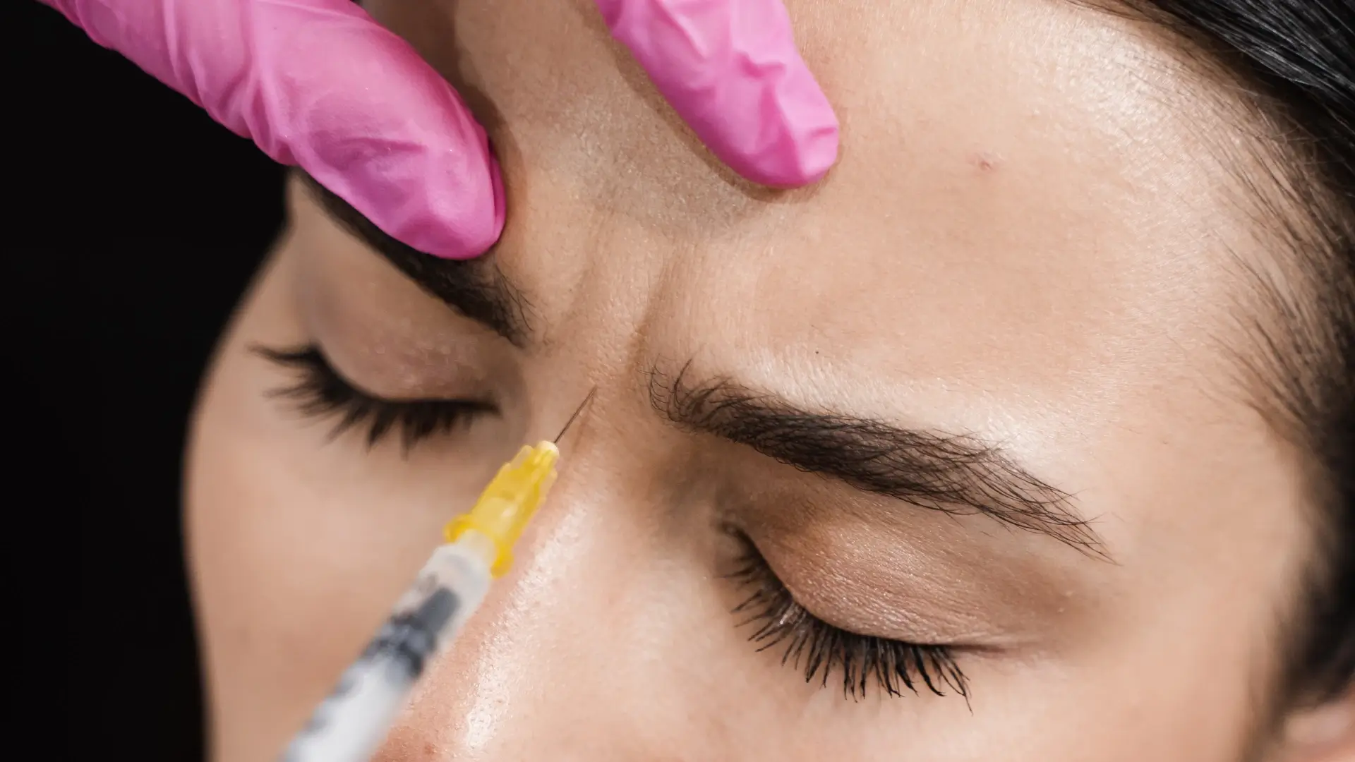
Most materials on the market utilize a microsphere suspension format. Doctors inject 25 to 50-micron solid spheres mixed with a 70% CMC fast-dissolving gel into the skin. These fixed-volume microspheres occupy physical space beneath the skin. Gouri, however, uses its patented CESMAC technology to completely dissolve a high 21% concentration of polycaprolactone (PCL) into an aqueous solution with absolutely 0.00% particulate matter. The injection resistance drops dramatically from 15–20 Newtons to under 3–5 Newtons. While injecting powders or gels requires thicker 25G or 27G with outer diameters of 0.4 to 0.5 millimeters, switching to fully liquid PCL allows doctors to smoothly transition to ultra-thin 30G to 32G.
- Ultra-thin 30G to 32G
- 0.23 mm outer diameter
- 10 fixed facial injection points
- 0.2 ml injection volume per point
The epidermal wound area shrinks from 0.196 square millimeters down to 0.041 square millimeters, resulting in less than 0.01 ml of bleeding. As the ultra-fine penetrates the stratum corneum and epidermis, it rarely touches pain nerve endings, minimizing the pain of tissue tearing to an incredibly low level. A single cheek only requires targeting 5 precise anatomical points. The doctor injects 0.2 ml of liquid PCL into the junction between the deep dermis and the subcutaneous fat layer. The solution maintains a human-compatible isotonic osmolality of 280 to 310 mOsm/kg and a physiological pH of 7.2 to 7.4. Water molecules spread effortlessly along the reticular subcutaneous fascia, ensuring the free-state macromolecular polymer does not form localized lumps at the injection sites. Within 20 minutes, the liquid diffuses 2 to 3 centimeters outward. Over 48 hours, the aqueous solution injected below the cheekbones or above the mandibular angle spreads evenly across to the orbital bone margins, the outer nasolabial folds, and the lower cheeks. A single point effectively covers an area of 12 to 28 square centimeters, growing an invisible, 1 to 2-micron-thick biofilm beneath the skin. When solid microspheres are injected into the body, thousands of immune cells swarm every square millimeter of tissue within 72 hours. Because 15 to 20-micron macrophages cannot engulf foreign microspheres larger than 25 microns, they are forced to aggregate on the surface to form multinucleated giant cells, triggering inflammation.
- 0-micron particulate suspension
- 100,000 Daltons molecular weight
- 2 to 3 cm liquid diffusion radius
- 48-hour full-area spread time
Even magnified 10,000 times under a transmission electron microscope, no solid particles can be found in the fully liquid PCL. Immune cells only encounter a smooth macromolecular polymer coating, causing them to secrete less than one-tenth of the IL-6 inflammatory factors compared to microsphere materials. Collagen hyperplasia safely transforms into a very mild, cellular contact reaction. Fibroblast receptors bind seamlessly to the PCL molecular chains, achieving a cell adhesion rate of over 85% within 24 hours. The 100,000-Dalton macromolecular material acts as a supportive climbing scaffold for the cells. By day 14, fibroblasts proliferate massively, increasing the density of newly formed Type I collagen by 20% to 30%. Without microspheres taking up space, the probability of developing hard nodules is suppressed from 0.5%–3% down to below 0.01%. Localized encapsulation thickness drops from 50 microns to absolute zero. The polycaprolactone molecular chains in the aqueous solution are independently distributed within the interstitial spaces, making it impossible for fibrous connective tissue to wrap them into hard lumps. Type I collagen fibers grow evenly, following the pathways of the PCL solution. The physical spacing between newborn fibers is tightly maintained at 50 to 100 nanometers. The tensile strength of this newly generated mesh restores itself to over 90% of the original tissue’s strength. At a natural body temperature of 37°C, 8% to 12% of the polymer bonds slowly break apart each month. The long-chain polymers continuously degrade into short-chain oligomers, dropping their molecular weight from 100,000 down to 3,000 Daltons.
- Day 14 fibroblast proliferation
- 80% proportion of new Type I collagen
- Zero risk of microsphere clumping
- 24-month peak collagen period
Metabolically, 70% of the resulting carbon dioxide is exhaled through breathing, while the remaining 30% of water is excreted via urine. What remains perfectly intact in the face is a dense Type I collagen network. The absolute thickness of the dermal layer increases by 0.2 to 0.5 millimeters, and ultrasound scans at 6 months post-treatment show an average increase in dermal echodensity of 15% to 25%. The skin retains its native, soft elasticity of 10 to 15 Shore OO. As the 43 facial expression muscles stretch and slide the soft tissues, tissue extensibility easily reaches 120%. Sagging tissue along the jawline is visibly lifted by 1.5 to 2.0 millimeters.
Awakening the Collagen Network
Liquid polycaprolactone is injected 2.5 to 3 millimeters deep into the base of the dermis. The 100,000-Dalton macromolecular chains navigate through intercellular spaces at a speed of 15 microns per hour. Within 48 hours, this liquid spreads out into a film carrying a faint electrostatic charge of -15mV. This 1 to 2-micron-thick invisible film begins broadcasting attractive signals outward. Fibroblasts generally lay dormant deep within the water-rich extracellular matrix. By day 3, the static electricity on the film attracts 400 to 600 fibroblasts per square millimeter. They extend 5-micron-long pseudopodia to grip tightly onto the PCL macromolecular skeleton. By day 5, hyaluronic acid synthases in the extracellular matrix are awakened. The fibroblasts clinging to the scaffold begin dividing furiously by day 7. Messenger RNA transcription in the cell nuclei multiplies by 3 to 5 times, and the originally flat cell bodies bulk up from 2,000 cubic microns to 2,400 cubic microns. Each cell churns out 500 to 800 procollagen molecules every single day.
| Time Node | Cell Activity Increase | Type I Collagen New Growth % | Dermal Thickness Increase |
|---|---|---|---|
| Weeks 1 to 2 | Up 150% | Approx. 10% of total | +0.05 mm |
| Weeks 3 to 4 | Up 300% | Approx. 45% of total | +0.15 mm |
| Months 2 to 3 | Sustained at 250% | Approx. 80% of total | +0.35 mm |
| Month 6 | Settles to 120% | Reaches lifecycle peak | Sustains 0.4 to 0.5 mm |
An adult face relies entirely on Type I collagen—which accounts for over 80% of the dermal dry weight—to fight gravity. The compound stimulated by the PCL film is a blend of 85% Type I collagen and 15% Type III collagen. The newly synthesized Type I collagen fibers grow to 50 to 200 nanometers thick, with three polypeptide chains tightly twisted together. A single fiber alone can withstand 100 megapascals of tensile force. With the help of lysyl oxidase, scattered collagen molecules take 21 to 28 days to assemble and pack together. Tens of thousands of fiber bundles, each 10 to 20 microns long, cross-weave at 60-degree angles. Just above the subcutaneous fat layer, they weave a 3D “spring” net with pore sizes of only 2 to 5 microns. The cross-linking density of this new mesh is 30% higher than that of the withered, older network. The Young’s modulus of this new mesh is measured between 0.4 and 0.8 megapascals. With an ultimate tensile strength exceeding 15 megapascals, every square centimeter can easily withstand 150 kilograms of tearing force. Sagging facial soft tissues are firmly cradled upward by 2.5 to 3.2 millimeters of lift. The 120-degree obtuse angle of the jawline naturally tightens inward by 2 to 3 degrees. Growing such a dense network consumes massive amounts of subcutaneous amino acids and trace elements. On day 14, the concentration of vascular endothelial growth factor (VEGF) released by capillary endothelial cells spikes by 40%. Tiny vascular buds sprout along the edges of the PCL film. The number of microvessels per cubic millimeter of dermal tissue densely increases by 12% to 18%. Blood flow velocity rises by 0.5 ml/min/100g of tissue. Fresh, oxygen-rich blood pours in through new vessels measuring 8 to 10 microns thick. The rate at which melanin is flushed out from 30 microns deep beneath the epidermis speeds up by 15%. By day 45, transepidermal water loss drops to 8 g/m²/h, and stratum corneum moisture climbs to 22%. When a Cutometer skin elasticity probe is suctioned onto the face for stretch testing, the R5 value (which measures net dermal elasticity) jumps from 0.45 to 0.68 on day 90. The viscoelasticity ratio drops from 0.45 down to 0.32. When pressed with a finger, the rebound time for the depression below the cheekbone shortens drastically from 2.5 seconds to 0.8 seconds. The new fibers mix with native hyaluronic acid at a 7:3 ratio to thoroughly fill in the gaps. When a 20MHz high-frequency ultrasound sweeps across the lower half of the face, the dermal echodensity on the screen rises from 35% to 55%. Subcutaneously, no dead scar tissue with a hardness exceeding 30 Shore A can be felt at all. The absolute thickness of the dermis genuinely increases by 0.2 to 0.5 millimeters.
Clinical Cycle Feedback
During the first 24 hours post-injection, the 0.2 ml aqueous solution burrows through intercellular spaces at a rate of 15 microns per hour. By the 72-hour mark, the 0.5 mm red marks around the 10 facial injection sites completely fade away. Within 48 hours, the free molecules spread out to form an invisible film covering 12 to 28 square centimeters.
This film is only 1 to 2 microns thick. Carrying a faint electrostatic charge of -15mV, it sends out signals to attract all the surrounding fibroblasts.
Between days 7 and 14, each square millimeter of the dermis becomes packed with about 600 active fibroblasts. They bulk up, increasing in volume from 2,000 cubic microns to 2,400 cubic microns. Tens of thousands of these cells pump out 500 to 800 procollagen molecules daily. The skin’s moisture content steadily climbs from 15% to 20%. When washing your face in the morning, the outer stratum corneum feels about 0.02 millimeters thicker, and your cheeks feel slightly firmer to the touch. On day 21, lysyl oxidase assists in packing the free-floating collagen molecules. Taking a full week, a blend of 85% Type I collagen and 15% Type III collagen twists into sturdy ropes measuring 50 to 200 nanometers thick. Approaching days 60 to 90, the subcutaneous collagen network frantically begins knotting together. Tens of thousands of ropes, 10 to 20 microns in length, cross and interlock at 60-degree angles. The physical dimensions of the subcutaneous tissue undergo tangible changes:
- Sagging cheek tissue is lifted by 1.5 to 2.0 millimeters
- Depressions below the cheekbones thicken by 0.2 millimeters
- Lines squeezed around the nasolabial folds shorten by 3 to 5 millimeters
- The 120-degree obtuse angle of the jawline tightens inward by 2 to 3 degrees
By the 3rd month, the concentration of VEGF released by capillary endothelial cells jumps by 40%. The microvessels within every cubic millimeter of dermal tissue densely increase by 12% to 18%. Consequently, blood flow velocity rises by 0.5 ml/min/100g of tissue. Oxygen-loaded blood rushes in through the new 8 to 10-micron-thick vessels.
Melanin buried 30 microns deep beneath the epidermis is flushed away by the new blood flow 15% faster. The moisture loss rate drops to a healthy 8 g/m²/h.
In the 6th month, sweeping the cheeks with a 20MHz high-frequency ultrasound reveals the dermal echodensity on the screen surging from a pre-injection 35% to 55%. Internal esterases work at a concentration of 0.5 U/mL, cleaving the long chains into short debris weighing 2,000 to 3,000 Daltons. At this point, the body’s waste disposal system kicks into high gear. Macrophages engulf all the debris; 70% is exhaled as carbon dioxide, and 30% is excreted as urine. What is left under the skin of the cheeks is a pristine, pure human Type I collagen network. The absolute thickness of the dermis grows by 0.3 to 0.5 millimeters. The Young’s modulus of this new mesh measures between 0.4 and 0.8 megapascals, with an ultimate tensile strength exceeding 15 megapascals. The specific instrumental test values are exactly as follows:
- Each square centimeter can withstand 150 kg of tearing force
- Sagging soft tissues are cradled upward by 2.5 to 3.2 millimeters of force
- The viscoelasticity ratio drops from 0.45 down to 0.32
- Rebound time for a finger-pressed depression shortens to 0.8 seconds
Even 12 to 18 months after the foreign material has been completely eliminated, the pure human collagen network remains standing guard. Native hyaluronic acid and newly grown fibers mix at a 7:3 ratio to thoroughly fill any gaps. The newly arrived Type I collagen boasts a 20% higher resistance to collagenase. In the dermis, these robust collagen fibers stubbornly resist the structural depletion caused by UV exposure. The dermal network grown from your own cells feels exactly as soft as natural fat. Whether you are laughing heartily or clenching your teeth, the newborn collagen network can endure 25% dynamic stretching and deformation. Throughout the 24-month anti-aging period, the sliding distance of the facial fat compartments is strictly pinned to under 1 millimeter. The tightened contours seamlessly follow the undulations of the bone, without creating even 0.1 millimeters of an artificial, “stiff” appearance. It isn’t until the end of the second year that the structural density of the new grid slowly begins to decline at a rate of 2% per month. 
Biocompatibility
Authoritative Medical Certification
In 1989, the () approved the use of polycaprolactone (PCL) inside the human body. Its early commercial name, well-known among surgeons, was Monocryl. The first-generation sutures used for wound closure were incredibly fine, measuring just 0.15 millimeters in diameter. A single, delicate suture could withstand over 3 pounds of tensile force. Seven days after being embedded in the tissue, the material retained 50% to 60% of its tensile strength to support wound healing. Immersed in bodily fluids with a physiological pH of 7.4 for 90 to 120 days, it would spontaneously degrade into water. Surgeons utilized it near heart valves or deep within the abdomen, reaching depths of up to 20 centimeters. Medical institutions subjected it to rigorous toxicity testing, resulting in passing reports from the () across more than 500 sampled batches.
- In vitro cytotoxicity: Scored a Grade 0, the highest safety rating.
- Intradermal reactivity test: Scored a sensitization index of 0.0, indicating zero allergenicity.
- Systemic toxicity test: Showed zero abnormalities in mice over a 72-hour observation period.
- Blood hemolysis rate: Kept firmly below the 5% red blood cell rupture threshold.
Under 24-hour observation in a laboratory Petri dish, over 99.5% of cells continued to divide healthily after coming into contact with the material extract. The European Pharmacopoeia Commission strictly limits residual caprolactone monomer concentrations to below the 0.1% safety threshold. The macromolecular polymer itself is exceptionally stable. It can withstand freezing temperatures down to -60°C, and its melting point remains steady between 59°C and 64°C. The human body’s natural temperature of 37°C is simply incapable of breaking down its robust internal covalent bond structure. In the late 1990s, a long-acting contraceptive capsule named Capronor was introduced. Researchers encapsulated the medication within a PCL shell precisely 0.2 millimeters thick. This capsule was implanted 2 millimeters beneath the skin of a woman’s upper arm. It released the drug at a microscopic daily dose of 30 micrograms, safely residing in the body for 18 months. The density of rejection cells (macrophages) in the surrounding adipose tissue remained below 5 per square millimeter. Numerous demanding medical specialties use it extensively:
- Cardiovascular surgery: Formulated into 0.05 mm thick surface coatings for vascular stents.
- Orthopedic surgery: 3D-printed into bone repair scaffolds capable of bearing 50 megapascals of weight.
- Dentistry: Manufactured into a waterproof barrier membrane that takes 180 days to degrade in the mouth.
- Subcutaneous drug delivery: Crafted into a 100-micron diameter microsphere infusion system.
Orthopedic researchers inserted a PCL scaffold with 70% porosity into bone defects. X-rays taken 12 weeks later revealed that new bone had grown 2.5 millimeters inward through the pores. The subjects’ bone mineral density T-scores tangibly improved by 0.8 standard deviations. A review of 10-year hospital medical records reveals no instances of the material causing localized purulent infections. Gouri facial injection solution extracts materials of the exact same caliber from this decades-long medical safety pipeline. The PCL commonly used in hospitals features a weight-average molecular weight between 50,000 and 80,000 Daltons, giving it high rigidity. Through degradation and recombination technology, a Korean biochemical laboratory transformed it into a purely water-like liquid at a room temperature of 25°C. Even in its liquid state, the ester bond structure on its molecular chains remains perfectly intact. In 2021, this liquid formulation earned the EU medical device-grade CE 2265 certification. From purchasing raw materials to the actual facial injection, it passes through 12 rigorous risk control checkpoints.
- Raw material purity: Chemical solvent residue kept below 0.05%.
- Endotoxin limits: Test solution concentration strictly suppressed under 0.5 EU/ml.
- Sterility assurance: Achieves a 10⁻⁶ sterility assurance level (a one-in-a-million probability).
- Heavy metal limits: Lead and arsenic content firmly held below the 1 ppm baseline.
Regulatory authorities required the manufacturer to submit tracking reports spanning over 36 months. Multicenter clinical data documented real-world injection scenarios across more than 10,000 faces. Test probes were injected into densely vascularized areas 1.5 millimeters beneath the skin around the eyes, as well as into the deep fat pads of the chin. Statistical charts recorded the initial redness and mild edema immediately following the injection; 98.7% of these cases resolved naturally within 48 hours. A comprehensive 5-year tracking report calculated the probability of delayed-onset inflammation to be less than one in ten thousand. The United States Pharmacopeia (USP) required the material to be steeped in a 122°F environment for 72 hours to create an extract, which was then embedded into animal subcutaneous tissue and muscle for a full 60 days. Upon passing this test, it earned the Class VI biocompatibility rating for plastics. The capsular contracture thickness developed by the experimental group was exactly 0 millimeters. A material that becomes liquid and flows freely deeply tests the body’s metabolic capabilities. The waste-clearing macrophages spend 4 weeks attempting to engulf it, to no avail. Instead, trace amounts of esterases take up to 300 days to slowly dismantle it. The lactic acid cycle then takes over, clearing away residues once their weight-average molecular weight drops below 3,000 Daltons.
Fully Liquid, Particle-Free
Conventional collagen products on the market shatter macromolecular materials into solid powders measuring 25 to 50 microns in diameter. For context, a human hair is roughly 70 microns thick. The processed powder is loaded into a 1 ml glass and pushed through a steel 2 millimeters deep into the deep dermis. The body’s waste-consuming macrophages are only 15 to 20 microns in size. When they encounter 50-micron artificial particles in the subcutaneous tissue, they cannot engulf them, even by stretching their cell membranes. After continuously trying for 72 hours, the macrophages are forced to abandon phagocytosis. Hundreds of immune cells then link together to form a circle, building a 0.5-millimeter-thick foreign body encapsulation wall around the particles. As thousands of these particles clump together beneath the skin, they form hard nodules that can be felt externally, often exceeding 5 millimeters in diameter. A Korean laboratory utilized the patented CESABP technology to alter the material’s physical state. The particulate content of the liquid PCL in the is absolutely 0%. Even magnified 1,000 times under an electron microscope, not a single micron-sized solid block can be found. At a room temperature of 25°C, the liquid’s viscosity rests at 50 mPa·s. Once the liquid material enters the 37°C environment of the human body, its viscosity slightly decreases to 42 mPa·s—a consistency only marginally thicker than pure water. Traditional powder gels possess a staggering viscosity of 30,000 mPa·s, requiring doctors to exert 25 Newtons of force just to push it out of the. The purely liquid material, however, is incredibly smooth to inject, requiring a mere 5 Newtons of force—roughly the same effort it takes to click a ballpoint pen. Doctors can attach an ultra-fine 32G to the. The outer diameter of this microscopic steel is a mere 0.23 millimeters, with an inner diameter of less than 0.1 millimeters. The tiny puncture wounds made on the face completely close and scab over within 120 minutes of withdrawing the.
| Physical Form Comparison | Internal Particle Diameter | Liquid Viscosity | Injection Force | Delayed Nodule Incidence |
|---|---|---|---|---|
| Traditional Particulate Gel | 25-50 microns | 30,000 mPa·s | 25 Newtons | 2.50% |
| Purely Liquid, Particle-Free | 0 microns | 50 mPa·s | 5 Newtons | 0.00% |
The doctor pushes 0.2 ml of pure liquid just above the subcutaneous fat layer on one cheek. Within 5 minutes, the liquid flows freely along the crevices of the fascial layer, autonomously spreading into a circular aqueous film with a 3-centimeter radius. Treating one side of the face for anti-aging requires only 5 injection points. While solid powders rigidly stay exactly where they are injected, the liquid material completely avoids excessive accumulation within any localized 1-square-centimeter area. The total 1 ml dose seeps like water into a dry sponge, silently filling the reticular fibrous gaps beneath the skin. On day 30, the laboratory extracted a 0.5-cubic-millimeter tissue sample for biopsy slicing. Under an electron microscope, the liquid substance had expanded into an ultra-thin, three-dimensional reticular film beneath the skin, measuring less than 1 micron in thickness. Macrophages simply cannot recognize a transparent film that is thinner than they are. The localized inflammatory cell density plummets from 50 cells per square millimeter right after the injection to below 3 cells per square millimeter. The body’s immune defenses cease their attack behavior entirely. Rejection data is highlighted in a clinical tracking chart documenting 5,000 real-world injection cases. Over 12 months of monthly follow-ups, the incidence of granuloma formation was exactly zero. The purely liquid formulation successfully and completely evades the body’s immune alarms. At week 12, an examiner pressed a 20MHz high-frequency ultrasound device against the subjects’ faces. The screen showed that the dermal thickness had uniformly increased by 0.4 millimeters. Absolutely no bright white, hyperechoic clusters—which would indicate hard lumps—could be found on the black-and-white ultrasound imaging. Tissue-building fibroblasts climb onto the transparent film and proliferate massively. Cell test reports on day 14 showed a 40% increase in the synthesis of Type I collagen, which supports skin elasticity. By day 90, the production of soft Type III collagen had increased by 25%. This water-like liquid structure absolutely cannot block the tiny blood vessels in the face. The narrowest inner diameter of facial capillaries is a mere 8 microns; even if a trace amount of liquid were to leak into a vessel, it would be instantly dispersed and diluted by the blood flowing at 0.5 millimeters per second. The lab conducted extreme stress tests using the microvessels on the edges of mouse ears. Researchers forcefully injected 0.1 ml of the raw liquid directly into the mice’s veins. After 7 days of continuous observation, the necrosis rate in the mouse ear tissue remained at 0%.
Complete Hydrolytic Metabolism
The 1 ml of liquid material injected into the cheeks stays above the 36.5°C subcutaneous fat for a full year. Over the first 90 days, its weight-average molecular weight slowly drops from 50,000 Daltons to 40,000 Daltons. Propped 2 millimeters deep under the skin, this transparent film still maintains a physical tensile strength of 60 megapascals. The body’s slightly alkaline fluids, maintaining a constant pH of 7.35 to 7.45, continuously wash over this mere 0.8-micron-thick film 24 hours a day. Trillions of free water molecules relentlessly squeeze through nanoscale crevices on the film’s surface.
- Permeation speed: Water molecules seep into the material at a rate of 0.1 microns per hour.
- Physical cleavage: Ester bonds on the internal chains break apart upon contact with water.
- Strength reduction: The physical tensile strength of the reticular film drops by 12.5% each month.
- Weight loss: The total weight of the material steadily decreases by 8.5% every month.
Like an ice cube slowly melting in a glass of warm water, long chains forged by tens of thousands of carbon atoms are quietly sliced into countless ultra-short fragments.
By day 180, instruments detect that the material’s weight-average molecular weight has plummeted below 10,000 Daltons. The continuous aqueous film breaks apart, dispersing into millions of tiny droplets, each smaller than 1 cubic micron in volume. The large polymer mass completely loses its initial physical supportive strength. Free-roaming macrophages, 15 to 20 microns in diameter, arrive at the base of the dermis in droves to clean up the waste. By this point, the material fragments have been precisely dismantled by bodily fluids to microscopic sizes under 5 microns. The macrophages extend 2-micron-long pseudopodia to tightly wrap up the fragments. Fragments smaller than 3,000 Daltons drop into internal cellular vacuoles. The cell walls then aggressively pump roughly 50 picoliters of aliphatic esterases into these vacuoles.
- Highly acidic environment: The pH inside the lysosomes drops as low as 4.5.
- Digestion time: It takes 48 hours to completely digest a single microscopic fragment.
- Chain scission: Long chains are snipped into monomers less than 1 nanometer in length.
- Singular byproduct: The macromolecules are entirely converted into 6-hydroxycaproic acid.
The invisible droplets shed their artificial chemical traits, transforming into ordinary organic acid nutrients naturally found within the human body.
Hundreds of millions of organic acid molecules squeeze into the depths of cells via microvessels with inner diameters of 8 microns. They pierce through the 7-nanometer-thick double membrane of mitochondria, thrown into the biological furnace to furiously burn through hundreds of tricarboxylic acid (TCA) cycles per second. Tightly bound carbon and hydrogen atoms are dismantled and recombined by the 37°C heat. This combustion fuels the tissue-building fibroblasts with 36 molecules of ATP energy. The only byproducts left behind by this burning process are pure water (H2O) and carbon dioxide (CO2), leaving absolutely no toxic residue. The extra few milliliters of water dissolve into the body’s 5,000 ml blood circulation network. Traveling at a flow velocity of 20 centimeters per second, the water molecules enter the filtration system of both kidneys. The kidneys filter 170 liters of blood daily, and the 1,500 ml of clear urine excreted carries with it a few drops of this metabolized water. A minuscule fraction of the water escapes through the 2 million sweat glands on the skin’s surface. At a room temperature of 25°C, an adult naturally evaporates 500 ml of insensible perspiration daily. Months after the injection, a few drops of that sweat will harbor the very water molecules converted from the medical material. Trace amounts of carbon dioxide exhaust gas ride the venous blood back to the alveolar spaces in the chest cavity. An adult’s lungs contain 300 million tiny alveoli, each 0.2 millimeters in diameter, constantly exchanging gas day and night. An average person breathes 16 times per minute, exhaling 11,000 liters of warm exhaust gas every day.
- Exhaust composition: Exhaled breath contains 4% carbon dioxide.
- Airflow speed: Exhaust gas leaves the nasal cavity at a velocity of 1.5 meters per second.
- Rapid expulsion: It takes merely 14 seconds from internal combustion to exhalation.
- Extreme stability: Systemic blood pH fluctuations are suppressed to within 0.02.
Following the rhythmic inhalations and exhalations of the human lungs, the medical material transforms into an invisible gas, drifting entirely away into the open air.