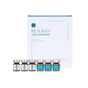
Rejuran functions fundamentally through its proprietary active ingredient, Polynucleotide (PN), a heat-resistant biological polymer derived from wild salmon germ cells that exhibits a 98% DNA base pair similarity to humans. Instead of relying on artificial cross-linking agents, the treatment utilizes DNA Optimizing Technology (DOT™) to create a viscous, three-dimensional scaffold within the dermis that physically supports fibroblast proliferation and collagen production. This long-chain DNA structure degrades via the metabolic salvage pathway, supplying cells with pure nucleotide raw materials to fuel deep tissue repair, a mechanism clinically proven to increase dermis density by 14.66% and significantly enhance elasticity after four weeks.
Mechanism
The mechanism of Rejuran originates from its main ingredient Polynucleotide (PN). After entering the dermis, PN is decomposed by nucleases, providing ready-made nucleotide raw materials for cells through the Salvage Pathway, so that DNA synthesis does not require a large amount of ATP (Adenosine Triphosphate) consumption. PN molecules also act as ligands, specifically binding to the A2A adenosine receptors on the surface of fibroblasts, activating the cAMP signaling pathway and promoting the secretion of Type I and Type III collagen. Clinical histological data shows that after 4 weeks of treatment, dermal density increased by 14.66%, stratum corneum thickness increased by 20%, and the expression of VEGF (Vascular Endothelial Growth Factor) was significantly upregulated, improving microcirculation.
Salvage Pathway
Cellular Materials
When human skin cells undergo division, proliferation, or self-repair, they must first complete the replication of DNA (Deoxyribonucleic Acid). This process requires a large amount of raw materials—nucleotides. There are only two ways for cells to obtain nucleotides: De Novo Synthesis and Salvage Pathway. In young, metabolically active organisms, de novo synthesis dominates. Cells use small molecule compounds such as amino acids (e.g., glutamine, aspartic acid), formate, and carbon dioxide to assemble nucleotides bit by bit through more than ten steps of complex enzymatic reactions. It is like starting from mining ore and smelting metal to eventually manufacture a screw. Although self-sufficient, this process consumes a lot of energy (ATP) and is very slow. With age, mitochondrial function in skin cells declines, and ATP generation efficiency decreases. Aging fibroblasts are often in a “state of energy deficiency,” making it difficult to support high-energy-consuming de novo synthesis. At this time, the speed of cellular DNA repair and replication slows down, directly manifesting as slower skin healing and reduced collagen synthesis.
Energy Conservation
It does not manufacture from scratch but utilizes ready-made bases (Base) or nucleosides (Nucleoside)—these usually come from DNA/RNA fragments degraded within the cell or from exogenously ingested substances. In this pathway, cells only need to undergo simple phosphoribosyl transfer reactions to convert these “semi-finished products” into usable nucleotides.
- De Novo Synthesis: Synthesizing each nucleotide molecule typically consumes 6 to 7 ATP equivalents of energy.
- Salvage Pathway: Utilizing ready-made bases to synthesize nucleotides typically consumes only 1 ATP equivalent.
For aging cells with insufficient energy reserves, salvage synthesis is not only a shortcut but also a necessary means to maintain normal physiological functions. However, aging tissues often face an awkward situation: although they have enzymes (processing machines), they lack sufficient free bases and nucleosides (raw materials).
Dismantling PN Molecules
The long chains of PN (Polynucleotide) after Rejuran injection are gradually degraded by endonucleases in the body within a few hours to a few days after entering the dermis. After the PN chains break, a large amount of Purines and Pyrimidines and their derivatives are released.
- Adenine and Guanine: Through the action of hypoxanthine-guanine phosphoribosyltransferase (HGPRT), they are rapidly converted into AMP (Adenosine Monophosphate) and (Guanosine Monophosphate).
- Thymine and Cytosine: Through the action of enzymes such as thymidine kinase (TK), they are converted into TMP (Thymidine Monophosphate) and CMP (Cytidine Monophosphate).
Since the base composition ratio of salmon DNA is highly consistent with human DNA (similar ratios of adenine, guanine, cytosine, and thymine), the utilization rate of these degradation products is extremely high, with almost no metabolic waste produced.
Accelerating Enzymatic Reactions
When the dermis is filled with nucleosides produced by PN degradation, the activity of salvage synthesis enzymes within the cell is activated by the increased substrate concentration. Under normal physiological conditions, the speed of DNA synthesis is often limited by the activity of Ribonucleotide Reductase (RNR) and the supply of substrates. The exogenous nucleosides provided by Rejuran directly bypass multiple rate-limiting steps in de novo synthesis.
- S-phase Acceleration: The S-phase in the cell cycle is the stage where DNA replication occurs. An adequate supply of nucleotides can shorten the residence time of cells in the S-phase, allowing fibroblasts to complete division faster.
- Repair Efficiency: The skin is exposed to ultraviolet rays every day, which cause adjacent pyrimidines on the DNA chain to form Pyrimidine Dimers. Cells need to excise this damaged DNA and refill it. This process (Nucleotide Excision Repair, NER) is highly dependent on whether there is an adequate reserve of dNTPs (Deoxyribonucleoside Triphosphates) in the nucleus. The metabolic products of PN quickly fill this reserve.
Shifting Energy Allocation
When cells no longer need to deplete ATP for synthesizing DNA raw materials, the saved energy is redistributed to other physiological activities. For fibroblasts, this means more ATP is available for:
- Protein Translation: Synthesizing macro-molecular Type I collagen and elastin.
- Secretion Activity: Excreting synthesized collagen into the extracellular matrix.
- Cell Migration: Around micro-trauma wounds (such as holes), fibroblasts need to consume energy to migrate to the damaged site for repair.

Activating Adenosine Receptors
Release of Signaling Molecules
After Rejuran enters the human body, its long-chain Polynucleotides (PN) do not exist merely as passive fillers. As the phosphodiester bonds are cleaved by enzymes in the body, PN gradually decomposes into smaller fragments, eventually releasing a large amount of free nucleosides, the most active signaling molecule being Adenosine. In the skin microenvironment, adenosine is usually a “distress signal” released by cells under hypoxic or damaged stress. However, in aging skin, this signal is often weak and insufficient. Rejuran injection artificially creates a high concentration of adenosine in the local environment. This does not mean the cells are truly damaged; rather, it simulates a biochemical environment requiring urgent repair, tricking the cellular sensing system into initiating coping mechanisms.
Switches on the Cell Membrane
Released adenosine molecules wander in the interstitial spaces until they collide with specific receptors on the surface of fibroblasts or immune cells. The main target of Rejuran’s action is the A2A adenosine receptor. The A2A receptor belongs to the G protein-coupled receptor (GPCR) family. It is a protein structure that spans the cell membrane seven times. When the adenosine molecule precisely fits into the binding site on the exterior of the receptor, the receptor undergoes a conformational change (shape change).
- Conformational Change: The part of the receptor on the inner side of the cell membrane twists.
- G Protein Activation: This activates the Gs protein (stimulatory G protein) connected to the inside of the receptor.
- Enzyme Activation: The Alpha subunit of the Gs protein detaches, moves, and activates another enzyme on the membrane—Adenylyl Cyclase.
Through this series of precise mechanical transmissions, chemical signals from outside the cell successfully cross the cell membrane and are transformed into internal instructions.
Intracellular Relay Race
Once adenylyl cyclase is activated, it begins to manufacture a large amount of the “second messenger”—cyclic Adenosine Monophosphate (cAMP). The concentration of cAMP surges within the cytoplasm, acting as an internal “amplifier.” High concentrations of cAMP seek out and activate their downstream target: Protein Kinase A (PKA). Activated PKA is a multi-functional “foreman.” It can not only phosphorylate various proteins in the cytoplasm, changing their activity, but can also pass through the nuclear membrane into the nucleus.
- Entering Command: After entering the nucleus, PKA phosphorylates cAMP Response Element Binding Protein (CREB).
- Modifying Blueprints: Phosphorylated CREB binds to specific regions of DNA, initiating or turning off the transcription of specific genes.
This is the complete pathway by which Rejuran changes skin status from the molecular level: from extracellular adenosine all the way to DNA expression within the nucleus.
Closing Inflammatory Pathways
In many skins with spots, sensitivity, and persistent redness, the interior of the cells is typically in a state of chronic inflammation. In this state, the NF-kappaB pathway within the cell is abnormally active, continuously producing pro-inflammatory factors (such as TNF-alpha, IL-6). By activating A2A receptors and the subsequent cAMP signaling pathway, Rejuran can strongly inhibit the activity of NF-kappaB.
- Blocking Signals: High levels of cAMP can prevent NF-kappaB from entering the nucleus.
- Factor Switching: Experimental data shows that activation of A2A receptors can reduce the secretion of the pro-inflammatory factor TNF-alpha by 30% to 50% (depending on the degree of inflammation) while significantly upregulating the expression of the anti-inflammatory factor IL-10.
This mechanism is not simple “anti-inflammation”; it switches the tissue microenvironment from a “pro-inflammatory/destructive mode” to an “anti-inflammatory/healing mode” by adjusting the balance of cytokines.
Promoting Angiogenesis
Issuing Hematopoietic Instructions
As the capillary network thins and atrophies, skin cells are in a chronic state of “hypoxia” and “starvation”. The first step for Rejuran to reverse this process is to issue biochemical instructions for angiogenesis at the molecular level. The core of this instruction lies in the transcription and expression of Vascular Endothelial Growth Factor (VEGF). When PN (Polynucleotide) is decomposed into nucleosides and activates the A2A adenosine receptor, intracellular cAMP levels rise, thereby phosphorylating the transcription factor CREB. Phosphorylated CREB enters the nucleus, binds to the promoter region of the VEGF gene, and forces the initiation of that gene’s transcription. At this stage, fibroblasts act as “signal transmission towers.” They synthesize and release large amounts of VEGF-A into the extracellular matrix. These soluble protein molecules diffuse in the interstitial fluid. Once they contact the VEGFR-2 receptors on the surface of nearby vascular endothelial cells, the cascade reaction of angiogenesis is immediately initiated. Data shows that within 48 to 72 hours after PN intervention, VEGF concentrations in local tissues reach a significant peak, providing sufficient signal concentration for subsequent physical construction.
Endothelial Cells Set Out
Vascular endothelial cells (the cells forming the inner walls of blood vessels) that receive VEGF signals undergo dramatic morphological changes. Originally tightly connected, resting endothelial cells begin to loosen, and the connection bonds between them (such as VE-cadherin) are temporarily unfastened. This process involves not only cell separation but also local degradation of the basement membrane. Endothelial cells secrete enzymes called Matrix Metalloproteinases (MMPs), which dissolve the basement membrane wrapped around old vessels. Subsequently, endothelial cells differentiate into two different roles:
- Tip Cells: These are like pathfinders. They extend slender filopodia to sense VEGF concentration gradients in the environment, leading rear cells to migrate towards high-concentration Rejuran injection areas.
- Stalk Cells: Following closely behind tip cells. Their main task is to proliferate rapidly to extend newborn vascular buds, forming the trunk of the blood vessel.
This clear division of cellular labor ensures that blood vessels do not grow in an orderly fashion but extend precisely to areas with the highest metabolic demand (i.e., highest PN concentration).
Building Transport Pipelines
With the guidance of tip cells and the proliferation of stalk cells, the newborn solid cell cords gradually deepen into the dermis. The next task is to turn this “solid column” into a “hollow tube” to allow blood passage. This process is known as Lumen Formation. Endothelial cells create a tiny cavity in the center of the cell cord through their own morphological changes. Cavities from multiple cells merge to eventually form a continuous pipeline. At this point, the newborn vessels also need to be “networked.” New vascular buds will merge with other new vessels or existing vascular networks (Anastomosis) to form a closed-loop circuit. Only when a circuit is formed can blood truly begin to flow. The vascular network induced by Rejuran is highly organized, capable of forming dense capillary loops in the Dermal Papillae, which are the sole source of nutrients for epidermal basal cells.
Reinforcing Newborn Vessel Walls
Newly formed vessels are fragile and highly permeable (Leaky). If vessels remain in this state, fluid from the plasma will leak into tissues, causing edema or erythema. To establish a long-term stable microcirculation, vessels need to “mature”. In this stage, the role of PN is reflected in the regulation of Angiopoietin-1 (Ang-1). Ang-1 can recruit auxiliary cells called Pericytes.
- Pericyte Wrapping: Pericytes tightly wrap around the exterior of newborn vascular endothelial cells.
- Physical Reinforcement: This wrapping provides physical support, preventing vascular collapse.
- Signal Stability: Signaling between pericytes and endothelial cells inhibits excessive endothelial cell proliferation, bringing vessels into a stable functional state.
The vascular network reinforced by pericytes is no longer composed of pathological vessels under inflammatory conditions (prone to rupture and bleeding), but structural, functional, healthy physiological vessels. This microcirculation improvement resulting from structural optimization usually takes 2 to 4 weeks to be fully established clinically.
Ingredients Explained
Rejuran’s formula is extremely streamlined, its main active substance being Polynucleotide (PN), with a concentration of 20mg/ml (2%) in the classic black box (Healer). PN is extracted from the germ cell DNA of wild salmon (Salmon), purified via the DOT™ patented technology, removing proteins that trigger immune reactions, achieving extremely high purity. The base sequence of PN has a similarity of up to 98% with human DNA and possesses extremely high heat resistance (does not degrade at 135°C). Different from ordinary PDRN, PN is a long-chain polymer, capable of building 3D physical scaffolds in the dermis. The entire formula is presented as an isotonic gel and is completely free of BDDE cross-linking agents, rarely causing allergic or granulomatous reactions.
What is the PN Ingredient
Specific Structure of Long Molecular Chains
The basic unit of PN is a nucleotide. Each nucleotide consists of three parts: a phosphate group, a five-carbon sugar (deoxyribose), and a nitrogenous base.
- Chain Length: Different from ordinary PDRN (low molecular weight DNA fragments), the molecular weight of PN undergoes specific screening, with the average molecular weight typically being above 1000kDa (kilodaltons), and some long chains even higher. In comparison, the molecular weight of PDRN is usually only around 350kDa. This huge difference in molecular weight determines that PN is more viscoelastic in terms of physical properties.
- Base Composition: Four types of bases—adenine (A), guanine (G), cytosine (C), and thymine (T)—are arranged on the PN chain. The arrangement order and ratio of these bases come from the original extract (salmon germ cells), but in the final product, they exist in the form of double-helix structure fragments.
- Chemical Bond Connection: Monomers are extremely stably connected via 3′-5′ phosphodiester bonds. This connection method makes PN very stable under human physiological conditions, preventing it from easily breaking unless specific enzymes are encountered.
How Physical Scaffolds are Built
Once PN is injected into the dermis, it does not rapidly diffuse and disappear like water, thanks to its rheological properties (Rheology).
- 3D Network Structure: Due to the very long PN molecular chains, they naturally entangle and fold in solution. When the concentration reaches 20mg/ml, these long chains form a three-dimensional network gel with a certain spatial structure in water.
- Viscosity and Support: This network structure possesses high viscosity and high water retention. After injection into the skin, it can temporarily replace the originally collapsed or lost extracellular matrix (ECM), forming a physical “scaffold” within the dermis.
- Cell Attachment: Fibroblasts within the skin need to attach to a certain matrix to function properly. The scaffold formed by PN provides attachment points for fibroblasts, allowing cells to “make this home” and subsequently start secreting collagen.
Entering Cells to Become Raw Materials
PN in the skin is more than just physical filling; it is eventually decomposed and utilized by cells. This process involves a specific biochemical pathway: Salvage Pathway. Human cells synthesize DNA in two ways:
- De Novo Synthesis: Utilizing small molecules like amino acids and sugars to synthesize nucleotides from scratch. This process is highly energy-consuming (ATP) and time-consuming.
- Salvage Pathway: Utilizing ready-made nucleotide fragments or bases to directly recycle and synthesize DNA.
Once PN enters the body, it is gradually hydrolyzed into nucleotide monomers and bases by nucleases in the interstitial fluid. Skin cells (especially damaged or aging cells) will prioritize the Salvage Pathway to directly ingest these ready-made “parts” to repair their own DNA or undergo cell division.
| Synthesis Method | Energy Consumption | Source of Raw Materials | Role of PN |
|---|---|---|---|
| De Novo Synthesis | Extremely High | Amino acids, formate, etc. | No direct correlation |
| Salvage Pathway | Extremely Low | Free bases, nucleosides | Directly providing high-purity raw materials |
This mechanism saves the cells a significant amount of metabolic energy, allowing more energy to be used for secreting collagen and self-repair.
Purification Process and Heat Resistance
The patented technology DOT™ (DNA Optimizing Technology) used by PharmaResearch is the means to obtain high-purity PN. Original biological extracts contain large amounts of proteins, lipids, and peptides, which are the main sources of allergies in the human body.
- Impurity Removal Rate: This process can reduce residues of proteins and lipids to extremely low levels, almost undetectable. This allows the PN solution to remain undenatured even at high temperatures (as DNA itself is much more heat-resistant than protein).
- Heat Resistance Data: Experiments show that the PN solution can maintain structural integrity without degradation or breakage in a high-temperature and high-pressure sterilization environment of 135°C. This means the product can achieve the highest level of sterility without the need for any bacteriostatic agents or preservatives.
Receptor Activation Promoting Circulation
Besides serving as a physical scaffold and metabolic raw material, the decomposition products of PN (mainly adenosine and nucleotides) can also play a signaling role at the molecular level.
- Adenosine A2A Receptor: Released adenosine can bind to adenosine A2A receptors on the cell surface. This binding triggers intracellular anti-inflammatory cascades, inhibiting the release of inflammatory factors (such as IL-1, IL-6, TNF-alpha).
- Vascular Endothelial Growth Factor (VEGF): The presence of PN has been proven to upregulate the expression of VEGF. VEGF is a signaling molecule that promotes microvascular generation. The regeneration of microvessels means an improvement in skin microcirculation, thereby bringing more oxygen and nutrients to the skin, which is the physiological basis for Rejuran to improve dull skin color.
Reasons for Choosing Salmon DNA
Similarity to Human DNA
- Base Pairing Overlap: Laboratory sequencing data shows that the base arrangement and combination of salmon DNA have a similarity of up to 98% with human DNA.
- Double Helix Structure: The double helix structure (Double Helix) of salmon DNA is almost identical to human DNA in geometric size and helical spacing. When these fragments enter the human dermis, immune cells in the human body (such as macrophages) find it difficult to distinguish them from the body’s own DNA.
- Low Immunogenicity: Due to this high level of similarity, when PN is injected into the body, it rarely triggers IgE-mediated allergic reactions. In contrast, if plant DNA or other species with distant genetic relations were used, the probability of the human body producing a rejection reaction would increase exponentially.
Avoiding Viruses and Plagues
In the history of medical aesthetic fillers and biological preparations, cows (Bovine) and pigs (Porcine) were once the main sources of raw materials (for example, early collagen). However, salmon was chosen to avoid the risk of zoonotic diseases in mammals.
- Interspecies Infection Risk: Cows and pigs are closer to humans on the evolutionary tree, which means many viruses and pathogens can spread across species. For example, the risk of prions (Prions) from bovine spongiform encephalopathy (mad cow disease) and swine flu viruses. These pathogens are extremely difficult to thoroughly remove through conventional sterilization methods.
- Species Barrier: Fish are cold-blooded animals, evolutionarily distant from warm-blooded humans. Currently known fish pathogens (bacteria and viruses that primarily infect fish gills or skin) are almost impossible to survive in the human environment of 37°C, let alone infect human cells.
- Sterile Raw Material Reservoir: Salmon used for extraction are usually caught from deep-sea cold-water regions (such as the North Pacific), where the living environment is relatively pure. Compared to livestock raised in crowded environments, wild salmon have an extremely low probability of carrying drug-resistant bacteria or complex pollutants.
Exclusively Extracting DNA from Milt
The raw material for Rejuran is not taken from the skin or muscle of salmon but is exclusively extracted from the milt (Milt) of male salmon.
- Extremely High DNA Content: Sperm cells can be viewed biologically as a ‘DNA carrier rocket.’ Unlike somatic cells (Somatic Cells), which contain large amounts of complex organelles like cytoplasm, mitochondria, and Golgi apparatus, and have a small proportion of DNA, sperm cells have eliminated most organelles and are almost packed with high-density DNA.
- Minimal Impurities: Extracting DNA from the liver or skin requires using a large number of chemical reagents to dissolve fats, degrade proteins, and strip away sugars. In contrast, extracting from the milt is simpler and gentler due to its single composition (mainly nucleoprotein).
- Fewer Chemical Residues: Because the initial raw material is already “clean” enough, the amount of organic solvents and chemical processing agents required in subsequent purification steps is significantly reduced. This directly minimizes the risk of chemical residues (such as phenol, chloroform, etc.) in the final product.
Difference Between PN and PDRN
Difference in Cutting Precision
Although both PN (Polynucleotide) and PDRN (Polydeoxyribonucleotide) are extracted from salmon milt DNA, their processing methods on the production line are completely different. Processing of PDRN: Engineers cut this rope into very small pieces. PDRN is positioned as a “tissue regeneration catalyst,” requiring rapid penetration and quick action. Processing of PN: Longer rope segments are preserved. PN is positioned as a “skin structural scaffold,” requiring sufficient length to entwine and form physical volume. The difference between the two is very intuitive when looking at the key indicator of Molecular Weight: PDRN: The average molecular weight is usually controlled around 350 kDa (kilodaltons). This size is small enough to easily penetrate cell membranes or be rapidly absorbed by microvessels. PN: The average molecular weight is typically above 1000 kDa or even higher. This large molecular weight endows it with completely different physical properties; it is too large to traverse freely like water and can only remain at the injection site until it is slowly decomposed by enzymes.
Difference in Physical Form
PDRN is liquid: If you shake a PDRN solution (usually used for standard mesotherapy or as medication), it is almost indistinguishable from water, with high fluidity. Once injected into the skin, it rapidly diffuses in all directions. PN is gel: Due to the very long molecular chains, physical entanglement occurs between PN molecules, forming a high-viscosity gel state. When you point the downwards, the PN solution does not immediately drip out. After injection into the dermis, it can form a mass similar to a “water ball.” This high visco-elasticity allows it to support collapsed skin textures, “pushing” wrinkles up from the bottom.
How Long It Stays Subcutaneously
Metabolic Timeline of PDRN: Because of its small molecules, it is easily hydrolyzed by nucleases in the body or carried away and metabolized by the blood circulation. Typically, within 24 to 48 hours after injection, the physical presence of PDRN disappears, leaving behind the biological signaling effects it triggered. Metabolic Timeline of PN: It takes more time for the human body to dismantle such long-chain polymers. Nucleases can only begin to “nibble” bit by bit from the ends of the long chains. The long-chain structure of PN can stay in the dermis for 3 to 4 weeks, or even longer. During this lengthy period, it continuously releases decomposition products (nucleotide raw materials), ensuring long-lasting repair effects.