Hyaron is a classic South Korean medical-grade hydro-lifting injection, whose core ingredient is high-purity non-cross-linked sodium hyaluronate (a single typically contains 25mg/2.5ml of hyaluronic acid). Through micro-injections performed by professional doctors into the dermis, it directly replenishes moisture to the deep layers of the skin, thereby producing a significant “water-light” brightening effect and effectively relieving dryness and superficial fine lines. The most common injection sites cover areas prone to visible aging, such as the face, neck, and back of the hands. Since non-cross-linked hyaluronic acid is very easily metabolized by the human body, it is generally recommended to have 3 to 4 sessions as a foundational treatment course (with intervals of approximately 2-3 weeks) to consolidate the hydrating effect.
Sodium Hyaluronate
The sodium hyaluronate in Hyaron hydro-lifting injections is extracted from non-animal source bio-fermentation. A single 2.5ml specification contains 25mg of active ingredients, with a concentration of 10mg/ml. Its molecular weight is distributed around 1,000 kDa, the pH value is stable at 6.8-7.5, and the osmotic pressure (approximately 300 mOsml/kg) is isotonic with human physiological tissues. This non-cross-linked polysaccharide molecule contains no BDDE cross-linking agent. Its initial half-life after entering the dermis is 24-48 hours, and it is usually decomposed into water and carbon dioxide through tissue metabolism within 1 to 2 weeks.
Molecular Physical Properties
In a 2.5ml pre-filled, 25mg of sodium hyaluronate that has not undergone chemical cross-linking treatment is dissolved, with the concentration set at 10mg/ml. Data from High-Performance Liquid Chromatography (HPLC) show that its weight-average molecular weight is maintained at around 1,000 kDa. Each long polymer chain is composed of approximately 2500 to 3000 alternating disaccharide repeating units. Long-chain polysaccharides exhibit a random-coil free conformation in aqueous solution, with a radius of approximately 200nm derived from hydrodynamic testing. The specific spatial folding morphology dictates the three-dimensional spatial occupancy of the components within the extracellular matrix (ECM).
- Molecular weight of disaccharide units: approx. 400 Da
- End-to-end chain length: between 15 microns and 25 microns
- Physical spatial conformation: flexible single-helix or double-helix structure
- Polydispersity Index (PDI): range controlled between 1.2 and 1.5
The closer the PDI is to a value of 1, the more consistent the length of the polysaccharide molecular chains in the solution becomes. Non-cross-linked polymer chains undergo physical entanglement in phosphate-buffered saline, exhibiting the viscoelastic characteristics of a non-Newtonian fluid. Tested by rheological equipment, the storage modulus (G’) of the sodium hyaluronate solution in the formula is much lower than its loss modulus (G”). The fluid maintains a high viscosity state under static atmospheric pressure; however, once subjected to the squeeze of the plunger, the viscosity value drops sharply in an instant.
| Physical Rheological Parameters | Standard Test Conditions | Instrument Quantitative Readings |
|---|---|---|
| Zero-shear Viscosity | 25°C environment, 0.01 s⁻¹ shear rate | 50 – 80 Pa·s |
| Storage Modulus (G’) | 1 Hz oscillation frequency | Less than 10 Pa |
| Loss Modulus (G”) | 1 Hz oscillation frequency | 30 – 50 Pa |
| Shear Thinning Magnitude | When 100 s⁻¹ shear rate is applied | Value decreases by more than 90% |
The physical phenomenon of shear thinning significantly reduces the frictional resistance when the medicinal liquid penetrates extremely fine. During clinical application, when paired with fine of 30G (internal diameter 0.15mm) or 32G (internal diameter 0.09mm) specifications, the injection force applied to the plunger only needs to be maintained in the range of 10 Newtons to 15 Newtons. Once the medicinal liquid penetrates the epidermis and reaches the papillary dermis at a depth of 1.5mm to 2.0mm, the externally applied physical shear force immediately disappears. The viscosity data of the fluid will return to the initial static high-viscosity state in less than 0.1 seconds.
- Applicable thrust for 30G: test average is 12 Newtons
- Applicable thrust for 32G: test average is 15 Newtons
- Viscosity recovery time after injection: less than 0.1 seconds
- Initial physical morphology in dermis: forms small papules approximately 2-3mm in diameter
The osmotic pressure balance of local subcutaneous tissue is entirely influenced by the physical and chemical indicators of the injected liquid. At the formulation level, Phosphate-Buffered Saline (PBS) is used as the liquid base, locking the overall pH parameter within the range of 6.8 to 7.5. 0.9% Sodium Chloride (NaCl) is added proportionally to the solution ingredients. Approximately 154 mmol of sodium and chloride ions are dissociated per liter of medicinal liquid, stabilizing the total osmotic pressure index between 280 mOsm/kg and 320 mOsm/kg.
| Microenvironment Physicochemical Indicators | Laboratory Testing Standard Range | Human Dermis Physiological Comparison Value |
|---|---|---|
| Liquid pH Range | 6.8 – 7.5 | 7.35 – 7.45 |
| Osmotic Pressure Parameters | 280 – 320 mOsm/kg | 285 – 295 mOsm/kg |
| Sodium Ion Molar Concentration | 140 – 160 mmol/L | 135 – 145 mmol/L |
| Bacterial Endotoxin Limit | Less than 0.05 EU/mg | Requires absolute sterility |
The 25mg of macromolecules injected into the superficial dermis immediately initiate a physical migration process within the three-dimensional collagen fiber grid of the extracellular matrix. Monophasic polysaccharides with a molecular weight as high as 1,000 kDa lack the constraints of chemical bonds and follow the fluid pressure distribution under the physical flushing of interstitial fluid. After a 0.05ml dose is injected at a single point, the liquid will diffuse radially along the tissue gaps within 48 hours.
- 2 hours after single-point injection: physical diffusion radius reaches approximately 2 mm
- 24 hours after single-point injection: physical diffusion radius expands to approximately 4-5 mm
- 72 hours after single-point injection: physical diffusion radius extends to approximately 6-8 mm
- Dermal penetration coverage: 100% coverage of the 8mm microenvironment around the injection point
The reticular polysaccharide structure within the tissue gaps will soon enter the body’s endogenous enzymatic metabolic program. Fibroblasts and macrophages distributed in the dermis secrete hyaluronidase (Types HYAL1 and HYAL2) at a fixed rate. HYAL1 and HYAL2 enzymes can specifically recognize and hydrolyze the β-1,4-glycosidic bonds on the polysaccharide molecular backbone. After hydrolysis occurs, the 1,000 kDa long-chain polymer first breaks into medium-length molecular fragments of 200 kDa to 500 kDa. No 1,4-butanediol diglycidyl ether (BDDE) cross-linking agent is added to the ingredients, so the degradation process is not physically protected by steric hindrance. The enzymatic reaction reaches its peak rate within 24 hours after subcutaneous injection, with the half-life measured between 24 and 48 hours.
| Enzymatic Metabolism Timeline | Metabolic Physiological Stage | Molecular Weight Numerical Change Process |
|---|---|---|
| 0 – 6 Hours | Initial free stagnant phase | Maintains 1,000 kDa original weight |
| 12 – 24 Hours | Primary glycosidic bond hydrolysis phase | Breaks into 200 – 500 kDa fragments |
| 48 – 72 Hours | Deep continuous hydrolysis phase | Fragments into oligosaccharides smaller than 100 kDa |
| 7 – 14 Days | Microcirculatory system metabolic phase | Completely decomposed into water and carbon dioxide substances |
Oligosaccharide fragments with molecular weights dropping below 20 kDa break free from the physical constraints of the extracellular matrix and, following the fluid pressure of microvessels, seep into the micro-capillary lymphatic network at the base of the dermis. The fluid within the capillary lymphatics converges into local lymph node areas, where most free oligosaccharides are captured and undergo secondary degradation by LYVE-1 receptors on lymphatic endothelial cells. Approximately 10% of the remaining tiny molecules flow into the systemic circulatory system through the thoracic duct. Trace carbohydrate derivatives within the blood circulation system ultimately undergo oxidation reactions in the liver via the biochemical pathway of the Tricarboxylic Acid (TCA) Cycle. The daily amount of carbon dioxide and water excreted through respiration and urine achieves mass conservation with the 25mg of polysaccharide component initially injected.
Dermal Hydration Mechanism
After 25mg of non-cross-linked sodium hyaluronate is injected into the papillary dermis at a depth of 1.5mm to 2.0mm, the D-glucuronic acid units of the monomer molecules are completely exposed. The densely arranged hydroxyl groups (-OH) and free carboxyl groups (-COO-) on the long polymer chains enter a physical hydrophilic state within the extracellular matrix (ECM). Negatively charged carboxyl groups repel each other in interstitial fluid at pH 7.4, forcing the coiled molecular chains to stretch out with a radius of 200nm under the skin. The stretched polysaccharide backbone utilizes hydrogen bonds, allowing each hyaluronic acid molecular network to capture and fix approximately 1000 polar water molecules. Physically, the 25mg of solute contained in a 2.5ml can adsorb and bind as much as 25 grams (equivalent to 25ml) of free water within the dermal interstices.
- Monomer water-carrying ratio: 1 gram of solute binds 1000 grams of H2O
- Molecular chain expansion radius: 150nm to 250nm range
- Physical state of bound water: non-Newtonian fluid gel
- Dermal fluid increase: volume expansion of 15% to 25%
The gel-like matrix fills the spatial gaps between collagen and elastic fibers, causing the local tissue water content readings to climb within the first 24 hours after injection. Data monitored by a Multi-frequency Bioelectrical Impedance Analyzer (BIA) show that the absolute water content of the target area’s dermis rises from a baseline of 60% to over 75%. The aggregation of water molecules changes the local osmotic pressure gradient, causing the osmotic pressure around the injection area to briefly deviate from the physiological baseline of 290 mOsm/kg. The physical concentration difference prompts plasma components at the arterial end of capillaries to undergo unidirectional osmosis through the vessel walls toward the hypertonic extracellular matrix. The blood filtrate increases the hydrostatic pressure of the interstitial fluid; when the interstitial fluid hydrostatic pressure rises to near the capillary internal pressure of 25 mmHg, transmembrane fluid exchange reaches a dynamic equilibrium.
- Initial baseline skin thickness: average 1.5mm to 2.0mm
- 24-hour post-injection increment: thickness increase of 0.2mm to 0.3mm
- Capillary hydrostatic pressure: maintained at an average of 25 mmHg
- Local interstitial fluid pressure difference: approaches 0 mmHg equilibrium state
A cross-sectional echogram recorded by an ultrasonic thickness gauge (frequency 20MHz) documents the vertical displacement trajectory of subcutaneous tissue. The thickened dermis exerts upward physical thrust, lifting the recessed dermal-epidermal junction (DEJ), and the wavy rete ridge structure of the epidermis is mechanically flattened. Measurements from images captured by an optical 3D skin surface profilometer indicate that the skin surface roughness index (Ra) decreased by 15% to 22% 48 hours after injection. With the support of water pressure from below, the tightness of stratum corneum cells increases, and the physical gaps between cells narrow to below 20nm. The improvement in stratum corneum smoothness is accompanied by changes in the Transepidermal Water Loss (TEWL) value. Determined by an evaporimeter in a laboratory environment at a constant 22°C and 50% relative humidity, the TEWL value of the tested area dropped from 15 grams to within 10 grams per hour per square meter. The physically expanded extracellular matrix exerts three-dimensional mechanical stretching force on the fibroblasts distributed in the reticular layer. Integrin receptors on the cell membrane surface perceive a change in matrix stiffness rising from 1.0 kPa to 3.0 kPa. Mechanical deformation triggers intracellular signal transduction pathways, causing the physical volume of fibroblasts to expand by approximately 15% under tension. Fluorescence quantitative analysis by flow cytometry measured that the expression level of type I procollagen mRNA within the cytoplasm was upregulated by 1.5 to 2.0 times within 72 hours after mechanical stretching.
- Cell volume tension increase: approx. 15% expansion
- Perceived matrix stiffness range: 1.0 kPa rising to 3.0 kPa
- Peak mRNA expression level: 72nd hour after injection intervention
- Newly synthesized fiber diameter: 50nm to 200nm
The stretched fibroblasts simultaneously increase the secretion of endogenous hyaluronic acid synthases (HAS1 and HAS2). The natural daily synthesis of endogenous hyaluronic acid rises slightly from its routine 3 grams, establishing a second hydration cycle before the externally injected components degrade. Testing the diffuse reflectance of the dermis to 550nm wavelength visible light with an integrating sphere device recorded an increase of 8% to 12% on the 14th day. The dermis medium with high water content reduces scattering losses when light penetrates the collagen network, and most of the light rays return to the epidermis as regular refraction. In a follow-up test on the 28th day using a Corneometer, capacitance readings 10% higher than the original baseline were measured in the stratum corneum of the epidermis. Although the 25mg of macromolecular free sodium hyaluronate has been metabolized, the cellular regulation triggered by physical stretching causes a temporal shift in the subcutaneous hydration effect. Doppler blood flow imaging shows that microcirculatory blood flow in the superficial dermis returns to near baseline at 10 ml/100g tissue per minute on the 30th day. To maintain an extracellular matrix fluid hydrostatic pressure higher than the baseline, a second 2.5ml dose of isotonic medicinal liquid enters a repeated replenishment phase within a window of 21 to 28 days. Three consecutive grid-like injections at the same depth transform the physical hydration state of the papillary dermis from a single pulse type into a stable high value at the plateau phase. On Magnetic Resonance Imaging (MRI) T2-weighted maps, the signal intensity representing free water proton density on the 90th day is still 35% higher than the scan data before the initial injection.
Subcutaneous Diffusion Performance
2.5ml of non-cross-linked sodium hyaluronate is typically delivered via mesotherapy hydro-lifting equipment or manual, adapted with extremely fine of 30G or 32G specifications. The injection depth is mechanically limited to within the papillary dermis at 1.5mm to 2.0mm. Each mechanical thrust or manual drug release accurately releases 0.02ml to 0.05ml of 10mg/ml concentration medicinal liquid at specific points under the skin. The initially injected liquid forms a visible hemispherical papule on the skin surface, with a measured physical radius between 1.5mm and 2.5mm. The fluid hydrostatic pressure generated inside the droplet far exceeds the original -2 mmHg negative pressure baseline of the interstitial fluid.
The negative pressure state of the interstitial fluid and the 0.05ml volume of liquid injected at a single point produce a transient pressure difference of up to 15 mmHg under the skin, physically driving the medicinal liquid to migrate toward surrounding low-pressure areas.
Fluid displacement follows Darcy’s law for fluid seepage in porous media. Due to the lack of steric hindrance from BDDE cross-linking bonds, the long macromolecular chains exhibit isotropic physical diffusion within the collagen fiber grid. Within 10 minutes after injection, as the internal liquid seeps outwards, the vertical height of the hemispherical papule rapidly drops by 40% to 50%.
- 10 minutes after injection: vertical height of the papule estimated to drop by 40% to 50%
- 60 minutes after injection: liquid moves laterally into the surrounding dermal tissue by 1.5mm
- 120 minutes after injection: local fluid hydrostatic pressure data falls back to 5 mmHg
- 240 minutes after injection: surface physical protrusion completely flattens in most tested areas
In a free state, long polymer chains with a weight-average molecular weight of 1,000 kDa continuously travel through fiber gaps with widths of only 50nm to 100nm. Free molecules with a hydrodynamic radius reaching 200nm must undergo constant physical deformation and folding to cross anatomical barriers much smaller than their own volume. Confocal Laser Scanning Microscopy (CLSM) tracks the path of fluorescently labeled molecules, recording their movement rate within the extracellular matrix maintained at 0.5 to 1.0 microns per second. At the 24-hour mark after injection, a single 0.05ml dose of medicinal liquid has covered a circular area of 1.2 to 1.5 square centimeters in the superficial dermis.
The physical diffusion radius of the medicinal liquid expands non-linearly over time, with the area coverage 24 hours later reaching 25 to 30 times the cross-sectional area of the initially injected volume.
The standard spacing for facial grid-like administration is usually set at a physical span of 1.0cm to 1.5cm. The physical diffusion edges of adjacent droplets begin to overlap spatially between the 12th and 18th hour after injection. The intersection of free molecular chains within the interstitial fluid stitches together a continuous high-hydration physical layer in the superficial dermis. In areas where diffusion edges overlap, the local concentration of exogenous sodium hyaluronate stabilizes at approximately 2.5mg per cubic centimeter of tissue.
- Clinical injection grid spacing: physical span set at 1.0cm to 1.5cm
- Edge liquid network overlap: occurs between the 12th and 18th hour post-injection
- Concentration at diffusion junction: stabilizes at an equivalent of 2.5mg/cubic centimeter of tissue
- Full-face fluid coverage: physical stitching is completed in approximately 24 to 48 hours
In a severe photoaged skin model with epidermal water content lower than 10%, the osmotic displacement speed of interstitial fluid produces a mechanical rise of 15% to 20%. The extremely dry extracellular matrix exhibits higher values of colloidal osmotic pressure, physically pulling the 10mg/ml concentration free moisture outward. Ultrasonic imaging at 20MHz confirms that under hypertonic conditions, the liquid anechoic band breaks through downwards, seeping into the reticular dermis at a depth of 3.0mm.
The colloidal osmotic pressure of dry tissue exerts a strong sponge-like physical adsorption on moisture, forcing some liquid to seep to a depth of 3.0mm along the vertical axis.
The local temperature of dermal tissue also regulates the kinetic energy of free molecules. Minor mechanical trauma from microneedle punctures usually causes local skin surface temperature to rise slightly from 36.5°C to 37.5°C. The upward trend in temperature data leads to a physical decline in the dynamic viscosity of the medicinal liquid by 8% to 10%. The physical frictional force experienced by the macromolecular network sliding on the surface of collagen bundles weakens, broadening the limit diffusion radius of single-point fluid to nearly 8.5mm.
- Local wound surface temperature: maintained at 36.5°C to 37.5°C during the micro-trauma phase
- Dynamic fluid viscosity variable: 8% to 10% decline due to thermal energy
- Macromolecular movement rate: displacement increases by 0.1 to 0.2 microns per second
- Single-point limit expansion radius: maximum push to physical boundary of 8.5mm
At the physical end-point of 48 hours, the concentration gradients across the injection plane tend to equalize, and the mechanical diffusion phase stops. Long-chain molecules of 1,000 kDa transition from an active physical migration state to a static reticular hydration phase within a subcutaneous depth of 1.5mm to 2.0mm. The liquid initially allocated in the 2.5ml has now completely penetrated and covered a facial dermal area of 350 to 400 square centimeters. The uniform distribution of free molecules eliminates local fluid accumulation, avoiding physical compression of more than 10 mmHg on the epidermal capillary loops. 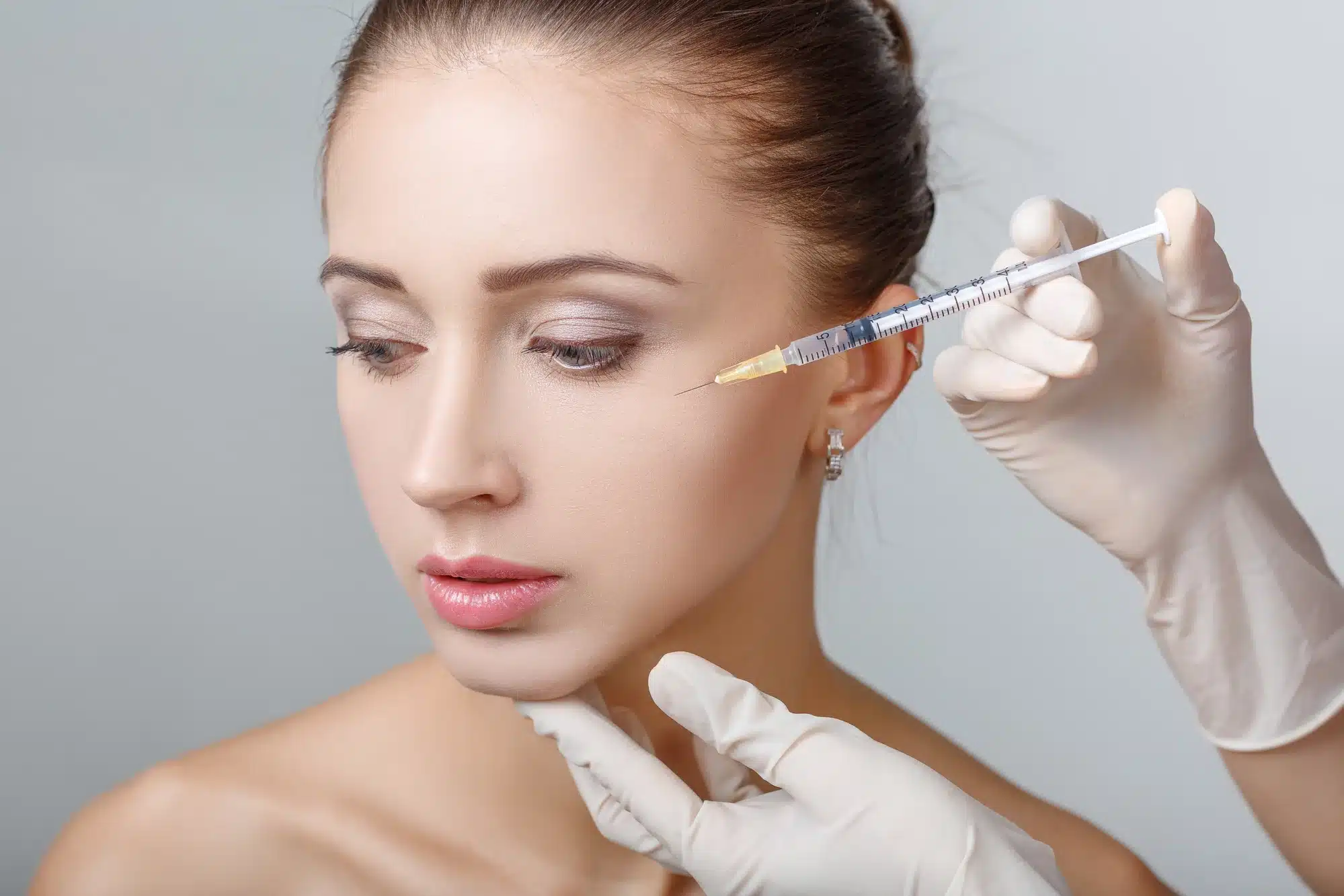
Glow Effect
The “Glow Effect” of Hyaron comes from 25mg/2.5ml of non-cross-linked sodium hyaluronate per. After being injected into the dermis at a depth of 1-2 mm, hyaluronic acid with a molecular weight of approximately 1000 kDa can bind 1000 times its own weight in moisture. Within 72 hours, the moisture content of the skin’s stratum corneum can increase by 30%-40%, and the transepidermal water loss (TEWL) rate decreases by about 15%. Cellular filling improves epidermal micro-smoothness and optimizes the light refractive index, thereby producing a hydrated mirror-like reflection on the surface that lasts for 14 to 21 days.
Visibly Identifiable Luster
After sodium hyaluronate molecules are injected into the dermal mesoderm, the physical light reflectivity changes. 25mg of non-cross-linked sodium hyaluronate spreads at a depth of 1.5 to 2.0 mm, adsorbing free moisture from surrounding capillaries and interstitial spaces. Hyaluronic acid with a molecular weight of 1 million Daltons can expand the volume of local interstitial fluid by 15% to 20% within 24 to 48 hours. The increase in moisture fills the tiny gaps beneath the epidermis, and the papillary dermis is pushed outward, altering the micro-topography of the skin surface. Human facial skin surfaces possess natural furrows (sulci cutis) with a depth of approximately 10 to 50 microns. A large amount of moisture penetrates toward the stratum corneum, causing shriveled keratinocytes to absorb water and swell, increasing their individual volume. The epidermal stratum corneum consists of 15 to 20 layers of dead cells; after water absorption, the overall thickness slightly increases by 0.1 to 0.2 mm. The depth of tiny skin furrows is thus reduced, and uneven skin texture is physically smoothed out. When light hits the skin surface with increased smoothness, the optical reflection path shifts.
- The proportion of specular reflection of incident light on the smooth epidermis rises from 15% to about 40%.
- Diffuse reflection, which causes a visually dull complexion, is significantly reduced.
- Natural light or indoor lighting forms distinct mirror-like reflection spots on the high points of the face.
- The light transmittance of the stratum corneum increases, allowing light to penetrate and reflect the microvascular blood color of the dermis.
Dermatology clinics in Europe and America use a Glossmeter to quantitatively test the optical performance of the skin after sodium hyaluronate injection. Gloss Units (GU) are an objective indicator for measuring the reflective capacity of a surface. In typical dry and aging skin samples, the baseline glossiness of the cheek area fluctuates between 15 and 20 GU. On the 7th day after injecting 2.5ml of Hyaron, the Glossmeter reading for the same test area climbs to 30 to 35 GU. The polarized light mode of the Visia skin analyzer records an expansion of the facial reflective area by 20% to 25% during the same period. When the probe of a stratum corneum moisture tester (Corneometer) contacts the skin surface on the 14th day after injection, the capacitance reading rises from 30 units before injection to 50 to 60 units. A transepidermal water loss meter (Tewameter) measures a decrease in water evaporation rate from 20 g/h/m2 to 14 g/h/m2.
- Day 3 post-injection: Dermal hydration initiates, and stratum corneum moisture content rises by 5%.
- Day 7 post-injection: Moisture permeates outward, and skin furrow depth decreases by about 15 microns.
- Day 14 post-injection: Glossmeter readings reach their peak, with the highest surface reflectivity.
- Day 21 post-injection: As hyaluronic acid naturally metabolizes, glossiness decreases by about 1 to 2 GU per day.
When liquid foundation spreads on a stratum corneum containing 30% moisture, microscopic particles with a diameter of 5 to 10 microns can be distributed more evenly. Tiny keratin flakes of about 0.5 mm in size in dry areas are suppressed by moisture, reducing the probability of foundation cakeyness. Makeup wear tests in cosmetic laboratories in Europe and America show that on skin injected with sodium hyaluronate, the time for foundation oxidation and dullness is delayed from 4 hours to 8 hours after application. The foundation adheres closely to the smooth stratum corneum, preserving the refractive index of the base makeup products and keeping the skin bright while wearing makeup. A moisture reserve that adds 0.15 mm of thickness to the dermis is the physical foundation of surface translucency. The sodium hyaluronate molecules contained in a single Hyaron exist as a loose network structure in the dermis.
- Flowing free moisture is evenly distributed over the 0.5 square meter epidermal area of the face.
- Local interstitial fluid pressure is maintained at a healthy level of 15 to 20 mmHg.
- Compression on microcirculatory vessels in the dermis with a diameter of about 10 microns is reduced, increasing the blood perfusion rate by 5%.
Hyaluronidase decomposes non-cross-linked hyaluronic acid molecules at a specific rate per hour within the dermis. After 28 days, the adsorbed free moisture is gradually released and excreted from the body. The dermal thickness drops back by 0.1 mm, and skin furrow depth returns to above 30 microns as it was before injection.
Improvement in Tactile Texture
25mg of non-cross-linked sodium hyaluronate is distributed in micro-droplets within the dermal mesoderm at a depth of 1.5 mm. The polymer network with a molecular weight of 1000 kDa unfolds in the tissue gaps, intertwining with collagen and elastic fibers. Skin biopsies show that hyaluronic acid expands the volume of the local extracellular matrix by 15% within 48 hours. When pressing the cheek with a finger, the free moisture within the dermis buffers the mechanical pressure. Dermatologists in Europe and America often use a Cutometer to quantify changes in physical touch based on the principle of negative pressure suction. The probe adsorbs a 2 mm area of the epidermis under a constant negative pressure of 300 mbar. The hydrated dermis exhibits a higher resistance to deformation. Measurement data show that the R0 parameter, representing skin firmness, decreases by an average of 12% to 18% on the 14th day after injection. A decrease in the R0 value indicates that the skin becomes softer when subjected to vertical external forces. The increase in interstitial fluid reduces the friction between fiber bundles within the dermis. The rheological properties of non-cross-linked hyaluronic acid molecules allow the skin to rebound quickly at a speed of 0.5 mm per second after being pressed. The R7 parameter, representing biological elasticity in elasticity tests, shifts simultaneously. The R7 value of healthy young skin is usually maintained between 0.4 and 0.5. Two weeks after a single injection of 2.5ml Hyaron, the R7 value of shriveled and aging skin climbs from 0.3 to around 0.42. The climb in data corresponds to a noticeable rebound in fingertip touch. Free moisture permeates from the dermis to the basal layer of the epidermis at a speed of 0.5 mm per hour. Stratum corneum moisture content increases from 15% to 30%, and dry, exfoliating keratinocytes are physically softened under moisture infiltration. The skin roughness meter (Visioscan) captures the improvement in microscopic smoothness brought about by the softening of the stratum corneum. A high-resolution camera records 2D images of surface fine lines and desquamation. Software analysis indicates that epidermal roughness parameters decreased by 20% to 25% under hydration.
| Test Item | Baseline Before Injection | Day 14 Post-Injection | Tactile Feedback |
|---|---|---|---|
| Stratum Corneum Moisture | 12% – 15% | 28% – 32% | Reduced Roughness |
| Roughness (SEr) | 4.5 – 5.0 | 3.5 – 3.8 | Disappearance of Graininess |
| Desquamation Index | 15.2 | 8.5 | Increased Smoothness |
| Skin Thickness | 1.2 mm | 1.35 mm | Increased Fullness |
The rotating probe of a skin friction meter tests surface smoothness under a pressure of 50 grams. Due to tiny peeling phenomena on un-intervened dry skin, the torque value of the probe’s rotation remains at a high level. The epidermal microenvironment absorbing moisture reduces the sliding friction coefficient of the probe by 15%. The fingertip no longer feels a noticeable resistance when gliding across the cheek. The 0.1 mm thick stratum corneum becomes densely arranged, and corneodesmosomes complete their degradation process normally under the action of enzymes. Beyond smoothness, sodium hyaluronate also affects skin fullness in three-dimensional space. The mechanical tension exerted by the papillary dermis upward onto the epidermis increases by approximately 10 kPa. Fibroblasts in the reticular layer undergo passive stretching in a moisture-filled environment.
- Collagen fiber bundle spacing increases by 2-5 microns
- Local interstitial fluid pressure rises by 2 mmHg
- Superficial fine line depth decreases by 15-20 microns
- Cheek compression rebound time shortens by 0.5 seconds
- Skin fold thickness increases by 0.2 mm
The viscoelasticity of the extracellular matrix reaches a dynamic equilibrium between Day 7 and Day 14. 25mg of sodium hyaluronate forms a uniform gel-like buffer layer within the 50 square centimeter injection area. The buffer layer alters the way the skin folds during dynamic facial expressions. When smiling or frowning, the fluidity of moisture within the dermis dissipates local stress caused by muscle contraction. The superficial epidermis no longer produces sharp creases during facial movements like dry paper does. High-frequency skin ultrasound (20 MHz) detects changes in the dermal echogenic band. The originally densely packed high-echogenic collagen bands are separated by a low-echogenic free moisture network. The low-echogenic band thickness expands from 0.8 mm to 1.0 mm. Black anechoic areas on ultrasound images represent pure liquid retention. The density of the dermis is physically diluted temporarily, bringing a soft tactile sensation similar to a sponge after absorbing water. Anti-aging laboratories in Europe and America conducted a 28-day blind evaluation on 50 subjects. Professional evaluators touched the subjects’ faces wearing standard latex gloves. At the 14-day mark, 85% of the evaluators were able to accurately identify the injected area of the cheek based on touch. The duration of texture improvement is limited by the natural degradation rate of hyaluronidase. Approximately 3% to 5% of non-cross-linked hyaluronic acid molecules are hydrolyzed into oligosaccharides every day. The bound water then slowly enters local capillaries and lymphatic vessels for blood circulation. Biological elasticity parameters decrease at a rate of 0.02 per day on the 21st day. Stratum corneum thickness recedes by 0.05 mm, and the surface torque recorded by the friction meter begins to rise slowly. Tactile sensation at the fingertips basically returns to the baseline level of dryness around the 28th day.
Luster Maintenance Cycle
After 25mg of non-cross-linked sodium hyaluronate is injected into the dermis at a depth of 1.5 to 2.0 mm, its maintenance cycle is limited by the metabolic rate of endogenous hyaluronidase in the human body. Polymers with a molecular weight of 1000 kDa are hydrolyzed in interstitial fluid at 37 degrees Celsius at a rate of about 3% to 5% per day. Within the first 72 hours after injection, 2.5ml of gel-like liquid completes its physical diffusion within the 50 square centimeter cheek area. The hyaluronic acid molecular network begins to capture moisture leaking from surrounding capillaries, and local interstitial fluid pressure rises by about 2 mmHg. Stratum corneum moisture content slowly climbs from 15% to 20%, and epidermal microvascular dilation brings a slight increase in blood flow over a 0.5 mm range. Observations from confocal microscopy in European and American dermatology records show that Day 3 to Day 7 is the peak period for water binding. Hyaluronic acid molecules swell to 1000 times their original volume, pushing the entire 0.1 mm thick epidermal layer upward by about 15 microns. Glossmeter readings increase from a baseline of 18 GU at a rate of 2 GU per day during this phase. By Day 14, the optical reflectivity of the facial skin reaches its peak, and the Corneometer shows capacitance values stabilizing between 55 and 60 units. A high-density hydration zone about 1.2 mm thick forms in the dermis, smoothing out skin furrows with a depth of 30 microns. The proportion of specular reflection of incident light on the epidermis maintains its highest position at 40%.
- Local tissue moisture content increases by 30% compared to baseline
- Peak glossiness reading reaches 35 GU
- Transepidermal water loss (TEWL) drops to 12 g/h/m2
- Epidermal roughness (SEr) parameter decreases by 25%
Biopsy tracking reports from the Los Angeles Anti-Aging Research Center show that the half-life of non-cross-linked hyaluronic acid in the dermal matrix is about 3 to 5 days. It typically takes 21 to 28 days for the polymer to be completely metabolized into oligosaccharides and excreted with water.
The decline of physical mirror reflection becomes visible to the naked eye on the 21st day. As macromolecules are cut into oligosaccharides, free moisture in tissue gaps enters the facial lymphatic circulation at a rate of 0.5 ml per hour. Stratum corneum thickness shrinks by about 2 microns per day, and the epidermis re-develops depressions with depths exceeding 15 microns due to the loss of underlying support. Glossmeter readings begin to drop at a rate of 1.5 GU per day. Diffuse reflection regains optical dominance, and the Visia polarized light analyzer records a 15% reduction in the area of facial highlight reflection. The skin elasticity R0 parameter of the test subjects rebounds to above 0.3 (pre-injection level), and cheek compression rebound time extends from 0.5 seconds to 0.8 seconds. By Day 28, 95% of the exogenous sodium hyaluronate has been hydrolyzed and metabolized. Stratum corneum capacitance drops back below 30 units, and the physical luminescence on the skin surface completely disappears. Standard medical aesthetic procedures in Europe and America require a second supplementary injection of 2.5ml dose at this point to maintain a healthy hydrostatic pressure of 15 mmHg in the dermis.
- Days 1 to 3: Epidermal moisture content rises by 2% per day
- Days 7 to 14: Specular reflectivity maintains its peak at 40%
- Days 15 to 21: Physical degradation rate of hyaluronic acid reaches 60%
- Days 22 to 28: Surface glossiness decreases by 1.5 GU per day
Long-term tracking data from dermatology clinics in New York show that after completing 3 injection sessions spaced 28 days apart, the activity of local fibroblasts increased by 12%. The efficiency of cellular synthesis of endogenous hyaluronic acid increases, extending the half-life of non-cross-linked components in the body by about 48 hours. In the fourth injection cycle, the time that stratum corneum moisture stays above 45 units extends from the original 7 days to 12 days. High-frequency skin ultrasound (20 MHz) detects that the 1.0 mm thick low-echogenic hydration zone in the dermis is still clearly visible on Day 35.
Medical aesthetic standards from the European Committee for Standardization (CEN) suggest using a single injection containing 25mg of non-cross-linked hyaluronic acid as a monthly basic maintenance project. The cumulative physical effect of 3 consecutive injections can permanently raise the overall stratum corneum moisture baseline by 15%.
The depth of tiny skin furrows is maintained below 20 microns for a long time, making it difficult to form a rough keratin interface that generates diffuse reflection. In a routine follow-up 40 days after injection, the Glossmeter can still measure an average reading of 25 GU, which is higher than the baseline of 18 GU for shriveled states. The 25mg of sodium hyaluronate provided by a single 2.5ml solution cannot withstand the continuous degradation by human enzymes. Maintaining a moisture reserve of 0.2 mm thickness in the epidermis requires strict periodic physical supplementation. For every 1 ml of interstitial fluid lost in the dermis, surface optical reflectivity will simultaneously drop by 5%; the persistence of luster depends entirely on the dynamic balance of local substance concentration. 
Injection Areas
Hyaron injections must be precisely localized in the superficial to mid-dermis (depth of approximately 1.0mm to 1.5mm). Clinical practice in Europe and the US often utilizes multi-mesotherapy (31G to 34G) or the single-wheal injection technique. The injection volume per point is controlled between 0.02ml and 0.05ml, with a point spacing maintained at 1cm to 1.5cm. These parameters ensure that the non-cross-linked hyaluronic acid (25mg/2.5ml) diffuses uniformly into the surrounding tissues within 24 to 48 hours, achieving large-scale hydration.
Facial Dermal Layer
There are physical discrepancies in histological thickness across different anatomical regions of the face, ranging from 0.4mm to 2.0mm. Performing Hyaron injection procedures requires adjusting parameters with vernier caliper-level precision. Physicians in Europe and the US commonly use ultra-thin wall (32G or 34G, 4mm length) to complete dermal punctures. The total dose for a standard full-face treatment is fixed at 2.5ml, containing 25mg of non-cross-linked sodium hyaluronate. The solution is introduced above the reticular layer of the dermis through 100 to 120 uniformly distributed puncture points. The operator must maintain a constant injection pressure, controlling the release volume per point between 0.01ml and 0.05ml. Once the fluid enters the tissue, it encounters entirely different physical resistance to diffusion, limited by the variations in tension of the dense connective tissue in each anatomical region.
| Facial Anatomical Region | Mean Epidermal + Dermal Thickness | Recommended Puncture Depth | Angle | Volume per Point |
|---|---|---|---|---|
| Forehead and Glabella | 0.9mm – 1.2mm | 1.0mm | 15° – 30° | 0.02ml |
| Periorbital and Tear Trough | 0.4mm – 0.6mm | 0.5mm – 0.8mm | 10° – 15° | 0.01ml |
| Lateral Mid-Cheek | 1.2mm – 2.0mm | 1.5mm | 45° | 0.05ml |
| Perioral and Chin | 1.0mm – 1.5mm | 1.2mm | 30° | 0.03ml |
The forehead area is covered by the frontalis muscle and has an extremely thin subcutaneous fat layer, with vascular networks mostly distributed parallel to the skin surface. If the solution is injected too deeply, it can easily enter the muscle layer and be rapidly metabolized; if injected too superficially, it may cause epidermal papules that persist for several days. During the procedure, a very small angle must be strictly employed.
- Bevel of the fully facing upward, piercing 1.0mm
- Inject a constant volume of 0.02ml per point
- Maintain horizontal point spacing at 10mm
- Set vertical row spacing at 12mm
- Avoid the area where the frontal branch of the superficial temporal artery travels
The tension of forehead tissue is relatively high, resulting in greater physical diffusion resistance for sodium hyaluronate molecules here. Local pale wheals with a diameter of about 2.5mm and clear edges will form. At normal body temperature, a solution with a molecular weight of 1 million Daltons takes 18 to 24 hours to complete horizontal physical penetration. The skin tissue thickness around the eye sockets is only one-fourth of that of the cheeks, and it extremely lacks sebaceous gland distribution. The very thin stratum corneum results in a transepidermal water loss (TEWL) rate in this area that is 30% higher than the facial average. When placing a solution with a concentration of 10mg/ml here, the osmotic pressure gradient must be strictly controlled to prevent local edema.
- Switch to a 34G ultra-fine for micro-droplet operation
- Reduce puncture depth to 0.5mm – 0.8mm
- Limit release volume per point to 0.01ml
- Expand point spacing to 15mm
- Reserve a 5mm safety buffer zone from the lower eyelid margin
The micro-dose administration method can control the volume of single hydration expansion in periorbital tissues, reducing the drainage pressure on the lymphatic system. The tiny bumps formed by the solution under the skin usually flatten within 12 hours and complete preliminary binding with the native collagen fiber network in the extracellular matrix. The mid-cheek area possesses the thickest dermal reticular layer of the face, serving as the optimal matrix water reservoir for accommodating fluids. Fibroblast distribution density is highest in this region, allowing it to withstand large doses of physical fluid expansion. The should be inserted at a 45-degree angle to the skin surface to a depth of 1.5mm.
- Use a dense grid-like cross-point technique
- Inject a maximum dose of 0.05ml per point
- Form full wheals with a diameter of 3mm to 4mm
- Reduce point spacing to 10mm to facilitate overlapping diffusion
- Increase delivery density by 15% on the lateral edge of the nasolabial fold
A single cheek administration consumes approximately 1.2ml to 1.5ml of solution, accounting for more than 50% of the total facial dose. High-purity hyaluronic acid binds water molecules from surrounding interstitial fluid through hydrogen bonding within the dermis. The rich blood supply of the cheek microvascular bed accelerates the uniform spreading of the solution within 24 hours. The perioral and chin areas are attached to a large number of high-frequency expression muscles; muscle contraction can accelerate the mechanical degradation of the fluid. The perioral dermis thickness is moderate, within the range of 1.0mm to 1.5mm, but the tissue density is extremely high. Injections should maintain a 30-degree angle, delivering 0.03ml of solution to the mid-dermis at a depth of 1.2mm. The reticular structure of the facial dermis exerts uniform physical centripetal pressure on the non-cross-linked macromolecular solution. Free hyaluronic acid molecules radiate outward along tissue gaps at a rate of 0.5mm to 1.0mm per hour. After 72 hours, the original wheal morphology completely subsides, and the water content in the target tissue increases by 15% to 20%. Clinical high-frequency ultrasound (HFUS) monitoring images show that the dermal thickness increases by an average of 0.12mm on the 7th day after administration. A single molecule of hyaluronic acid binds 1000 times its own weight in water molecules within the tissue. The local matrix hydration state can be maintained for 21 to 28 days, subsequently returning to physiological baseline levels following the half-life degradation of endogenous hyaluronidase.
Superficial Neck Area
The skin tissue in the anterior neck region has unique anatomical characteristics, with an average dermal thickness of only 1.1mm to 1.5mm. The stratum corneum in this area is extremely thin, and the distribution density of sebaceous glands is 40% to 50% lower than that of the cheek area. The weakness of the physical barrier leads to a consistently high transepidermal water loss (TEWL) rate. When treating this anatomical partition, European and American dermatologists usually select 34G ultra-thin wall with a 4mm length. The insertion angle must be strictly controlled between 10° and 15°, ensuring the bevel always faces upward. The operator must precisely control the puncture depth, smoothly delivering the to 1.0mm in the superficial dermis. The dose of non-cross-linked hyaluronic acid (10mg/ml) per injection must be limited within the micro-droplet range of 0.01ml to 0.015ml.
- Control horizontal injection spacing between 8mm and 10mm
- Arrange in a bead-like pattern along the physical direction of neck horizontal lines
- Maintain total dose per session usually between 1.5ml and 2.0ml
- Keep injection pressure constant at 0.5kg to 1.0kg
- Avoid the external jugular venous network superficial to the sternocleidomastoid muscle
Continuous micro-droplet operations will form a row of hemispherical pale wheals on the skin surface with a diameter of about 1.5mm to 2.0mm. The immediate whitening reaction of the epidermis confirms that the hyaluronic acid solution has been accurately deposited within the dense dermal connective tissue. Microcirculatory blood flow in the anterior neck region is relatively slow, and local skin temperature is usually 0.5°C to 1.0°C lower than that of the face. Hyaron solution with a molecular weight of up to 1 million Daltons faces strong physical diffusion resistance upon entering this matrix. Complete absorption of macromolecular liquids by the extracellular matrix requires a long cycle of 48 to 72 hours. During this period, the wheals will flatten uniformly at a rate of approximately 0.2mm every 12 hours, while single hyaluronic acid molecules continuously bind surrounding water molecules in the physical space. The platysma is an extremely superficial expression muscle, located only 2.0mm to 3.0mm beneath the epidermal surface. If the puncture depth exceeds the 1.5mm warning line, the solution will rapidly penetrate the reticular layer at the base of the dermis. Hyaluronic acid entering the myofascial layer will be rapidly metabolized within hours by the rich microvascular bed. The dermis will fail to obtain the expected physical hydration, and operators must establish a physical isolation barrier through specific techniques.
- The non-holding hand must apply 0.5 Newtons of lateral skin tension to both sides
- Mechanical resistance of the penetrating the epidermis must be below 0.2 Newtons
- If 0.01ml of blood backflow appears in the, the tip must be withdrawn immediately by 0.5mm
- Reduce release volume per point by 20% in the weak midline area of the anterior neck
- Set a fan-shaped high-risk avoidance zone 2cm above the cricoid cartilage
Some clinical institutions in North America use a hybrid formulation for intradermal spreading for deeper horizontal neck lines. Standard operation involves mixing 1.5ml of Hyaron preparation with 10 to 15 units of Botulinum Toxin Type A. The prepared composite solution is uniformly distributed among 80 to 100 superficial puncture points. Trace neurotoxins are responsible for relaxing the superficial muscle fibers of the platysma, while hyaluronic acid molecules simultaneously increase the volumetric capacity of the extracellular matrix by 12% to 15%. High-frequency ultrasound (HFUS) imaging shows an average increase of 0.08mm in local dermal thickness on the 14th day after administration. The reconstructed high-hydration collagen network structure can maintain physical integrity under the skin for 21 to 28 days. European medical aesthetic guidelines suggest that a basic treatment cycle should include 3 consecutive standardized operations at intervals of 14 to 21 days. The fixed physical interval precisely matches the biological stimulation cycle of fibroblasts. Local erythema caused by physical puncture usually subsides spontaneously within 90 to 120 minutes. Subjects must avoid exposing the 150 square centimeter anterior neck area to warm water above 38°C for 24 hours to prevent accelerated capillary dilation.
Back of Hands and Décolletage
The back of the hand lacks subcutaneous fat tissue as a physical buffer layer, with mean dermal thickness fluctuating between 0.8mm and 1.2mm. A natural loss rate of about 1% per year causes the subcutaneous extensor tendons and venous networks to gradually approach the 1.0mm free boundary of the epidermis. Long-term UVB exposure leads to an abnormal thickening of the stratum corneum by 20% to 30%. In North American and European clinical standards for this severe photoaging area, the standard dose of Hyaron for a single back of hand is strictly set within the 1.0ml to 1.5ml range. Physicians often use 25G (0.5mm outer diameter) or 22G (0.7mm) to complete deep dermal spreading. The entry point is selected 1.5cm distal to the transverse wrist crease, or at the proximal junction of the 2nd and 4th metacarpal spaces.
Guidelines from the American Society for Dermatologic Surgery suggest that after insertion, the must be advanced steadily along the superficial subcutaneous fascia at a horizontal angle of less than 10°, avoiding the puncture of superficial venous trunks with a diameter exceeding 2.0mm.
- Control unidirectional advancement depth between 4.0cm and 5.0cm
- Implement a retrograde injection technique to slowly release hyaluronic acid
- Maintain single linear injection volume at 0.05ml to 0.08ml
- Distribute points uniformly in a fan-shaped radial pattern at a 30-degree angle
- Keep the solution distributed between the deep dermis and superficial fascia gaps
The high-molecular-weight non-cross-linked preparation remains in the weak 0.5mm to 1.0mm subcutaneous gap on the back of the hand. Physical compression massage is a standard operation after hand administration, applying a uniform pressure of about 1.5 kPa for 3 to 5 minutes. The kneading action encourages 1 million Dalton free molecules to complete horizontal physical diffusion within the dense connective tissue. Within 48 hours after injection, the water content of the local extracellular matrix achieves an inverse increase of 15% to 20%. The décolletage area extends from the lower edge of the clavicle to above the cleavage, forming an inverted triangle of about 150 square centimeters, where the dermal thickness is maintained at 1.0mm to 1.4mm. The density of hair follicles and sebaceous glands in this anatomical region is extremely low, and the surface lipid film thickness is only 33% of that of the cheek. Physical folding tension generated by side sleeping is as high as 3.5 times the natural tension of the face. The skin compliance of the chest is poor, and the mechanical resistance to vertical sharp puncture is greater than 0.3 Newtons. Clinical practice often utilizes 32G or 34G ultra-thin wall with a 4mm length, with the insertion angle fixed between 30° and 45°.
European medical aesthetic injection guidelines suggest that chest operations must avoid the hypersensitive nerve distribution areas 1.5cm on both sides of the sternal midline, blocking the risk of local cardiovascular vagal reflex reactions caused by deep puncture.
- Use a grid-like cross-layout to plan the operation area
- Set horizontal and vertical injection intervals at 1.0cm to 1.5cm
- Inject a constant volume of 0.01ml to 0.02ml per point
- Lock the insertion depth at 1.0mm in the superficial to mid-dermis
- Avoid the superficial microvascular bed of the subclavicular artery branches
High injection resistance causes the immediate formation of pale hemispherical wheals with a diameter of 2mm to 3mm and a height of about 1.0mm at each puncture point. The total drug consumption for a single full décolletage treatment should reach 2.0ml to 2.5ml, covering 120 to 150 puncture sites. Non-cross-linked hyaluronic acid molecules rely on concentration gradients to physically penetrate surrounding tissue gaps within a 1.0cm radius at a speed of 0.3mm per hour. The complete flattening cycle for wheals is often extended to 48 to 72 hours, with free water molecules simultaneously captured by high-density hydrogen bonds. Local erythema caused by micro-puncture accounts for about 15% of the operation area and subsides spontaneously within 120 minutes post-procedure alongside capillary contraction. Subjects must endure a slight physical expansion pressure of less than 0.5 kPa under the skin for 48 hours.
- Stratum corneum moisture content increases by 12% on the 7th day post-operation
- Transepidermal water loss rate decreases by 25% on the 14th day
- Dermal ultrasound echogenicity increases by 10% to 15%
- Physical depth of fine lines is reduced by up to 0.2mm
- The physical maintenance cycle of the high-hydration microenvironment is 21 to 28 days
Tracking records from European and American clinical databases indicate that following a 0.01ml micro-droplet or 0.05ml linear administration every 21 days for 3 consecutive sessions, significant physical reconstruction of the collagen fiber network density occurs. The degradation curve of a single hydration treatment shows a clear numerical inflection point on the 30th day.