For those looking to eliminate localized fat, fat-dissolving injections are a popular choice. Lipo Lab and Kabelline are two common fat-dissolving products that both help break down fat using deoxycholic acid (DCA).
Fat-Dissolving Mechanism
Adipocyte Cytolysis
A injects 0.2 mL of the solution into the subcutaneous fat layer at a depth of 2 to 4 mm, where it spreads outward in a disc-like shape for about 1 cm. The body’s interstitial fluid, maintaining a constant temperature of 37°C and a pH of around 7.4, perfectly neutralizes the slightly alkaline solution. With a molecular weight of only 392.57 g/mol, the small deoxycholic acid molecules can easily penetrate the intercellular spaces to approach the target fat cells. Mature white adipocytes are exceptionally large, with a single cell volume reaching 0.6 to 0.8 nanoliters. Over 90% of their internal space is filled with liquid fat. These cells are enclosed by an extremely thin membrane, only 5 to 10 nm thick. Deoxycholic acid molecules act as nanoscale wedges, forcefully squeezing into the gaps of the membrane’s bilayer structure. The membrane’s tension is instantly disrupted, exceeding its physical tolerance limit of 8 mN/m. The tightly packed membrane molecules are forced apart, tearing open microscopic pores of 20 to 50 nm within 30 to 120 seconds. The cell membrane’s normal resting membrane potential completely collapses from its baseline of -70 mV, and the protective barrier fails. Water and high concentrations of sodium ions from the outside violently rush into the cell through these microscopic pores:
- Internal osmotic pressure surges from 280 to over 320 mOsm/kg.
- The tubulin cytoskeleton supporting the cell’s shape completely collapses.
- Mitochondria, which provide energy, lose their internal potential.
- Internal lysosomes rupture, leaking potent digestive enzymes.
- The protein shells encapsulating the lipid droplets undergo severe deformation.
The rapid influx of water causes the cell to swell rapidly within 5 to 8 minutes, expanding to 1.2 to 1.5 times its original volume. Pushed beyond its elastic limit, the membrane bursts like an overinflated balloon. Instantly, 85% of the internal liquid, butter-like substance is expelled into the surrounding interstitial space. Local concentrations of lactate dehydrogenase—a marker for cell death—skyrocket to dozens of times the normal baseline of 450 IU/L. The escaped free fatty acids temporarily lower the pH of the surrounding microenvironment to around 6.8. The rupture site is left as a microscopic biochemical wasteland, containing:
- Large yellow lipid droplets 10 to 50 μm in diameter.
- Long-chain free fatty acids with 16 to 18 carbon atoms.
- Shredded cell membrane fragments.
- Fragmented DNA remnants of 180 to 200 base pairs.
- Non-functional, discarded mitochondrial waste.
Macrophages deployed by the body secrete digestive enzymes at concentrations exceeding 200 ng/mL. The massive fat chunks are meticulously chopped into smaller fatty acids and monoglycerides by invisible biochemical scissors. Water-soluble glycerol, with a molecular weight of just 92.09 g/mol, quietly seeps into the capillary network along the concentration gradient to be transported away. The scattered lipid debris triggers the microenvironment’s alarm system. Vascular endothelial cells detect the high-fat signal, and their surface distress receptors more than triple within 2 hours. Patrolling monocytes in the bloodstream abruptly decelerate from a velocity of 1 mm/s to 0.1 mm/s, sticking to the blood vessel walls as if hitting the brakes. Between 400 and 600 mononuclear leukocytes, 20 μm in diameter, emerge per square millimeter, squeezing through the capillary walls. Within 12 to 24 hours, they rapidly engorge themselves, transforming into 50 μm macrophages that devour the lipids. The local temperature consequently rises by 0.5 to 1.2°C, and microcirculatory blood flow surges by approximately 30%. Inside the macrophages, an incredibly complex processing assembly line begins:
- Engulfed vesicles fuse with digestive sacs to form a super processing plant.
- Superoxide anions are massively released to forcefully degrade stubborn lipids.
- Pro-inflammatory signals are fired outward at 5 to 10 pg/mL.
- Multiple cells clump and fuse into a giant 100 μm scavenger.
- Highly acidic, concentrated digestive fluids are frantically produced and secreted.
The pH inside this processing plant plummets from a neutral 7.0 to a highly acidic 4.5. Hydrolases activated under these extreme acidic conditions shred large fat debris into nanoscale particles. Lipid residue left unprocessed within 7 to 14 days attracts a second wave of immune cells, clustering to form hypoechoic nodules 2 to 5 mm wide that are visible under ultrasound. The engorged macrophages are packed with transparent lipid droplets 1 to 2 μm wide, appearing under a microscope like dense foam. A single cell can ingest up to 2.5 times its own volume in debris at once. Carrying semi-digested lipid remnants, they retreat at a snail’s pace of a few micrometers per minute toward the nearby micro-lymphatic network. Within the injection area, 35,000 to 50,000 target adipocytes per square centimeter undergo this explosive rupture. The space previously occupied by the swollen cells collapses and contracts once cleared. Subcutaneous fibroblasts receive signals and, by day 14, vigorously begin producing nascent type I collagen filaments about 50 nm in diameter.
Immune Phagocytosis and Clearance
The ruptured adipocytes leave behind a subcutaneous mess of organelle fragments and triglycerides. The concentration of adiponectin in the surrounding interstitial fluid plummets by more than 40% within just 15 minutes. This abnormal debris field becomes a chemical signal source that triggers the body’s alarm. Capillary endothelial cells within 50 μm of the rupture epicenter are the first to react. Within 45 minutes of stimulation, they express numerous protein “anchors” called P-selectin and E-selectin on their surface. Neutrophils, which typically race through the bloodstream at 1 to 2 mm/s, are firmly snared. The snared neutrophils roll and decelerate along the vessel wall, flattening into sheets just 2 μm thick within minutes. They desperately squeeze through 0.5 to 1 μm gaps in the vascular wall into the lipid-filled interstitial fluid. In the first 6 to 12 hours post-injection, up to 15,000 neutrophils flood in per cubic millimeter. Neutrophils have a pitifully short lifespan, surviving at most 24 to 48 hours in the interstitial fluid. They are responsible for frantically secreting superoxide dismutase and myeloperoxidase. Local oxidative stress markers peak at the 12-hour mark, prematurely breaking down large cell membrane fragments. Monocytes, the true scavengers, follow closely behind, crossing the vascular wall en masse 24 hours post-injection. Originally small cells about 15 μm in diameter, monocytes begin to grow wildly upon entering the lipid-contaminated fluid. Expanding like balloons, their volume balloons to 50 to 80 μm within 48 hours, fully transforming into macrophages. These transformed macrophages possess an astonishing appetite, with a single cell capable of consuming about three times its volume in debris per day. They extend pseudopodia up to 20 μm long to net loose, small yellow lipid droplets. The engulfed lipid remnants form a phagosome about 5 to 10 μm in diameter inside the macrophage.
| Phagocytic Stage | Timeframe | Cellular Changes | Microenvironment Temp Change |
|---|---|---|---|
| Vascular Extravasation | 2-12 hours post-injection | Mass influx of neutrophils | Increases by 0.3°C – 0.5°C |
| Volume Expansion | 24-48 hours post-injection | Monocytes swell up to 80 μm | Increases by 0.8°C – 1.2°C |
| Acidic Degradation | 3-7 days post-injection | Lysosomal pH drops to 4.5 | Increases by 1.2°C – 1.8°C |
| Tissue Repair | 14-28 days post-injection | Phagocytes exit, collagen secreted | Returns to normal |
The phagosome physically fuses with a primary lysosome packed with highly acidic digestive fluids. Driven by rapidly acting proton pumps, the pH inside the phagolysosome drops drastically within 30 minutes. The initially balanced pH of 7.2 is violently driven down to an intensely acidic 4.5. Over 50 acidic hydrolases operate at full capacity in this 4.5 pH environment. Bulky triglycerides are ruthlessly shredded into small free fatty acids by molecular scissors. To tackle tough protein remnants, the macrophage injects internal reactive oxygen species at concentrations up to 100 μmol. These fierce microscopic biochemical clashes manifest to the naked eye as redness, swelling, heat, and pain. Subcutaneous thermometry reveals that the average temperature of the injection site at the 48-hour mark is 1.2 to 1.8°C higher than the surrounding skin. Extreme capillary dilation nearly doubles blood flow, and the resulting interstitial fluid exudation creates a subcutaneous lump. Faced with large, stubborn fat debris over 20 μm that cannot be swallowed individually, macrophages employ swarm tactics. Five to ten macrophages link their membranes and forcibly fuse into a giant cell exceeding 150 μm. These foreign body giant cells act as massive furnaces, continuously dripping digestive enzymes into their center. High-frequency color ultrasound probes applied to the skin clearly show dense, abnormal dark areas 3 mm below the surface. Anechoic inflammatory granulomas, 1 to 3 mm in diameter, serve as records of the phagocytic army’s brutal dismantling site. Loaded with semi-digested lipid waste, the macrophages’ bellies swell to become “foam cells” packed with transparent vesicles. Guided by lymphatic chemokines, they crawl at a sluggish pace of about 2 to 3 mm per hour, struggling as they slowly inch toward the nearest micro-lymphatic network to retreat. The gaps between lymphatic endothelial cells act like one-way trapdoors. Macrophages squeeze into lymphatic capillaries just a few micrometers wide, merging into the pale yellow lymph fluid. Approximately 60% of primary lipid metabolites pass through this one-way street and are transported to nearby regional lymph nodes over the next 7 to 14 days. The remaining 40% of free fatty acids, possessing stronger penetrating power, seep straight into the microvascular network. In the bloodstream, long-chain fatty acids quickly bind to albumin, which acts as a carrier; every single albumin molecule firmly tethers seven fatty acid molecules. The rushing bloodstream sweeps these free fatty acids into the liver’s biochemical processing line at a flow rate of several liters per minute.
Lipid Metabolism and Excretion
Macrophages stuff leftover fat debris into lymphatic vessels a mere 10 to 50 μm thick. The pale yellow lymph fluid flows sluggishly through layers of lymph nodes at a rate of 0.5 to 1.5 mL/min. Glycerol escaping the capillaries is tiny, with a molecular weight of just 92.09 g/mol, and is highly water-soluble. Traveling along tens of thousands of kilometers of venous channels, it circulates throughout the entire body in just 45 to 60 seconds within the blood plasma, which accounts for 55% of total blood volume. Free fatty acids that fail to enter the lymphatics force their way through capillary walls just 1 μm thick to join the bloodstream. Every 100 mL of normal blood plasma contains 4 to 5 grams of albumin, which has a molecular weight of about 66.5 kDa. Albumin, the blood’s natural transporter, immediately gets to work. Utilizing small surface grooves, a single albumin molecule tightly grips up to seven long-chain fatty acid molecules.
Dragged along by albumin, the fatty acids join the torrent of blood surging into the hepatic portal vein, which is about 6 to 8 cm long and 1.2 cm thick. A full 1,500 mL of blood washes through the 1.5 kg liver every minute, accounting for 75% of its local blood supply and delivering over 80% of the fatty waste to its final destination in one go.
The liver is densely packed with around 250 billion hepatocytes. As blood enters the hepatic sinusoids, which are 9 to 12 μm in diameter, its velocity plummets to 0.1 mm/s. Specialized fatty acid transport proteins on the surface of hepatocytes act like miniature cranes. Within just a minute, they actively haul over 30% of the free fatty acids from the blood into the hepatocytes. Once inside, the fatty acids are hurriedly dispatched to the mitochondria—the cell’s miniature power plants. A single hepatocyte is packed with 1,000 to 2,000 of these power plants. Here, long-chain fatty acids undergo the rigorous process of beta-oxidation. Stearic acid, containing 18 carbon atoms, is relentlessly chopped up, with each precise cut removing exactly two carbon atoms. The complete disassembly line follows four steps:
- Forcefully breaking the carbon-carbon single bonds spaced 1.54 Å apart.
- Forcibly inserting a new oxygen atom within milliseconds.
- Twisting the molecule’s original spatial angle of 109.5 degrees.
- Spitting out an acetyl-CoA molecule containing 2 carbon atoms.
After surviving 8 successive rounds of rapid cleavage, a single 18-carbon long-chain fatty acid yields 120 ATP energy packets. About 60% of the excess energy dissipates as heat, slightly raising the temperature of local liver tissue by 0.2 to 0.5°C. The thoroughly depleted carbon and hydrogen atoms, mediated by enzymes, are converted into carbon dioxide exhaust and wastewater. The carbon dioxide, bearing the legacy of the digested fat, travels back to the pulmonary capillaries via venous blood. With every breath, a person inhales and exhales about 500 mL of air. The 300 to 400 million alveoli, boasting a total surface area of 70 square meters, naturally expel the carbon dioxide exhaust into the air. Through 28,000 breaths over the course of a day, nearly 84% of the fat’s carbon waste is silently exhaled. Water generated from this breakdown flows down the large abdominal aorta into the two kidneys. Roughly 180 liters of primary urine pass through the fine filters of 2 million glomeruli across both sides daily. The excess wastewater mixes into the 1.5 to 2 liters of yellow urine produced each day, flowing down the 25 to 30 cm long ureters into a bladder capable of holding 500 mL, and is finally flushed out of the body.
Any unburned fatty acid residues migrate to the smooth endoplasmic reticulum, which has a tubular diameter of 50 to 100 nm, to be reassembled. Three fatty acids are paired with one glycerol to form a new lipid. These are packaged into very-low-density lipoprotein (VLDL) parcels with a diameter of 30 to 80 nm and a density of about 0.95 g/mL, then tossed back into the bloodstream for the muscles to consume.
The injected deoxycholic acid solution is precisely extracted by hepatocytes. The exogenous deoxycholic acid is bound to the body’s native glycine at a 3:1 ratio. It is then mixed in minuscule proportions into the 800 to 1,000 mL of slightly alkaline bile (pH 7.4 to 8.5) secreted by the liver daily. The dark green bile flows down the 0.8 cm thick common bile duct into the duodenum to digest food. The solution and small amounts of cholesterol waste mixed in the bile slosh through the 5 to 7 meter-long small intestine for 4 to 8 hours. The vast majority of useful components are reabsorbed by the body at a high rate of 95% at the terminal ileum for further use. The remaining roughly 5% of unwanted waste is swept into the large intestine, which is about 1.5 meters long. It remains in the colon for 12 to 24 hours while water is absorbed, blending into the approximately 200 grams of formed feces produced daily before being flushed down the toilet. At this point, the pulverized fat cells and fat-dissolving solution permanently exit the body’s internal circulation system. This precise waste disposal assembly line operates at full capacity starting from the third day post-injection. The concentration of related waste in urine and feces peaks between days 10 and 14. Abiding strictly by the physical rule of excreting 84% of carbon via respiration and 16% of water via urination, the fat debris vanishing from the injection site is completely cleared out within 21 to 28 days. 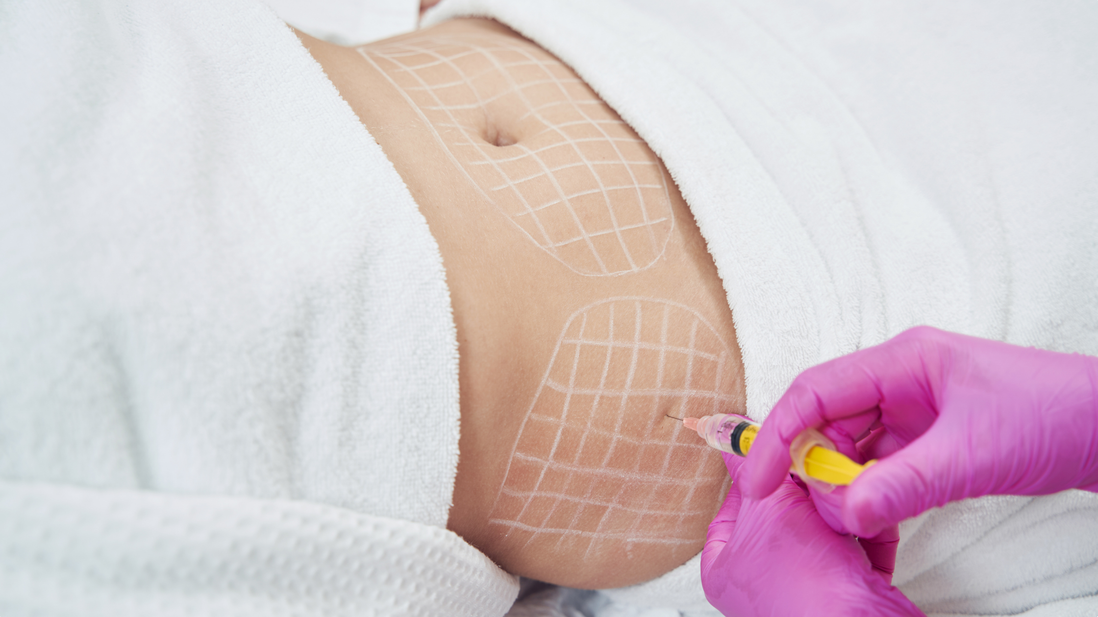
Deoxycholic Acid
Fat-Dissolving Mechanism
Doctors inject 0.2 mL of 99.8% pure deoxycholic acid solution at a 45-degree angle into the subcutaneous fat layer, 6 to 8 mm deep. The solution molecules, with a molecular weight of 392.57 g/mol, act like miniature icebreakers, crashing into the 7.5 nm thick outer lipid bilayer of the cells. The surface tension of 50 mN/m is forcibly snapped. The originally intact protective membrane is torn open, creating permanent, micron-scale holes within 120 to 180 seconds. The high intracellular pressure of 0.82 MPa, previously sealed by the 7.5 nm outer membrane, is completely exposed. Approximately 4.5 million adipocytes are packed along a single side of the jawline. The pure liquid triglycerides, which make up 87% of their volume, lose their containment and leak out at a rate of 0.1 microliters per second. The once plump, round fat cells, originally 120 μm in diameter, collapse into 20 μm protein husks in just 4 minutes. [Image of adipocyte structure] A massive influx of fluid containing 3.5 mmol/L of free fatty acids surges out, causing the surrounding pH to drop sharply from 7.41 to a slightly acidic 6.75. Mast cells in the deep dermis release 20 ng/mL of histamine within 15 minutes. Capillary wall permeability skyrockets by 300%, and blood plasma leaks out at a rate of 2 mL per hour. The skin surface temperature rises by 0.8 to 1.4°C within 4 to 12 hours. The protein-mixed exudate summons monocytes from the bloodstream. Guided by CCL2 chemokines, they transform into highly active macrophages within 24 hours. Once their CD68 receptors are activated, the cleanup process is extremely precise:
- Extending 5 μm pseudopodia to firmly grasp 10 μm lipid droplets.
- Spraying acid phosphatase to dissolve large chunks of fat.
- Secreting 40 pg/L of matrix metalloproteinases.
- Swallowing protein debris weighing about 15 pg in a single gulp.
A fully engorged macrophage, 18 μm in diameter, can hold 50 cubic micrometers of waste. Seventy-two hours after the injection, roughly 850,000 macrophages are densely packed into a 2-square-centimeter treatment area. Interleukin-6 concentrations spike to 15 pg/mL. When nerve endings 3 mm below the skin encounter these high concentrations of inflammatory particles, a mild stinging sensation, combined with the pressure from 5 mm micro-granulomas, is transmitted to the brain. [Image of macrophage phagocytosis] The water-soluble free glycerol, cleaved by enzymes, is squeezed into the venous blood by a capillary hydrostatic pressure of 15 mmHg. Once it reaches the liver, glycerol kinase uses 1 ATP to convert it into glycerol 3-phosphate. It undergoes dehydrogenation in the cytoplasm and joins the glycolysis pathway to generate energy for the body. For every 1 gram of free glycerol burned, the body actively expends 4.32 kcal of energy. Highly hydrophobic free fatty acids move at a sluggish pace of just 0.5 mm per second. Serum albumin in the interstitial fluid quickly approaches; a single 66.5 kDa albumin molecule can forcibly bind up to 7 fatty acid molecules. This water-soluble complex creeps forward through the one-way valves of the lymphatic capillaries, with over 300 lymph nodes in the neck acting as filters. After creeping along for 14 to 24 days, the complex drops into the bloodstream via the left jugular venous angle. Hepatocytes extract the complexes from the venous blood and toss the long-chain fatty acids onto the outer mitochondrial membrane. Carnitine palmitoyltransferase I pulls the 16-carbon palmitic acid into the inner membrane matrix for the beta-oxidation assembly line. Through four steps—dehydrogenation, hydration, a second dehydrogenation, and thiolysis—a 16-carbon chain is neatly sliced into eight acetyl-CoA molecules. Burning a single molecule yields 106 ATP, while the remaining carbon dioxide is exhaled through respiration. The half-life of the injected deoxycholic acid ranges between 32 and 43 hours. The remaining 2.5 mg of free molecules seep into the venules via the interstitial fluid, merging into the body’s natural enterohepatic circulation. An adult’s terminal ileum naturally absorbs 15 to 30 grams of bile acids daily. The tiny amount of drug introduced by the injection accounts for less than a 0.015% concentration, falling far below the threshold needed to trigger a systemic blood lipid alarm. The 0.5 cubic millimeter voids left by the ruptured fat cells stimulate neighboring fibroblasts. TGF-β1 awakens dormant stellate cells in the deep dermis, transforming them into myofibroblasts for repair work. This in-situ reconstruction relies on several strict parameters:
- Maintaining an inflammatory factor half-life of 5.5 days.
- Keeping peripheral capillary oxygen saturation strictly locked at 95%.
- Sustaining a constant temperature of 36.5°C in the deep dermis.
- Enduring a daily pressing force of 2 Newtons per square centimeter.
Every day, 1.5 micrograms of newly synthesized type I collagen fiber bundles weave a subcutaneous net, filling the 5 mm gaps left by the necrotic cells. Elastin chains pull against each other, retracting the jawline skin that was previously stretched by 40 grams of excess fat. By day 28, a scan with a 20 MHz high-frequency ultrasound probe will show a solid 18.5% increase in superficial fascia density, with the subcutaneous fat layer thinned by 2.4 mm.
Formulation Differences
Kabelline’s formulation takes a gentle approach. In the 8 mL ampoules straight from the factory, the absolute concentration of deoxycholic acid is strictly capped at 5 mg/mL. The measured osmotic pressure of this solution is 290 mOsm/kg. This perfectly matches the body’s own interstitial fluid, eliminating the stinging pain typically caused by hypertonic fluid absorption. The solution incorporates L-carnitine (molecular weight 161.2 g/mol) in specific proportions. Acting as chemical transporters, they roam among the fragmented fat, specifically dragging free long-chain acyl groups into the inner mitochondrial membrane. Suspended in the liquid is synthetic acetyl hexapeptide-11 at a concentration of 0.3 mg/mL. This small molecule, composed of 6 amino acids, intercepts acetylcholine signals at the neuromuscular junction. When the doctor pushes the solution down using an ultra-fine 32G, the burning sensation 6 mm below the patient’s skin is chemically blocked by 30%. Because mast cells are not triggered by high pressure, histamine release is extremely low. The mild formulation triggers an extremely restrained sterile inflammation. The increase in capillary permeability does not exceed 150%, and plasma exudation is limited to just 0.5 mL per hour. The peak accumulation of macrophages is capped at roughly 300,000 per square centimeter. For the patient, the period of redness and swelling on the face is drastically shortened from the 14 days typical of the original formula down to just 3 to 5 days. To compensate for the reduced fat excretion caused by the lower concentration, injections rely on high-frequency, grid-like overlapping. On the treatment table, the doctor draws a 1.0 cm equidistant grid, injecting 0.2 mL per point. The total volume consumed for a single facial session does not exceed 8 mL. Through a cycle of 3 to 6 sessions spaced 7 days apart, the 40 grams of fat debris accumulating along the jawline are slowly cleared out. Next door, Lipo Lab’s production line opts for a heavy-firepower blast approach. Its single 10 mL amber glass contains polyenylphosphatidylcholine (PPC), extracted under high pressure from soybean hypocotyls. Here, deoxycholic acid takes a backseat, tasked only with tearing open the 7.5 nm lipid bilayer. The true star of the show is the PPC base solution, acting as the absolute clearing force with a staggering concentration of 1,000 mg/mL. The tail end of a high-purity PPC molecule features an active, hydrophilic choline group, while the other end connects to lipophilic linoleic acid. It dives headfirst into the spilled triglyceride droplets, violently emulsifying them in the interstitial spaces. The originally massive free fat droplets are forcibly kneaded into oil-in-water nanoemulsion droplets less than 50 nm in diameter. The surface area of the fat droplets instantly multiplies by 6,000 times. This explosive increase in surface area exponentially expands the cutting contact surface for lipases. The fat excretion efficiency of a single-point injection is physically boosted by 38.5%. However, this exceptionally potent effect induces a high-intensity immune counterattack. Twenty minutes after the injection, the pH of the local microenvironment plummets past 6.5. Skin surface temperature rises by 1.5°C, and the gaps between vascular endothelial cells are forcibly widened. Within 48 hours, over 1.2 million macrophages flood into every square centimeter of subcutaneous tissue. This high-intensity phagocytic degradation leads to a massive exudation of interstitial fluid. The subcutaneous space fills with protein-rich exudate, resulting in visible congestive edema along the jawline that lasts 7 to 14 days. Pressing down with a fingertip yields a hard nodule tension of up to 4 Newtons.
| Physicochemical Parameters | Kabelline | Lipo Lab |
|---|---|---|
| Component Ratio | 5mg/ml Deoxycholic Acid + L-carnitine | 5mg/ml Deoxycholic Acid + 1000mg/ml PPC |
| Osmotic Pressure | 290 mOsm/kg (Isotonic) | 340 mOsm/kg (Hypertonic) |
| Lipid Excretion Pathway | L-carnitine transport to mitochondria | Potent PPC oil-in-water nanoemulsification |
| Peak Macrophages | 300,000/cm² | 1,200,000/cm² |
| Fluid Exudation | 0.5 mL/hour | 2.2 mL/hour |
| Injection Spacing | 1.0 cm | 1.5 cm |
The physical permeability of the two formulations shows visibly different results under the skin. When Kabelline’s isotonic solution is injected into the subcutaneous fat layer, its spherical diffusion radius strictly stops at 0.8 cm. The solution flows gently along the extracellular matrix grid at a speed of just 0.02 mm per second. The osmotic pressure of surrounding healthy cells remains undisturbed, leaving a necrotic boundary as clean as a knife cut. Lipo Lab’s measured hypertonic attribute of 340 mOsm/kg shatters the local fluid balance. Driven by a concentration polarization force of up to 50 mOsm, the viscous solution’s diffusion radius aggressively pushes out to 1.2 cm. During the first 4 hours, surrounding interstitial fluid rushes frantically into the injection zone along the pressure gradient, attempting to dilute that 1,000 mg of PPC. This massive influx of water causes the immediate post-operative swelling to double. The hypertonic microemulsion possesses extreme penetrating power within the 15 mmHg capillary network. To prevent the solution from indiscriminately dissolving hair follicles and blood vessel walls in the deep dermis, operational protocols have been heavily modified. The is swapped for a thicker 30G, and the insertion angle is adjusted to a vertical 90 degrees. Injection spacing must be strictly widened to 1.5 cm to leave a sufficient buffer zone to absorb the microscopic storm.
Post-operative Care and Expectations
The solution injected 6 to 8 mm beneath the skin triggers an intense rejection response within 48 hours. The hydrostatic pressure of the facial interstitial fluid violently spikes from -2.5 mmHg to +5.2 mmHg. Between 850,000 and 1.2 million macrophages cram into the 2-square-centimeter treatment area, gobbling up 30 cubic micrometers of waste fat per minute.
Coupled with the +5.2 mmHg water pressure and the crowding of millions of cells, the thickness of the soft tissue in the chin area will balloon by 35% to 55% within 24 hours.
To dilute the 7% protein fluid leaking from the blood vessels, drinking 2,500 to 3,000 mL of pure water daily is an absolute mandate. Drinking water continuously for two hours can lower the blood viscosity of 4.5 mPa·s by 12% to 15%. The 320 lymph nodes in the neck flush at a rate of 0.8 mL per minute, pushing the debris-filled cells into the veins. For the first 72 hours after the injection, the gaps in the blood vessel walls are stretched 2.5 times their normal width. Applying a 4°C medical ice pack to the chin for 15 to 20 minutes forcibly suppresses the epidermal temperature to 15.5°C. Once the dermis cools, the body delivers an incredibly precise response:
- The cross-sectional area of capillaries forcibly constricts by 42%.
- Mast cells release 25 to 30 ng less histamine per hour.
- Unmyelinated pain nerve conduction velocity slows by 15 meters per second.
After getting through the first three days, half of the 15 pg/mL of local inflammatory particles are consumed. Switching to a warm towel at 38.5 to 40.5°C dilates the subcutaneous blood vessels by 1.8 times, increasing superficial blood flow by 22% to 28%. For every 1°C increase in skin temperature, lymphatic smooth muscle contracts an additional 4 to 6 times per minute, accelerating fat removal. With 4.5 million fat cells dying off in clusters, pressing down 6 mm beneath the skin reveals a granular texture. Immune cells encapsulate any undigested 10 μm solid oil droplets, forming small hard lumps 2.5 to 4.5 mm in diameter. It is crucial not to rub blindly with more than 10 Newtons of force; tearing new blood vessels and leaking 5 mL of blood will result in bruises larger than 1 cm. On the 7th day post-injection, light pressing is introduced, using 1.5 to 2.0 Newtons of force with a single finger. Glide in a single direction at a speed of 2 cm per second, following the muscle behind the ear down toward the collarbone. This motion squeezes out residue stuck in the 0.2 mm lymphatic vessels, dropping the tension of the hard lumps from 4 Newtons to 1 Newton and cutting the resolution phase from 28 days down to 18 days. The 5 mg/mL solution only kills cells within a 0.8 cm radius; surviving adjacent cells still harbor 120 μm oil droplets. They are extremely gluttonous. If post-meal blood sugar crosses 7.8 mmol/L, high insulin levels will order them to absorb oil at triple the speed. Each injection effectively kills only 40 to 45 grams of fat.
If you consume an excess of 500 to 800 kcal a day during the 28 days after the procedure, the remaining cells will fatten from 120 μm to 300 μm within 14 days.
If the number on the scale creeps up by 1.5 to 2.0 kg, the newly formed oil droplets will seamlessly fill the 0.5 cubic millimeter tissue cavities. To achieve that crisp 120-degree jawline angle and 2.4 mm skin-to-bone contour, the patient’s BMI must strictly stay below 28.5, and female body fat percentage should consistently remain between 20% and 25%. The 290 mOsm/kg solution disperses in the fascial layer, bathing a cluster of fine 0.1 mm nerves. The solution strips away one layer of the 2 μm thick protective sheath around the nerves, causing a temporary, partial loss of sensation. Poking lightly with a sterile cotton swab will only register under a heavy pressure of 40 to 50 grams or more; a slight stinging lasting less than 300 milliseconds won’t be felt at all. The nerve, having lost its 2 μm protective shell, acts like a frayed wire, sending a faint 0.5 mV numbness signal to the brain every 4 to 6 hours. Schwann cells systematically secrete 0.5 to 0.8 micrograms of repair proteins daily to patch the holes. After waiting 14 to 28 days, the new shell regenerates at a rate of 1.2 mm per day, and the two-point discrimination sense within 5 mm on the face is completely restored. The chin skin, stretched by 40 grams of excess fat for 24 months, loses its support in just 4 minutes. Individuals aged 25 to 30, within the slightly acidic pH 6.75 environment, produce 1.5 to 1.8 micrograms of new type I collagen daily. The subcutaneous collagen network contracts and tightens over 30 days, as epidermal elasticity bounces back from 8 MPa to 15 MPa. Crossing the age of 35, the synthesis rate of type I collagen drops by 1.5% to 2.0% annually. To deal with soft tissue that sags by 3.5 mm, a 1 MHz radiofrequency device must be employed on the 10th day after the injection. The probe heats the 3 mm deep dermis to 42.5°C for 8 minutes, forcibly tightening the loose skin envelope. All the resulting 16-carbon fatty acid residues squeeze into the portal vein. The blood delivers the 106 ATP and coenzyme A generated from each molecule to the liver, whose maximum processing capacity for fat is rigidly capped at 15.5 grams per hour. After 14 days of continuous high-pressure operation, the ALT (alanine aminotransferase) reading on a blood test will quietly creep up by 10% to 15% from a baseline of 20 U/L.
Alcohol must be absolutely avoided during the 21 to 28-day lipid clearance window following the injection—even a 330 mL low-alcohol fruit wine at 3.5% ABV is strictly off-limits.
Upon entering the liver, the 46.07 g/mol alcohol molecules furiously compete for the enzymes responsible for breaking down fat. The lipid metabolism assembly line halts completely; serum LDL concentrations spike by 1.5 to 2.2 mmol/L within 72 hours, and the 18.5% tightening dividend period for the jawline contour is postponed indefinitely. 
Swelling Management
Differences in Clinical Manifestations
When injecting Lipo Lab, practitioners typically use a custom 13 mm or 25 mm blunt, inserting it vertically 8 to 12 mm deep into the subcutaneous fat. Between 0.2 and 0.5 mL of the solution is injected at each point, spaced 1.5 cm apart. When the high concentration of 1,000 mg of PPC encounters fat particles, large amounts of interstitial fluid begin to leak within 20 minutes of withdrawing the. The thickness of the subcutaneous fat layer along the jawline instantly balloons from its original 4 mm to over 9 mm. Touching the reddened skin reveals a temperature 1.2°C to 1.8°C higher than the surrounding untreated areas. Capillaries 3 mm below the skin fully dilate, allowing over 0.5 mL of fluid per minute to flood into the intercellular spaces. Looking in the mirror, the cheeks appear puffed up like rising dough, with a volume surge of 140% to 160%. The peripheral nerve network beneath the dermis is heavily compressed, triggering waves of dull, aching pain that last for up to 6 hours. The Visual Analogue Scale (VAS) pain score spikes to between 6 and 8. Swallowing just 20 mL of warm water or chewing food with a Shore hardness of 50 causes a sharp, pulling pain in the jaw. The swelling and exudation phase spans the entire 168 hours of the first week. Between the 3rd and 5th days, the subcutaneous texture changes drastically as large, hard nodules form within the fat layer, measuring 1.5 to 3.5 cm in diameter. Pinching the edge of a nodule with two fingers reveals a hard, immovable resistance. At a depth of 4 to 6 mm beneath the skin, clusters of macrophages are sweeping up the completely dissolved fat membrane debris, phagocytizing lipid droplets at a rate of 0.1 mL per hour.
- Local skin temperature rises by 1.5°C to 1.8°C.
- Superficial hard nodules grow up to 3.5 cm in size.
- Pain scores peak at 8.
- The congested, reddened area expands outward by 2.5 cm.
- The tissue swelling rate averages over 150%.
The injection technique for Kabelline switches to a 31G or 34G ultra-fine 4 mm short. Inserted at a 45-degree angle, it perfectly reaches the superficial fat layer 4 to 5 mm beneath the skin. The volume injected per point is reduced to 0.1–0.15 mL. The pH of the synthetic deoxycholic acid is strictly locked at 7.3 by the biochemical lab, perfectly matching the human body’s interstitial fluid pH environment of 7.35 to 7.45. When the 0.1 mL of solution is injected subcutaneously, there is barely any burning or stinging sensation. A full 120 minutes after removal, only a faint redness, 2 mm in diameter, appears around the 8 puncture sites. Interstitial fluid leakage is tightly confined to a 0.5 cm radius around each entry point. The skin thickness along the jawline increases slightly by 1 to 1.5 mm. Visually, the overall facial expansion ratio remains between 8% and 12%. Looking in the mirror, the appearance is equivalent to morning puffiness caused by drinking 800 mL of water the night before. By the morning of the 48th hour post-injection, about 85% of the local edema has cleared on its own. The VAS pain score plummets to below 1. The entire superficial subcutaneous fat layer retains its original soft texture. Pinching the cheek 2 cm from the jawline angle reveals no subcutaneous nodules or free-floating hard lumps that impede movement. A temperature gun reading shows a local epidermal temperature of 36.5°C, matching the untreated back of the hand exactly, with zero signs of abnormal heat generation throughout the process.
- The increase in fluid leakage peaks at less than 12%.
- Edema clears out on its own in just 48 hours.
- No hard nodules remain in the fat layer.
- The epidermal temperature difference is maintained at 0°C throughout.
- Pain scores drop below 1 within 24 hours.
At a depth of 5 mm below the skin, the absorption speeds of the two solutions are worlds apart. The 1,000 mg of complex macromolecules introduced by Lipo Lab linger in the interstitial fluid for 48 to 72 hours. These macromolecules constantly stimulate neighboring mast cells to release histamine. The subcutaneous histamine concentration surges to 40 ng/mL, forcing the gaps between the endothelial cells of the microvascular walls to stretch to three times their normal size. Consequently, large numbers of red blood cells, along with plasma proteins, spill out of the vessels. Light blue or dark purplish-red patches become clearly visible on the skin’s surface. A single bruise initially measures 8 to 15 mm across. Hemoglobin 3 mm below the skin slowly breaks down into bilirubin, turning the bruises a dark yellow hue by the 144th hour. It takes 14 to 21 days for them to fade completely and blend in with the surrounding skin tone. Going out generally requires applying a 2 mm thick layer of concealer, hiding the marks for a solid 336 hours. In contrast, the small single molecules in an 8 mL of Kabelline are completely absorbed by the dense surrounding capillary network within 12 hours. Histamine release from mast cells rises marginally to 12 ng/mL, keeping the gaps between microvascular endothelial cells tightly sealed. Lacking an exit pathway, red blood cells do not escape, resulting in zero subcutaneous bleeding spots in the dermis. The ultra-fine 34G tracks naturally close 4 hours after removal via platelet secretion, leaving only tiny red dots measuring 0.3 mm in diameter.
- Histamine concentration: 40 ng/mL vs. 12 ng/mL
- Bruise retention: 336 hours vs. 0 hours
- Puncture closure: 12 hours vs. 4 hours
- Cellular gaps: Expanded 3x vs. zero expansion
Consuming a massive 30 mL dose of Lipo Lab in a single session—injected into fatty areas over 3 cm thick, like the abdomen or outer thighs—triggers a release of free fatty acids reaching up to 5 grams per hour. The metabolic breakdown load on the liver is pushed to its absolute limit 48 hours after the injection. Blood chemistry tests will show a temporary spike in triglycerides by 15% to 18%. Thermometers reveal that some individuals experience a morning temperature rise to 37.3°C on the second day, alongside an increased heart rate of an extra 8 beats per minute, accompanied by bouts of weakness and fatigue. Kabelline’s maximum allowable systemic dose per session is strictly capped at 24 mL. The micro-dosing technique of 0.1 mL per point scatters the peak influx of fatty acids into the bloodstream. Blood lipid monitors record a lipid increase of less than 2.5% across the entire 28-day treatment course. A patient can casually drink a 350 mL iced Americano at 1 PM and easily handle 4 straight hours of intense mental work at their computer.
Early-Stage Cold Compress Protocol
Within the first minute of removal, microvessels 4 mm beneath the skin open entirely. Local interstitial fluid seeps out of the puncture sites at a rate of 0.3 mL per minute per square centimeter. You must absolutely keep your hands off that red ring. In a room at 26°C, simply pressing down with a finger—which carries a body temperature of 33°C—will instantly cause subcutaneous blood flow to spike by 40%. A medical ice pack must be frozen in a -18°C freezer for a solid 12 hours. Upon taking it out, it must be wrapped in a 2 mm thick dry cotton towel to block the surface frost and condensation. If ice colder than 4°C touches the skin directly, the nerve endings in the dermis will trigger a frostbite alarm within 30 seconds. Gently apply the wrapped ice pack to the swollen area, keeping the applied pressure under 50 grams. By the third minute of application, an epidermal thermometer reading will plummet from 36.8°C to 15°C. Sensing this cold stimulus, the capillary walls generate a potent contractile response. The inner diameter of the vessels, which had expanded to 0.05 mm, is forcefully shrunk back to its original state of 0.02 mm, slowing the blood flow rate by a full 60%. Within 15 minutes, fluid exudation is strictly suppressed to under 2 mL. As the subcutaneous tension drops, the dull, tugging pain on the nerves quickly transitions into a cold numbness. Immersed in a 15°C environment, the conduction speed of pain nerve fibers slows down by a solid 1.5 meters per second. The duration of a single cold compress session is strictly limited to 15 to 20 minutes—not a minute longer. If the subcutaneous tissue stays at 15°C for more than 25 minutes, the brain misinterprets the situation as a risk of local freezing tissue death. The body’s defense mechanism will reverse course, ordering microvessels to forcibly pump blood, causing interstitial fluid to pour out exponentially at a rate of 0.8 mL per minute. Remove the ice pack to give the skin a 45 to 60-minute room-temperature rewarming period. By the 10th minute away from the cold source, the epidermal temperature slowly climbs back to 30°C. This 45-minute breathing window prevents epidermal cells from developing necrotic purpura due to prolonged ischemia. Over 24 hours, this cycle of cooling and rewarming should be repeated 4 to 6 times. That final cooling session right before bed is especially effective. When lying flat, the resistance to facial venous return is naturally higher than when standing. Prop your pillow up by 8 to 10 cm to create a slight 15-degree incline between your head and body. By completing the last 15-minute compress right before shutting your eyes, you can reduce the probability of interstitial fluid flooding back into the fat crevices overnight by 70%. Waking up the next morning, use a soft tape measure to check the change in jawline width. If you strictly adhered to the 0-to-24-hour cooling standards, the measurement will increase from 11.0 cm to 11.3 cm at most. Skip just two compress sessions the day before, and the calipers will easily read a width approaching 12.5 cm. Comparison of penetration test data for different cooling materials during the first 48 hours:
| Material Type | Surface Contact Temperature | Subcutaneous Penetration Depth | Vasoconstriction Rate | Effective Duration |
|---|---|---|---|---|
| Silicone Medical Ice Pack | 2°C to 4°C | 6 mm to 8 mm | 65% | 20 mins |
| Ice-Water Mixture Bag | 0°C to 1°C | 5 mm | 50% | 12 mins |
| Constant-Temp Metal Cooling Device | 10°C fixed | 3 mm | 35% | Continuous |
| Wrung-Out Cold Towel | 12°C to 15°C | 1.5 mm | 15% | 3 mins |
A cold towel wrung out from the tap is useless. Slapping a 15°C wet towel onto a hot face means it will be neutralized and warmed by the 37°C body heat within 3 minutes. It can’t even penetrate the 1.5 mm dermis, let alone stimulate the microvessels 4 mm deep, and it won’t stop a mere 0.1 mL of exudate. A full 24 hours post-injection, the initial subcutaneous inflammatory storm weakens. The 6 mL of free fatty acids that ruptured and escaped are now resting in the tissue crevices. The local stress-induced exudation rate drops to 0.05 mL per minute. The frequency of applying ice packs to the face drops from 6 times to 3 times, with the applied pressure lightened by 20 grams. Once the 48-hour alarm rings, the ice pack must be ditched. The acute exudation phase triggered by the deoxycholic acid shuts down right on schedule. Stubbornly applying sub-zero ice into the 49th hour will turn those constricted capillaries into a solid wall. The 8 grams of dissolved fat particles will be deadlocked in place, unable to drain. Clamping down on the fluid leakage during the first 48 hours is what keeps the facial skin from stretching and sagging. It spares the collagen fibers in the dermis from soaking in 30 mL of interstitial fluid. As a result, the rebound index recorded by a skin elasticity tester remains firmly at a passing grade of 92% by the 50th hour after removal.
Late-Stage Warm Compress Protocol
A full 48 hours after the injection, the subcutaneous tissue stops weeping fluid. The tissue crevices are now packed with 8 to 10 grams of melted fat membrane debris and oil droplets. Soak a towel thoroughly in warm water at 42°C to 45°C and wring it out. Fold it into a 4 cm thick square and press it against the red, swollen jawline. Just 2 minutes into the application, the surface temperature gun reading will climb from a normal 36.5°C up to 39.5°C. The microvascular network 4 mm below the skin immediately senses the heat. Capillaries that were frozen down to 0.02 mm thick by ice over the past two days expand under the heat to 0.06 mm. The blood flow velocity triples, sending 1.2 mL of oxygen-rich blood per minute to flush through the subcutaneous crevices.
The working speed of macrophages depends entirely on local skin temperature. Basking in a warm 39.5°C environment, a single macrophage can devour 0.5 mL of fat particles per hour. Its current work rate rockets up by 150% compared to when it was idling at 36.5°C.
The thermal retention limit of a pure cotton wet towel is only 8 minutes. Once the towel’s surface temperature drops below 37°C, the blood-stimulating effect vanishes, and it instead begins drawing moisture away from the epidermis. Switching to a constant-temperature steam eye mask, set to 40°C and strapped to the jawline, can output a steady, continuous heat for 20 minutes. Liquefied fat metabolites cannot simply evaporate into thin air. They rely entirely on the dense subcutaneous lymphatic network to drain the waste. Warm compresses stretch the 0.1 mm diameter lymphatic vessel walls to 0.3 mm, literally widening the drainage channels by twofold. Wash and dry your hands, then apply 2 mL of additive-free baby lotion to your fingertips. Trying to forcefully rub dry skin yields a high friction coefficient of 0.8. Tugging and dragging will snap the newly repaired 1.5 mm long collagen fibers within the dermis. Group three fingers together and apply 150 grams of downward pressure. 150 grams is roughly equivalent to loosely holding a medium-sized apple. Following the contour line below the earlobe, glide smoothly down the edge of the neck toward the collarbone, taking 4 to 5 seconds per stroke.
| Action Type | Frequency Rule | Max Pressure | Glide Direction | Prohibited Actions |
|---|---|---|---|---|
| Warm Compress Massage | 1x Morning & Night | Strictly under 200g | 1-way towards collarbone | Reverse pushing/squeezing skin |
| Reps per Set | 15 to 20 times | Strictly under 200g | 1-way towards collarbone | Rubbing dry skin without lube |
The 1.5 cm diameter hard lumps left by Lipo Lab injections begin to soften at the edges under the 40°C heat flow and rhythmic massage. Keeping this routine up to the 168th hour will shrink the lump diameter measured by calipers down to 0.8 cm. For those who skip the warm compresses, the nodules remain rock-hard by day 15.
The lymphatic vessels dump the macrophages, which are stuffed with fat debris, into the bloodstream to be transported to the kidneys. Urine output between days 3 and 7 will be 400 to 600 mL higher than normal. A barely visible, ultra-thin 0.1 mm film of oil will float on the surface of the urine.
Patients treated with Kabelline do not experience residual subcutaneous nodules. A daily 20-minute, 40°C warm compress is more than enough to dry up the free fluid in the tissue crevices. The caliper reading at the jawline angle will reliably drop back to the pre-injection baseline of 11.0 cm by the 72nd hour. Absolutely never press a boiling hot water bottle over 60°C tightly against facial skin. The fat layer cell membranes are in an extremely fragile state, having just melted. Sustained high temperatures over 45°C will trigger protein denaturation in the dermis, leaving permanent, dark 3 cm spots on the face. This routine of warm compresses and lymphatic drainage must be rigorously performed for 10 to 14 days without exception. These 336 hours are the golden window for a true reduction in fat cell numbers. By day 14, the hyperechoic inflammatory plaques on an ultrasound monitor will completely disperse into evenly distributed dark areas.