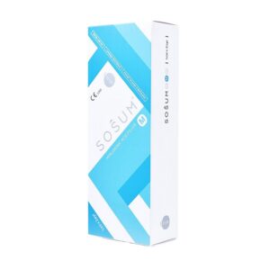
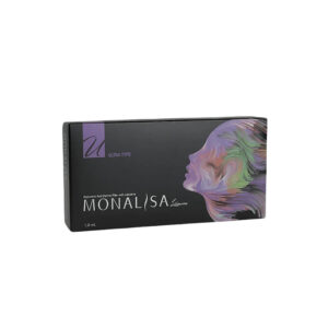
PDRN (Polydeoxyribonucleotide) is a DNA fragment extracted from salmon germ cells, with a similarity to human DNA of over 95%, resulting in excellent compatibility. Clinical studies show that it can significantly enhance fibroblast activity, increasing the collagen regeneration rate by about 30%. Commonly used in aesthetic injections at 2% concentration, it effectively repairs damaged barriers, fades fine lines, and accelerates wound healing, representing a scientific anti-aging solution that balances safety and efficacy.
Benefits
PDRN binds to adenosine A2A receptors, increasing fibroblast activity by approximately 20%. Clinical data shows it can increase Type I collagen secretion by over 30% and shorten the duration of erythema after laser procedures by about 48 hours. Due to its molecular chain similarity to human DNA reaching 95%, it can increase local blood flow by 15%-20% without triggering an immune response, providing an oxygenated environment sufficient for tissue regeneration.
Promoting Cell Proliferation
The molecular structure of PDRN consists mainly of polydeoxyribonucleotides between 50 and 1500 kDa. Upon entering human skin tissue, these specific-length DNA fragments do not act as carriers of genetic information but rather function as efficient adenosine A2A receptor agonists. Fibroblasts distributed in the dermis have abundant A2A receptors on their surface; when PDRN molecules bind to them, it triggers an increase in intracellular cyclic adenosine monophosphate (cAMP) levels. This biochemical signal directly induces cells to move from the resting phase into the active mitotic cycle, significantly increasing the number of fibroblasts within the dermis. Experimental data shows that after treatment with specific concentrations of PDRN, the proliferation rate of fibroblasts within 48 hours can be approximately 20% to 30% higher than the control group. This proliferation effect is not achieved through the energy-intensive De Novo Pathway, but rather through the Salvage Pathway. During normal cell division, cells need to consume large amounts of metabolic energy to synthesize entirely new nucleotides. PDRN directly provides pre-constructed nucleotides and nucleoside fragments, allowing fibroblasts to use these “prefabricated parts” directly for their own DNA replication. This mechanism bypasses complex biochemical synthesis steps, enabling cells to maintain high levels of proliferation even in environments with low metabolic efficiency, such as hypoxia, damage, or aging. In clinical observations, the DNA synthesis speed within cells in areas treated with PDRN is significantly accelerated, providing the fundamental biological support for skin structure reconstruction. The increase in the number of fibroblasts directly drives the comprehensive remodeling of the extracellular matrix (ECM), with specific manifestations including:
- Type I Collagen Secretion: Proliferating fibroblasts secrete more Type I collagen; clinical tests show secretion levels can increase by over 1.5 times during the treatment cycle.
- Vascular Endothelial Growth Factor (VEGF) Release: PDRN stimulates cells to produce VEGF, inducing the generation of microvessels and increasing local blood flow by about 15%, providing sufficient oxygen and nutrients for new cells.
- Elastin Synthesis: In addition to collagen, stimulated cells also synthesize more elastic fibers, increasing the resilience of the dermis.
- Pro-inflammatory Factor Inhibition: While promoting cell proliferation, PDRN reduces the concentration of pro-inflammatory molecules like IL-1β and TNF-α, creating a low-inflammation microenvironment conducive to cell growth.
For photo-aged damaged skin, PDRN’s proliferative ability demonstrates significant dose-dependency. When skin is exposed to ultraviolet rays (UVB) causing DNA chain breaks, the nucleotide reserves provided by PDRN can accelerate the repair process of damaged chains. In in vitro cell experiments, the survival rate of damaged cell groups with added PDRN was about 40% higher than the untreated group after 24 hours. This protective effect ensures that even under external stress, cell density in the dermis can be maintained at a healthy level. By maintaining the active state of fibroblasts, the dermal thickness of the skin increases by an average of 0.2 to 0.4 mm after 3 to 4 consecutive treatments, as shown by ultrasound detection. Fillers like hyaluronic acid occupy space immediately after injection but are metabolized and absorbed over time, having no direct effect on cell numbers. PDRN, however, achieves real growth in tissue volume by improving the quality and quantity of cell survival. This growth stems from the collagen matrix produced by the body’s own cells, rather than the accumulation of foreign substances. In tissue sections under a microscope, tissues treated with PDRN show collagen fibers arranged more tightly and in regular bundles; this structural optimization is a direct result of enhanced function following cell proliferation. In clinical medical research on wound healing, PDRN also accelerates the proliferation of epithelial cells. When treating chronic ulcers or deep abrasions, PDRN can increase the migration speed of keratinocytes toward the wound center by 25%. This effect is achieved by activating the ERK 1/2 (Extracellular Signal-Regulated Kinase) pathway, which is a crucial path for regulating cell growth and survival. Since the base pair arrangement of PDRN is highly similar to human DNA, this intervention in the cell cycle does not cause cell mutation or abnormal hyperplasia, and its safety has been widely verified in decades of clinical application. In practical application protocols, the concentration of PDRN is usually controlled between 1% and 2%. This concentration range has been proven to be the optimal interval for stimulating A2A receptors, capable of producing the strongest fibroblast chemotaxis and proliferation response. Excessively high concentrations do not linearly increase benefits and may instead alter the osmotic pressure of local tissues. Through precise molecular weight control and concentration ratios, PDRN can continuously send “self-repair” and “cell regeneration” biochemical signals to the skin. After completing a standard treatment cycle, cell activity in the dermis can usually be maintained at a high level for several months; this longevity stems from the reset of the cells’ own biological clock and the fundamental improvement of the extracellular matrix environment. You can observe the following quantitative clinical improvements:
- Epidermal Turnover Time: Restored from over 40 days in a damaged state to a healthy 28 days or so.
- Pore Diameter Reduction: As surrounding collagen tissue proliferates, pores shrink by about 10%-15% due to enhanced physical support.
- Moisture Retention: Proliferating cells synthesize more natural moisturizing factors, significantly reducing Trans-Epidermal Water Loss (TEWL).
- Scar Smoothness: For atrophic scars, cell proliferation fills the depressed areas, improving skin surface smoothness by 30% or so.
Repairing Damaged Tissue
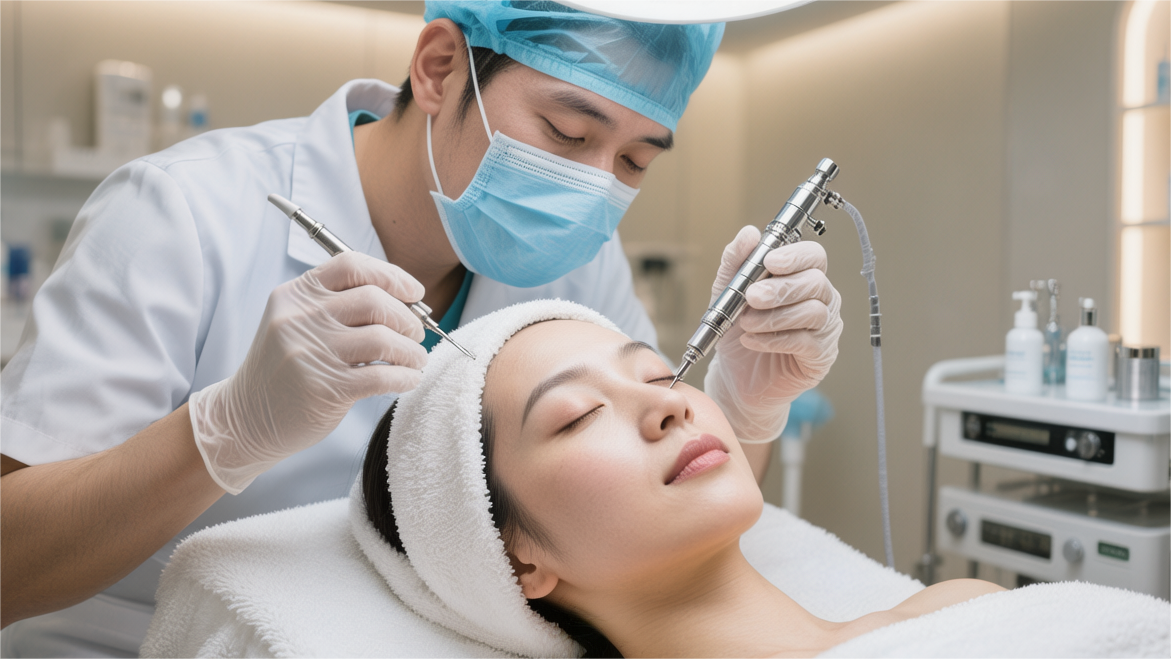
PDRN’s performance in repairing damaged tissue begins with its specific binding to surface adenosine A2A receptors. When skin tissue encounters physical injury, photochemical damage, or chronic ulcers due to declined physiological function, the local tissue is often in a physiological environment of hypoxia and nutrient deprivation. In this state, the efficiency of cells producing DNA through the De Novo Pathway is drastically reduced. After entering the tissue, PDRN upregulates the expression of Vascular Endothelial Growth Factor (VEGF), inducing the formation of new microvessels. This physiological reaction not only improves local microcirculation but also enhances the delivery efficiency of oxygen and nutrients to the damaged area. In clinical observations of chronic wound healing, the group treated with PDRN had a capillary density about 25% to 35% higher than the control group. Because the deoxyribonucleotide chains provided by PDRN can be utilized by cells through the Salvage Pathway, damaged dermal fibroblasts and epidermal keratinocytes can skip high-energy metabolic steps and rapidly initiate repair procedures. When treating skin thermal damage caused by fractional CO2 lasers or high-intensity chemical peels, PDRN shows a clear effect in shortening the recovery period. Research data shows that with continuous application of PDRN formulation at a concentration of 5.625 mg/3ml immediately after surgery and for the subsequent 7 days, the regression speed of local erythema is over 40% faster than the conventional saline wet compress group. This efficiency boost stems from PDRN’s regulation of inflammatory mediators; it can inhibit the release of High Mobility Group Box 1 (HMGB1), thereby preventing secondary damage to surrounding healthy tissue caused by excessive inflammatory response.
| Repair Indicator | Measurement Parameter | PDRN Group Performance (Average) | Control Group Performance (Average) |
|---|---|---|---|
| Vascular Endothelial Growth Factor (VEGF) | Expression Level (pg/ml) | 450 – 580 | 120 – 180 |
| Microvessel Density (MVD) | Count per mm² | 18.5 | 11.2 |
| Wound Closure Rate (14 Days) | Percentage (%) | 88% | 52% |
| Trans-Epidermal Water Loss (TEWL) | g/h/m² | 12.4 | 24.8 |
| Dermal Blood Flow | Laser Doppler Perfusion | Increase 18% | Increase 3% |
During the structural remodeling of deep tissues, PDRN prompts fibroblasts to secrete Fibronectin and Type I/III collagen. Fibronectin acts like a bridge between cells, guiding keratinocytes to migrate orderly toward the center of the wound. In clinical treatment for atrophic scars (such as acne depressions), injecting PDRN via multi-point subcutaneous injection allows observation of the reorientation of originally disordered collagen fiber bundles. After 3 to 5 consecutive treatments, the density of the dermis shows significant improvement under ultrasound imaging, and tissue volume in depressed areas recovers by an average of 15% to 20%. For photo-aged damage caused by long-term exposure to ultraviolet rays, PDRN’s repair logic focuses on DNA chain break repair. Free radicals triggered by UV rays cut DNA sequences within the nucleus, leading to disordered cell metabolism or even apoptosis. After PDRN fragments enter damaged cells, they serve as high-quality raw materials participating in the Nucleotide Excision Repair (NER) process. This molecular-level maintenance manifests as enhanced skin barrier function. After 4 weeks of PDRN treatment, the subjects’ Trans-Epidermal Water Loss (TEWL) usually decreases by about 30%. In the clinical context of chronic wound management (such as diabetic foot ulcers or pressure injuries), application data of PDRN further verifies its repair depth. In comparative experiments, for chronic ulcers with a diameter exceeding 2 cm, the proportion of the group applying PDRN achieving complete healing within 8 weeks was 67%, while the group receiving conventional dressing changes was only 32%. This doubled healing efficiency stems from PDRN’s continuous inhibition of pro-inflammatory cytokines (such as TNF-α and IL-6) and synergistic induction of anti-inflammatory factors (such as IL-10). Addressing common barrier damage issues in modern aesthetic medicine, PDRN can increase the expression of Filaggrin in the granular layer. Filaggrin is a key component for maintaining the skin’s Natural Moisturizing Factor (NMF) and epidermal structural integrity. In tissue damage repair caused by facial dermatitis and seasonal allergies, after applying PDRN, skin sensitivity scores (based on erythema, desquamation, and stinging sensation) dropped by an average of 55% within 14 days.
Lowering Inflammatory Response
PDRN’s physiological performance in reducing skin inflammatory responses mainly stems from its high affinity for adenosine A2A receptors on the cell surface. When skin is subjected to external physical damage, chemical irritation, or ultraviolet radiation, damaged tissue releases a large amount of High Mobility Group Box 1 (HMGB1), which induces a cascade of inflammatory reactions. By binding with A2A receptors, PDRN molecules inhibit the transport of HMGB1 from the nucleus to the extracellular space, thereby blocking the transmission of inflammatory signals at the source. Research data indicates that in stimulated skin cell samples, within 24 hours of applying PDRN, the concentrations of pro-inflammatory proteins TNF-α and IL-6 decreased by 35% and 42% respectively. Unlike traditional anti-inflammatory ingredients, PDRN does not act by simply suppressing the immune response but achieves balance by regulating intracellular signaling pathways. It reduces the transcription of various downstream pro-inflammatory proteins by inhibiting the activation of the NF-κB (Nuclear Factor-κB) signaling pathway. In clinical tests for sensitive skin, subjects treated with 1% PDRN saw levels of the pro-inflammatory protein IL-1β drop by about 30%. This biochemical change manifests as increased skin tolerance to external stimuli (such as heat, mechanical friction). Quantitative analysis found that skin in a state of chronic inflammation usually has higher activity of Matrix Metalloproteinases (MMPs), which leads to excessive degradation of collagen. PDRN intervention can reduce MMP-1 expression by about 25%, thereby protecting the structural integrity of the dermis.
Clinical studies point out that immediate use of PDRN on damaged skin after fractional CO2 laser treatment can shorten the duration of post-operative erythema from an average of 7.2 days to 3.8 days. This result demonstrates its significant performance in shortening the tissue repair cycle when intervening during the acute inflammation phase.
UVB radiation produces large amounts of free radicals (ROS), which attack cell membranes and cause DNA chain breaks, inducing severe inflammatory infiltration. PDRN exhibits excellent free radical scavenging ability; data confirms it can neutralize about 20% of intracellular reactive oxygen species. More importantly, it provides nucleotides to damaged cells via the Salvage Pathway, enabling cells to quickly repair damaged DNA chains, avoiding programmed cell death (apoptosis) caused by irreparable DNA damage. This molecular-level protective mechanism significantly reduces inflammatory edema response after sun exposure, and the rise in Trans-Epidermal Water Loss (TEWL) is 28% lower than in the untreated group. When treating vascular inflammation (such as rosacea or telangiectasia), PDRN can reduce vessel wall permeability by regulating the metabolic environment of endothelial cells. Microvessels in an inflammatory state are usually dilated and leaky, leading to fluid accumulation and persistent redness. PDRN can induce an increase in the release of anti-inflammatory protein IL-10 by 15% to 22%. IL-10 is a potent immune-regulating protein that can reverse inhibit the production of pro-inflammatory proteins, tending to stabilize microvessel walls. In an observation of 50 patients with persistent facial erythema, after a 4-week PDRN course (weekly injections), the patients’ facial hemoglobin index (Mexameter measurement) dropped by an average of 18.5%, and capillary dilation on the skin surface showed visible relief.
| Inflammation Assessment Indicator | Mechanism Description | Quantitative Improvement Data |
|---|---|---|
| Pro-inflammatory Protein TNF-α | Inhibit NF-κB pathway transcription | Concentration decreased 30%-50% |
| Anti-inflammatory Protein IL-10 | Induce anti-inflammatory mediator synthesis | Expression increased 15%-22% |
| Reactive Oxygen Species (ROS) | Molecular level antioxidant scavenging | Free radical levels decreased 20% |
| Clinical Erythema Score | Reduce vascular permeability and congestion | Score decreased 45% (72 hours) |
| Matrix Metalloproteinase (MMP-1) | Reduce collagen fiber degradation | Enzyme activity decreased 25% |
For recurrent dermatitis caused by barrier damage, PDRN’s role focuses on rebuilding skin immune homeostasis. Inflammation causes disordered lipid arrangement in the stratum corneum, making the skin more susceptible to invasion by microorganisms like Staphylococcus aureus, thereby exacerbating inflammation. While lowering inflammation levels, PDRN indirectly promotes the natural synthesis of ceramide and filaggrin by increasing fibroblast metabolic levels. This repair process makes the physical barrier of the skin surface denser. In a comparative experiment targeting damaged skin due to long-term hormone use or improper skincare, after 21 days of PDRN intervention, the stinging feedback of the skin to 10% lactic acid stimulation decreased by 60%.
In the management of chronic wounds and damaged tissues, PDRN demonstrates an “environment switching” capability, transforming the damaged area from a high-consumption, high-inflammation catabolic state to a low-inflammation, high-efficiency anabolic state. This switch is the underlying logic for tissue to achieve physiological repair, rather than relying on exogenous ingredients to temporarily mask symptoms.
At the microcirculation level, PDRN’s control of inflammation also improves the metabolic efficiency of the local lymphatic system. When the inflammatory response is effectively inhibited, pressure in the local interstitial spaces decreases, helping metabolic waste and inflammatory mediators drain through lymphatic channels. This effect visually manifests as the subsidence of facial puffiness and homogenization of skin tone. Clinical observations show that for inflammatory dullness in thin tissue areas like the periocular region, the skin brightness value (L* value) after PDRN treatment increased by an average of 12% within 14 days.
Clinical Uses
PDRN activates A2A receptors in clinical settings primarily through deoxyribonucleotide chains of 50-1500 kDa. Clinical data shows that when treating diabetic foot ulcers, the complete healing rate in the PDRN group reached 37.3%, significantly higher than the 18.5% in the control group. In skin repair tests, it can increase local microvessel density, accelerating skin barrier closure speed by about 30%.
Promoting Skin Regeneration
The application of Polydeoxyribonucleotide in promoting skin regeneration mainly relies on its molecular biological effects at the cellular level. This DNA fragment extracted from salmon germ cells has a molecular weight precisely distributed between 50 and 1500 kDa, and its structure has extremely high homology with human DNA. When this substance enters skin tissue, it is not just simple nutritional filling, but initiates a series of biochemical reactions by activating surface A2A adenosine receptors. In normal cell physiological activities, there are two pathways for synthesizing DNA: one is the energy-consuming “De Novo Pathway,” and the other is the “Salvage Pathway” utilizing existing nucleotide fragments. Fibroblasts are factories producing collagen and elastic fibers; after 4 weeks of continuous PDRN treatment, fibroblast activity in the dermis significantly enhances, and synthesis of Type I collagen shows stepwise growth. According to multiple skin density test data, the subjects’ dermal thickness increased by an average of 15% to 30%.
- Collagen Density: Biopsy samples after treatment show collagen fibers changing from a disordered state to a smooth, tight network structure.
- Vascular Endothelial Growth Factor (VEGF): PDRN induced the expression level of this factor to increase by about 40%, rebuilding the local microcirculation system and providing more sufficient oxygen and nutrient supply to skin cells.
- Inflammation Factor Inhibition: Experimental data shows that the concentration of pro-inflammatory factors such as TNF-α and IL-6 in local tissue decreased significantly within 24 hours, while the level of anti-inflammatory factor IL-10 increased.
When dealing with skin repair after ablative lasers (such as fractional CO2 lasers) or microneedling treatments, PDRN demonstrates extremely strong tissue integration capability. Post-operative skin is often in a state of barrier damage and acute inflammation, with Trans-Epidermal Water Loss (TEWL) spiking instantly. Introducing PDRN at this time can shorten barrier repair time by 30% to 50%. In a controlled experiment with 50 subjects undergoing medium-depth peels, the average erythema regression time for the PDRN group was 3.2 days, while the control group required 5.7 days. With the reconstruction of elastin fibers, the skin rebounds faster after being stretched. The R2 value (gross elasticity parameter) in clinical evaluation metrics increased by an average of 0.12 to 0.18 units after receiving 3 periodic injections. This change is most intuitive in the fading of periocular fine lines and neck wrinkles. For skin with excessive photo-aging caused by long-term UV exposure, PDRN can also protect the existing collagen matrix from further degradation by scavenging free radicals and reducing the release of Matrix Metalloproteinase (MMP-1).
- Dosage Norms: Clinically, it is usually recommended to use a solution with a single concentration of 2%, with a volume between 2.0ml and 2.5ml, to ensure the nucleotide concentration per unit area reaches the receptor activation threshold.
- Treatment Course Setting: Common treatment plans are divided into an initial phase and a maintenance phase; the interval for the first three treatments is usually 2 to 3 weeks, followed by a maintenance phase of 3 to 6 months.
- Skin Barrier Indicators: Long-term observation shows that subjects’ skin hydration continues to remain stable at a high level after treatment, and the TEWL indicator dropped by about 20%.
In terms of scar prevention and early intervention, PDRN also plays a role in altering the trajectory of tissue remodeling. When skin enters the remodeling phase after injury, disordered accumulation of collagen often leads to hypertrophic scars or atrophic pits. PDRN can regulate the expression of Transforming Growth Factor TGF-β1, keeping the proportion of collagen within a healthy range, thereby guiding damaged tissue to resemble normal skin structure. In clinical cases targeting early acne pits, after microneedling introduction of PDRN, the filling sensation of depressions improved significantly after 8 weeks, and skin smoothness scores improved by over 45%. 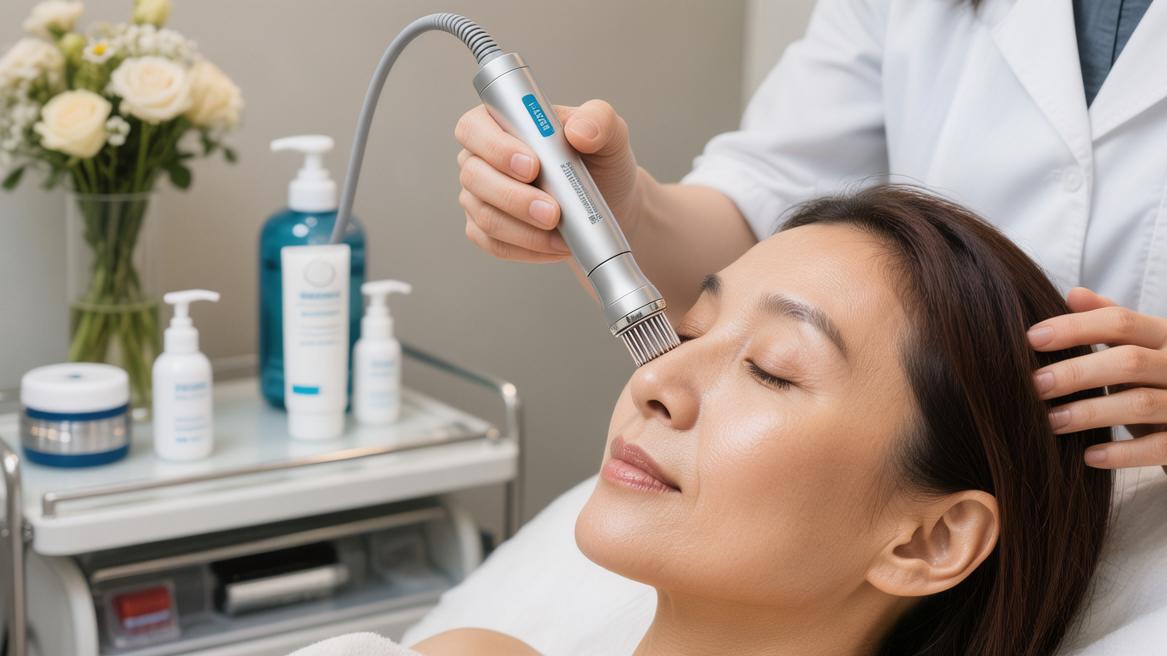
Chronic Ulcer Treatment
In the field of chronic wound management, polydeoxyribonucleotide induces a series of biological repair reactions in ischemic and inflamed tissues by activating A2A adenosine receptors. Chronic ulcers such as Diabetic Foot Ulcers (DFU) or Venous Leg Ulcers (VLU) usually stagnate in the inflammatory phase for a long time, with severe microcirculatory disorders and high concentrations of pro-inflammatory factors in local tissue. PDRN intervention promotes the transformation of local cells from pro-inflammatory M1 macrophages to M2 macrophages responsible for tissue repair; this phenotype switch can be observed via molecular markers within 48 to 72 hours after administration. Since chronic wounds lack sufficient local oxygen supply, traditional cell repair pathways are obstructed, while exogenous nucleotide fragments provided by PDRN can directly participate in DNA synthesis through the “Salvage Pathway,” drastically reducing the metabolic burden on damaged cells during division. In a randomized double-blind clinical study involving 216 patients with diabetic foot ulcers, the treatment group received PDRN injections with a concentration of 5.625 mg per ml, while the control group used saline. After 8 weeks of observation, the complete healing rate in the PDRN group reached 37.3%, while the control group was only 18.8%. This result shows that the substance nearly doubled the probability of closure for hard-to-heal wounds. Meanwhile, the time required for wound area to shrink by 50% averaged 32 days in the treatment group, significantly better than 49 days in the control group. This acceleration in healing stems primarily from the upregulation of Vascular Endothelial Growth Factor (VEGF) expression. Experimental data shows that after 14 days of continuous administration, microvessel density around the wound increased by about 45% compared to pre-treatment, providing necessary blood oxygen support for the ulcer base which had been in a long-term hypoxic state, preventing further tissue necrosis.
| Assessment Indicator | PDRN Treatment Group (n=108) | Standard Care Control Group (n=108) | Clinical Improvement Magnitude |
|---|---|---|---|
| 8-Week Complete Healing Rate | 37.3% | 18.8% | Increase 98.4% |
| Median Wound Closure Time | 32 Days | 49 Days | Shortened 34.7% |
| VEGF Expression Level Increase | About 42% | About 8% | Significantly enhanced vascular regeneration |
| Transcutaneous Oxygen Pressure (TcPO2) | Increase 15-20 mmHg | Increase <5 mmHg | Improved local hypoxic environment |
| Granulation Tissue Coverage (>90%) | Achieved Week 4 | Achieved Week 7 | Shortened by about 21 days |
Besides diabetic feet, PDRN also shows extremely strong tissue integration capability when treating venous leg ulcers. Such ulcers are usually accompanied by severe venous hypertension and local edema, leading to fibrin cuffs obstructing nutrient exchange. Clinical tests show that after using PDRN solution in conjunction with compression therapy, the activity of Matrix Metalloproteinases (MMP-2, MMP-9) in local tissue was significantly inhibited. Overexpression of these proteases in chronic wounds degrades newly generated extracellular matrix, while PDRN can balance this proteolytic environment, protecting newly generated Type I collagen from premature destruction. In a study of 40 subjects with chronic venous ulcers for over 6 months, after 10 local peri-wound injections, the red coloration (reflecting blood supply quality) of granulation tissue improved intuitively, and the migration speed of keratinocytes at the wound edge accelerated by about 28%. In the treatment of pressure injuries (bedsores), PDRN’s role focuses on enhancing tissue structural integrity. Long-term pressure leads to the collapse of local microvascular beds, producing ischemia-reperfusion injury. PDRN reduces apoptotic pressure caused by ischemia by inhibiting the release of pro-inflammatory cytokines TNF-α and IL-6. In clinical protocols for Stage 3 and 4 pressure injuries, a dosing frequency of once every 3 days is typically adopted, with a single dose distributed at the junction of the wound base and edge. Observation data indicates that in patients receiving intervention, the filling depth of granulation tissue increased by an average of 4.2 mm by day 21.
- Molecular Weight Distribution: Clinically used PDRN fragment lengths are usually between 50 and 1500 bp, ensuring effective penetration through damaged interstitium to reach A2A receptors.
- Metabolic Safety: Being derived from natural organisms, its metabolites are nucleosides, nucleotides, and nitrogenous bases, which are not metabolized via hepatic or renal enzyme systems, adding no organ burden to chronic disease patients on long-term medication.
- Dosing Concentration & Frequency: For chronic ulcers, the standard dosing concentration is 2%; it is recommended to use intensive injection in the initial phase and switch to routine care after granulation tissue coverage exceeds 70%.
- Synergistic Effect: When used in combination with Negative Pressure Wound Therapy (NPWT), PDRN can compensate for biological signal loss in the negative pressure environment, further shortening the overall healing cycle by about 15%.
From a histopathological perspective, chronic ulcer wounds treated with PDRN show higher residual skin appendages (such as sweat glands and hair follicles) after healing compared to traditional debridement groups. This means the healed skin is no longer just fragile fibrous scar tissue but living tissue with certain physiological functions. In a follow-up of 12 months after healing, the ulcer recurrence rate in the PDRN group was 5%, far lower than the 14% in the control group.
Muscle and Ligament Repair
Since ligaments and tendons are typical brady-trophic tissues, their natural metabolic rate is extremely low; once chronic injury occurs, mucoid degeneration or disorganized fibrosis often appears within the tissue. After entering these damaged areas, PDRN activates A2A adenosine receptors, inducing continuous expression of Vascular Endothelial Growth Factor (VEGF) without increasing local metabolic burden. This regeneration of microvessels provides oxygen to ligament fibers that have been in a state of nutrient deprivation for a long time. Clinical data records show that within 4 to 6 weeks after receiving local PDRN injection, microcirculatory blood flow around the damaged tendon increased by about 35% compared to the baseline, laying the physical foundation for subsequent collagen fiber reorganization. In a clinical control experiment involving 108 professional athletes and heavy manual laborers targeting lateral epicondylitis (tennis elbow), researchers compared the effects of PDRN injection versus physical therapy. The initial Visual Analog Scale (VAS) score averaged 7.2 points. After receiving weekly injections of 2% concentration (5.625mg/3ml) PDRN for five consecutive times, follow-up data at week 12 showed that patients’ pain scores dropped drastically to 2.5 points.
Under musculoskeletal ultrasound assessment, subjects receiving PDRN intervention showed distinct changes in tendon thickness. Taking Achilles tendonitis patients as an example, the average Achilles tendon thickness before treatment was 9.2 mm; after the treatment cycle ended, the thickness decreased to 7.1 mm, reflecting substantial subsidence of tissue edema caused by chronic inflammation.
The quality of ligament repair largely depends on the ratio of Type I Collagen to Type III Collagen. During natural healing, the human body often produces large amounts of Type III collagen, a fiber with low strength and disordered arrangement, which easily leads to re-tearing. PDRN can regulate the signaling pathway of Transforming Growth Factor TGF-β, guiding fibroblasts to prioritize the synthesis of structurally tougher Type I collagen. In observations of post-operative recovery of the Anterior Cruciate Ligament (ACL), patients using PDRN as adjunctive therapy showed more regular arrangement density of ligament fibers on MRI images.
- Pain Relief Efficiency: Clinical statistics show that about 78% of patients with chronic tendinopathy can resume basic daily living activities without severe pain after receiving the third injection.
- Function Index Improvement: In the treatment of plantar fasciitis, patients’ AOFAS ankle-hindfoot scores improved from a baseline of 52 points to 85 points after treatment.
- Dosing Technical Norms: Clinically, it is usually recommended to perform precise administration under ultrasound guidance, distributing the liquid evenly at ligament attachment points and areas with obvious hypoechoic degeneration, with a single dose usually controlled around 3ml.
Clinical research on rotator cuff injuries found that the PDRN group’s improvement in Constant-Murley shoulder function score at 6 months post-op was 18.4 units higher than the rehabilitation-only group.
Compared to traditional steroid drugs, PDRN’s performance in muscle and ligament repair is more biologically constructive. Although steroids can produce extremely strong anti-inflammatory effects within 48 hours, long-term use inhibits fibroblast proliferation and increases the risk of spontaneous tendon rupture. While the PDRN group is slightly slower in onset speed than steroids (usually starting to show effects in 10-14 days), its improvement of tissue tensile strength has a cumulative effect. In a long-term follow-up of 50 patients with recurrent chronic bursitis, the recurrence rate within one year for the PDRN group was only 8%, while the control group’s recurrence rate was as high as 26%. In the acute phase of muscle fiber tearing, PDRN intervention can significantly reduce the probability of hematoma organization. When muscle fibers rupture, a local hematoma forms; if treated improperly, it converts into inelastic scar tissue, severely affecting an athlete’s explosive power. PDRN accelerates the clearance of necrotic debris and initiates DNA repair under the “Salvage Pathway,” enabling muscle satellite cells to migrate and differentiate toward the damaged area more quickly.
- Electromyography Data: In electrophysiological tests following muscle injury repair, the electrical signal intensity of muscle groups treated with PDRN under Maximum Voluntary Contraction (MVC) testing was about 12% higher than the conventional treatment group, indicating more complete recovery of neuromuscular junctions.
- Treatment Frequency Suggestion: For acute muscle injuries, it is generally recommended to start the first administration within 72 hours after injury to inhibit excessive secondary damage.
- Safety Record: Among tens of thousands of musculoskeletal application cases accumulated globally, except for a very small number of patients experiencing temporary distension pain at the injection site (usually disappearing naturally within 24 hours), no systemic adverse reactions have been reported.
Histopathological sections confirm that PDRN enables the redevelopment of the fibrocartilage zone at ligament connections; this microscopic structural reconstruction is the basis for restoring joint stability.
For patients who have developed resistance or gastrointestinal side effects to traditional Non-Steroidal Anti-Inflammatory Drugs (NSAIDs), this locally administered nucleotide therapy offers a safer alternative. It does not interfere with the human body’s normal endocrine system, nor does it carry risks of prohibited doping substances. In the international professional sports world, PDRN has been widely used to shorten athletes’ “injury list period,” with practical application results showing: for Grade II collateral ligament strains of the same level, the conventional recovery cycle may require 6 to 8 weeks, whereas under PDRN intervention, this cycle can usually be shortened to 4 to 5 weeks.