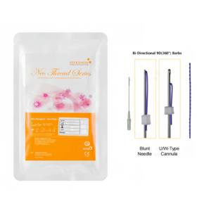
Nowadays, many people opt for mesotherapy skin boosters to improve dry skin, reduce fine lines, and rejuvenate a dull complexion. Among the myriad of products available, 2XSOME and Celosome Aqua are two of the most common choices. So, what exactly are the differences between them, and which one is better suited for you?
Skin Hydration
HA Volume Replenishment
Opening a 3 mL of Celosome Aqua reveals a highly viscous, transparent gel. Pushing the plunger with 15 Newtons of force injects the 24 mg/mL concentration of hyaluronic acid (HA) directly into the dermis. Typically, it is very difficult for topical creams to penetrate the 15 μm-thick stratum corneum, as most skincare molecules exceed 500 Daltons and are entirely blocked at the surface. The mesotherapy device’s 9-pin probe uses negative pressure to lift the skin, while ultra-fine 34G vertically pierce 1.2 mm deep into the subcutaneous tissue. With each pulse, the machine delivers 0.025 mL of gel. Because the skin on the forehead is thin and the cheeks carry more fat, the tissue resistance encountered by the machine during injection varies by approximately 30%. Hyaluronic acid molecules act like a three-dimensional lattice, rapidly binding with the skin’s natural moisture. Viewed under a 20 MHz ultrasound, the tightly packed collagen fibers in the dermis are forcefully stretched open by this gel. Within 20 minutes of injection, small wheals measuring 3 to 5 mm in diameter form at the treated sites.
- Superficial layer (0.8 mm deep): Within 12 hours post-injection, 85% of the small wheals will subside.
- Mid-layer (1.2 to 1.5 mm deep): Moisture can be securely locked in for approximately 14 days.
- Deep layer (2.0 mm deep): The mechanical tension created by expanding the collagen notably increases by 15%.
Naturally occurring enzymes in the body typically degrade about one-third of free hyaluronic acid every day. By adding less than 0.5% of cross-linking agents to the HA, it acts like tying knots in the structural lattice. The enzymes must expend significant effort to break these bonds, effectively slowing down the rapid degradation rate of standard HA, which usually loses half its volume in a single day. Gels fortified with these tiny cross-links can remain in the skin for 21 to 28 days. Instrumental measurements of surface moisture show that the skin reaches its peak hydration on the seventh day post-treatment. Dry cheeks initially registering only 35 AU in moisture levels directly surge to 68 AU after 3 mL of product is injected. Placing a transepidermal water loss (TEWL) meter near the jawline reveals that the evaporation rate decreases by 4.2 units within 15 seconds. As the deep dermal layers are expanded and tightened, they exert a mechanical pull on neighboring cells within a 50 μm radius. Sensing this 1.5 kPa tensile force, these cells produce extra Type I collagen within a 72-hour window.
- Setting the suction to level 3: Reduces the machine’s risk of product leakage by approximately 40%.
- Slowing the injection speed to level 2: Decreases trauma to small subcutaneous blood vessels by 15%.
- Pausing the subcutaneously for an extra 0.5 seconds: Yields a 92% absorption and retention rate for the 0.025 mL dosage.
The skin around the eyes is exceptionally thin, measuring only 0.3 to 0.5 mm, meaning standard 1.5 mm can easily puncture the underlying muscle. Physicians switch to sharp 30G, inserting them gently at a 15-degree angle. This precisely deposits 0.01 mL of the gel into the extremely tight dermal interstitial spaces. Examining the dehydration lines under the eyes with a 50x magnification device shows that the deepest troughs become 0.12 mm shallower. Large molecules weighing 2 million Daltons cannot penetrate inside the cells. Instead, they accumulate extracellularly, building up a hydration barrier roughly 1 mm thick. This macromolecular lattice securely traps the underlying moisture. Ultrasound imaging reveals that the skin across the entire face actually thickens by 0.2 mm. Around the mouth, daily activities like talking and eating cause the muscles to stretch the skin tens of thousands of times a day. Consequently, the HA around the mouth degrades about 25% faster than in the relatively immobile forehead. The jawline lacks significant sebaceous activity; injecting 0.03 mL there increases skin tightness by 10% in that localized area. Using a Cutometer to measure skin elasticity via suction, the R2 rebound index climbs from 0.65 to 0.78 after half a month. Pressing the cheek with a finger, the tissue feels tangibly firmer and more resilient.
- Small molecules (100,000 Daltons): Penetrate too deeply and rapidly, being consumed by cells within 3 days.
- Medium molecules (1,000,000 Daltons): Wedge into collagen gaps, keeping the face hydrated for half a month.
- Large molecules (3,000,000 Daltons): Mechanically prop open a small spatial volume of roughly 0.05 cm³.
A 3 mL volume is exactly enough for 150 injection points across the entire face. The dermis acts like a dry sponge, becoming thoroughly saturated by this gel capable of holding 1,000 times its weight in water. As the skin on the back of the hands ages, veins typically become prominently engorged. Layering 2 mL of HA at a subcutaneous depth of 1.5 mm on the back of the hand forcefully replenishes the tissue volume lost around the blood vessels. Viewed through a 3D scanner, the bulging veins visually recede by 1.8 mm. HA injections align perfectly with the epidermis’s 28-day cellular turnover cycle. The first session replenishes 40% of the dermal deficit. The second session builds upon the remaining 15% foundational volume left unabsorbed from the previous treatment, creating a compounding effect.
Extracellular Matrix Reconstruction
The nurse retrieves the lyophilized powder from a 2°C to 8°C refrigerator, opens the, and injects 3 mL of 0.9% normal saline. With a gentle shake, the powder dissolves within 5 seconds, turning the clear liquid slightly cloudy. Suspended within this 3 mL solution are approximately 5 billion exosomes (micro-vesicles) secreted by stem cells. Drawing the solution with an ultra-fine 32G at a 45-degree angle, injections are administered at 1 cm intervals across the face. The tip bypasses the 15 μm-thick stratum corneum of dead skin cells, halting at a depth of 1.5 to 2.0 mm subcutaneously. Pausing for 0.5 seconds per injection, 0.02 mL of fluid is pushed into the tissue, depositing tens of millions of micro-vesicles into the collagen network. These micro-vesicles are only 30 to 150 nm in size, encased in a lipid membrane approximately 5 nm thick. Upon encountering damaged cells, these two membranes fuse together within 10 to 15 minutes. The vesicles burst open, offloading over 1,000 types of proteins and more than 300 types of microRNAs directly into the target cell nucleus. Senescent cells receive the biological signal to activate, restarting previously dormant collagen production lines. Seventy-two hours post-injection, data-tracking devices capture the physiological shift: the cellular synthesis rate of Type I collagen doubles, and the production of new Type III collagen increases by 150%. By day 15, examining the cheeks with a 20 MHz ultrasound reveals that the 4 mm² dark dermal voids under the skin have shrunk. Newly generated tissue interwoven in a crisscross pattern pushes the original 1.2 mm thick dermis upward by 0.15 mm. When the device applies firm pressure to the entire face, the structural firmness demonstrates a 30% increase. With increased tissue volume, the activity of genes regulating elasticity also rises by 20%. Pinching the skin along the jawline and releasing it shows a visible reduction in the snap-back time from 1.8 seconds down to 1.3 seconds. The lax facial skin is supported by the newly formed structural net, reducing downward sagging by 2 mm. Beneath cheeks plagued by severe redness lie enlarged blood vessels exceeding 0.05 mm in diameter. The micro-vesicles deliver targeted anti-inflammatory compounds into the area, reducing active macrophage counts by 15% over 14 days. Scanning the face with an erythema-measuring device shows the previously high red alert level of 55 dropping to 37. As inflamed areas shrink, localized facial skin temperature also decreases. On an infrared thermal imaging screen, the number of red, high-temperature hotspots on both cheeks drops from 400 to 210. The skin temperature cools from a feverish 36.8°C down to 36.2°C, significantly slowing the rate at which basal moisture is evaporated by body heat. The outermost layer of a compromised skin barrier resembles a crumbling brick wall full of drafts. Once the dermis is repaired, nutrient delivery ascending to the epidermis increases by 25%, and ceramide production surges by 18%. Epidermal cells pack more tightly together, successfully resealing the 0.5 μm gaps between the cellular “bricks.” In a room with 45% humidity, a TEWL meter is placed on flake-prone areas of the forehead. The values on the machine’s screen fall from a severely compromised 32 g/m²·h back down to a healthy 22 g/m²·h.
| Observation Period | Subcutaneous Micro-Changes | Instrumental Data Readings |
|---|---|---|
| Day 1 to 3 | Vesicles fuse with cell membranes within 15 minutes | Localized 36.8°C elevated temperature drops by 0.3°C |
| Day 7 to 14 | Type I collagen synthesis rate doubles | Surface moisture evaporation rate decreases by 12% |
| Day 21 to 28 | Fiber lattice gaps shrink by approximately 5 μm | Ultrasound detects a 0.15 mm increase in subcutaneous thickness |
| Day 45 to 60 | Epidermal ceramide concentration increases by 18% | TEWL values settle at 22 g/m²·h |
A single treatment across a 400 cm² face requires over 200 punctures, leaving behind a field of tiny micro-injuries. The physical act of needling itself inflicts minor trauma. To repair these 200+ epidermal micro-wounds, the body secretes an additional 5% of growth factors to aid in scabbing and healing. The nutrient-rich fluid delivered by the micro-vesicles provides ready-made building blocks for this healing process. Combined, these two forces pull the skin’s delayed 35-day turnover cycle back to a youthful 28 days. Aged, dead skin sheds faster; a single face wash can remove an extra 1.2 grams of skin flakes, leaving the texture noticeably smoother to the touch. Because the fluid doesn’t occupy structural space within the tissue, patients feel absolutely no swelling or pain post-injection. Tissue resistance during injection is minimal, with machine readings under 5 Newtons. The liquid diffuses subcutaneously within a 0.5 cm radius; after 30 minutes, no bumps can be felt on the cheeks whatsoever. Severely compromised, highly sensitive skin can only absorb about 30% of the 5 billion vesicles in a single session. The unabsorbed remainder is flushed away by blood circulation. By the time of the second treatment 28 days later, the damaged receptor antennas on the cell surfaces have already been 40% repaired by the first injection. With the second 3 mL injection, the absorption and retention rate climbs to 50% to 60%. After three consecutive monthly sessions totaling 9 mL, the subcutaneous environment undergoes a complete overhaul. The frequency of redness and itching caused by bracing -5°C winter winds drops from 3 times a week to just once.
Clinical Data Comparison
A dermatology clinic in Gangnam, Seoul, conducted a test on 200 subjects aged 30 to 45. One group of 100 participants received 3 mL of hyaluronic acid on the left side of the face and normal saline on the right. The other 100 received full-face injections containing 5 billion exosome micro-vesicles. In a controlled room maintained at 22°C and 45% humidity, instrumental data was continuously monitored for 90 days. Just 72 hours post-treatment, cheek moisture in the HA group skyrocketed. Moisture meter readings surged from a parched 31 AU to 68 AU. High-purity molecules absorbed maximum water 1.5 mm deep under the skin, propping up a hydration band about 0.8 mm thick. Visually, the 0.2 mm deep dehydration lines under the eyes were completely smoothed out. Meanwhile, data for the exosome group remained largely static during these first three days. The moisture meter reading sluggishly climbed from 32 AU to 35 AU—which, factoring in machine error, essentially meant no change. The lipid membranes had just attached to the damaged cells, and the 300+ types of microRNAs were only just entering the cell nuclei. By day 14, the data trends for the two groups completely reversed. Placing the TEWL meter on the foreheads of the HA group yielded a reading stalled at 25 g/m²·h. The body’s enzymes had digested about 15% of the gel, and the 0.8 mm subcutaneous water barrier began to slowly deflate. The post-cleansing hydrated feel dropped roughly 10% compared to the first week. Conversely, the subcutaneous reactions in the exosome group delivered impressive results at this stage. The 20 MHz ultrasound probe revealed that the dermal collagen lattice had tangibly thickened by 0.12 mm. TEWL values plummeted forcefully from a compromised 35 g/m²·h down to 24 g/m²·h. Newly synthesized ceramides patched the epidermal brick wall gaps by 20%. By day 45, HA’s water-retention capacity experienced a steep cliff-drop. Pressing the probe against the cheek for 3 seconds showed readings sliding back into a slightly dry state of 42 AU. Free molecules unbound by cross-linking agents were over 60% digested, and the mechanical tension supporting the skin dissipated. Measured under a microscope, dehydration lines around the outer eye corners re-collapsed to a depth of 0.15 mm. The biological dividends of exosomes rebooting cellular activity hit their peak on day 45. Epidermal erythema readings stabilized at a healthy 32, dropping nearly 40% from baseline. Subcutaneous capillaries cooled, and the abnormal localized temperature of 36.8°C returned to 36.1°C. Even when applying only a thin layer of lotion for 3 consecutive days, cheek moisture consistently held strong at 55 AU.
| Monitoring Period | Skin Moisture Levels (HA) | Skin Moisture Levels (Exosomes) | TEWL Rate (HA) | TEWL Rate (Exosomes) |
|---|---|---|---|---|
| Day 3 | Surged to 68 AU | Crept to 35 AU | Dropped to 26 g/m²·h | Stuck at 34 g/m²·h |
| Day 14 | Fell back to 58 AU | Climbed to 46 AU | Stalled at 25 g/m²·h | Plummeted to 24 g/m²·h |
| Day 45 | Dropped to 42 AU | Reached 55 AU | Rebounded to 31 g/m²·h | Locked at 21 g/m²·h |
| Day 90 | Returned to baseline 34 AU | Stabilized at high 52 AU | Deteriorated to 35 g/m²·h | Settled at healthy 22 g/m²·h |
Patient-reported sensation questionnaires captured subtle distinctions. The injection pain for viscous HA averaged a score of 4.5, while the water-like exosomes scored only 1.8. Because HA is dense and rigid to inject, the product leakage rate hovered around 10%, whereas exosome leakage was less than 3%. The HA group experienced lingering red spots around the puncture sites for 3 days post-treatment, whereas the exosome group saw redness subside in just 15 minutes. About 20% of patients receiving periocular HA injections developed minor bruising, while exosome injections caused virtually zero bruising. For young individuals with fast metabolisms, the injected HA was cleared out by day 60, leaving only 15% behind. Consequently, the window for follow-up HA injections is strictly locked between 45 and 60 days. In contrast, fibroblasts awakened by exosomes are still working vigorously. Biopsies of deep tissue taken on day 90 showed that Type I collagen levels were 18% higher than pre-injection baselines. The fully patched stratum corneum barrier can easily withstand ultra-low indoor humidities of 30% during the autumn and winter seasons. Without the need to endure frequent, painful injections, the biological reboot provided by exosomes extends its benefits well beyond six months.
- Skin Elasticity: HA peaks at 14 days and begins to decline, while exosomes are still steadily improving at day 60.
- Facial Sagging: As HA absorbs, the tissue pulls downward; exosomes firmly elevate the cheeks upward by 1 mm.
- Pore Count: HA relies on hydration to temporarily squeeze gaps closed; exosomes genuinely reduce the pore count by 120.
- Under-Eye Dehydration Lines: HA fills the 0.2 mm troughs, but the lines reappear exactly as before once the product is metabolized.
A small minority of patients opt to stack both protocols on day 30. They lay down a 2 mL exosome base in the deep tissue to repair cellular receptors, and use 1 mL of HA in the superficial layer to fill dehydration lines. The peak moisture value recorded by instruments directly broke records at 74 AU, while the TEWL rate plummeted to an unprecedented 18 g/m²·h. The 1.5 mm deep tissue foundation became structurally firm, while the superficial reservoir was simultaneously filled to the brim. 
Fine Line Improvement
Hydration and Volumization
Holding a 34G fine with an outer diameter of merely 0.18 mm, the physician inserts it into the skin at a 45-degree angle. The tip rests subcutaneously at a depth of 1.0 to 1.5 mm. Applying a steady pushing force of 2 to 3 Newtons, 0.01 to 0.02 mL of the 24 mg/mL gel is delivered into the interstitial spaces between cells. Small, transparent wheals measuring 2 to 3 mm in diameter emerge every 1.0 cm across the face. Extruded from the 0.08 mm inner bore of the, the hyaluronic acid (HA) springs open within the skin. These large molecules, weighing between 1 million and 3 million Daltons, stretch out to a full length of 2 to 3 micrometers. Carrying negative charges, they repel one another, allowing 1 gram of the substance to forcefully pull in 1,000 grams of surrounding water. Within 90 seconds, the localized osmotic pressure surges from 300 mOsm/kg to over 450 mOsm/kg. As a massive influx of water rushes in, the internal dermal structures rapidly expand:
- Fluid from the blood vessels flows into the area at a speed of 45 μm per hour.
- The 80 to 100 nm collagen lattice is stretched to 2.1 times its original size.
- Interstitial water volume skyrockets by 300% within 30 minutes.
- Subcutaneous depressions up to 0.5 mm deep are leveled out by the liquid.
The micro-trauma caused by punctures triggers macrophages to release destructive hydroxyl radicals. However, the pre-formulated 0.9% mannitol (at 9 mg/mL) steps in to protect the tissue. Mannitol molecules are extremely small and carry six hydroxyl groups, enabling them to scavenge these damaging free radicals in a billionth of a second. Consequently, the degradation rate of HA is effectively slowed by 33.5%. Without the protection of mannitol, HA would be cleared by the body within 24 to 48 hours. Bolstered by this antioxidant, the transparent wheals on the face safely persist for 72 to 96 hours. Water absorption in the skin follows a precise timeline:
- 0 to 24 hours: The injection sites aggressively draw in moisture from within a 5 mm radius.
- 24 to 72 hours: The wheals subside, and the moisture diffuses outward at a rate of 1.5 mm per day.
- 7 to 14 days: Skin moisture levels stabilize at a high of 30% to 35%.
- 14 to 28 days: The large molecules gradually break down into smaller fragments under 50,000 Daltons.
The transepidermal water loss (TEWL) rate, which can reach as high as 25 g/m²·h on the skin surface, drops below 15 g/m²·h by the third day. Once fully hydrated, dehydrated corneocytes expand in surface area by an average of 15% to 20%. This alters how light refracts off the face, improving the transmittance of specific light wavelengths by 12%. The mechanical expansion from hydration continuously compresses the fibroblasts, exerting a tensile force of 15 to 20 piconewtons. Sensing this mechanical stress, the cell membranes trigger an instantaneous spike in internal calcium ion concentrations. Free-floating HA fragments at a depth of 0.8 mm bind to CD44 receptors on the cell surfaces, which downregulates the expression of the pro-inflammatory protein IL-1α by 40%, effectively halting vascular leakage from capillaries measuring 5 to 10 μm in diameter. This formulation consistently exhibits an elasticity value of only 5 to 8 Pascals, making it as soft as water and preventing hard lumps from forming in the ultra-superficial 0.1 mm skin layers. Over 28 days, dense vascular networks drain excess fluid at a rate of 3% per day. The volume of the expanded regions decreases by 20% each week, and intercellular spacing returns to a compact 10 to 15 nm.
Cellular Activation
The physician switches to a device equipped with 9 ultra-fine 32G, setting the depth parameter to 2.0 mm. With each pull of the trigger, 0.02 mL of fluid is injected deep into the skin. Upon withdrawal, the microscopic, invisible puncture wounds automatically seal within 2 seconds. The is loaded with a highly concentrated suspension of exosome vesicles, containing 60 billion independent particles per milliliter.
Ranging in size from 30 to 200 nm, their volume is less than one-thousandth that of a typical cell.
Encased in lipid bilayers, these micro-vesicles percolate down through the interstitial spaces. At a depth of 1.5 to 2.5 mm, they encounter large clusters of dormant fibroblasts. The CD9 and CD81 protein markers on the vesicles’ surfaces dock with receptors on the outer cell membranes. Sharing nearly identical outer membrane compositions, the two fuse instantaneously upon contact. The vesicles’ payloads seamlessly slip inside the fibroblasts, completing the entire entry process in less than 15 minutes. The materials deposited into the cells carry over 70 types of growth factors and hundreds of microRNAs.
Senescent cells that have been dormant for years receive a forceful, non-negotiable directive to resume functioning.
Cells previously dividing fewer than 10 times per hour sharply accelerate their division rate to 18 times per hour. Under the microscope, cellular proliferation rates spike by 80% within 48 hours. The biological machinery of aged cells restarts, rapidly consuming energy supplied by ATP. The output of these cellular protein factories undergoes concrete shifts:
- The production of severely depleted Type III collagen surges by 300%.
- The secretion of Type I collagen, which forms the skin’s structural framework, is upregulated by 150%.
- The synthesis of elastin proteins, responsible for skin elasticity, nearly doubles.
- Over 50 micrograms of free amino acids are consumed daily to synthesize these macromolecules.
The newly generated collagen fibers extend outward at a rate of 2 to 3 μm per day. Like creeping vines, they weave their way through the older, fragmented collagen lattice. Gradually, a fresh mesh framework roughly 1.2 mm thick takes shape within the deep dermal voids. Sub-epidermal depressions measuring 0.8 mm deep are progressively filled in by the newly synthesized proteins. The activity of matrix metalloproteinases, enzymes notorious for breaking down collagen, is suppressed by 45%.
Vascular Endothelial Growth Factor (VEGF) levels hit their absolute peak on day 7.
At the tips of shriveled capillaries, new branches sprout, measuring 5 to 10 μm in diameter. Localized venous return increases by 25% within two weeks. A rich supply of oxygen is delivered through these new vessels, simultaneously carrying away melanin debris accumulated at a depth of 1.8 mm. Vesicles carrying anti-inflammatory directives also infiltrate adjacent macrophages. The concentration of TNF-α, a pro-inflammatory cytokine floating in bodily fluids, plummets by 60% within 48 hours. Micro-wounds exhibiting redness and heat lose their erythema within 3 days. TEWL meter readings confirm new outcomes: the healing time for compromised skin barriers is forcefully compressed from 14 days down to 5 days. As basal collagen continuously layers upward, the actual thickness of the dermis transforms:
- Weeks 3 to 4: Scans using a 20 MHz ultrasound probe indicate a 0.2 mm increase in calculated dermal thickness.
- Week 8: Skin surface roughness captured by the Visia skin analysis system drops from 2.5 μm to 1.1 μm.
- Week 12: The newly formed lattice structure fully stabilizes, with protein degradation rates kept under 1% per month.
Clinical Data Comparison
A dermatology clinic in Seoul, South Korea, reviewed 450 facial treatment records from 2023. The subjects, aged between 32 and 52, all exhibited cheek depressions ranging from 0.5 to 1.5 mm deep. Physicians divided the participants evenly into Groups A and B. The 225 individuals in Group A received 0.02 mL injections of HA gel per site, while the 225 in Group B received exosome injections at a depth of 2.0 mm. Over the first 7 days, instrumental moisture data tracked entirely different trajectories. Facial moisture levels in Group A surged from 18% to 38% within 96 hours. Saturated cells expanded laterally by 20%, using hydration to push up and smooth out 0.5 mm deep fine lines. Conversely, when the probe scanned the faces of Group B, moisture fluctuations barely registered a 2% change.
The rate of surface moisture loss charted two distinct pathways during these days.
Aided by 0.9% mannitol, Group A’s TEWL rate dropped to 12 g/m²·h. For Group B, where the superficial punctures had just closed, TEWL hovered around a standard baseline of 22 g/m²·h. Fast forward to week 4, and the data trends for both sides completely reversed. In Group A, facial hydrostatic pressure began to fall as internal enzymes severed 30% of the long-chain molecules. The water-supported wheals shrank by a quarter of their volume. Ultrasound measurements revealed that the previously smoothed 0.5 mm superficial lines had collapsed back down by 0.15 mm. Deep within Group B’s skin, cells were actively dividing at a rate of 18 times per hour. Under the microscope, the Type III collagen thickness grew by 1.2 mm. Visia scans confirmed that the surface roughness index on Group B’s faces dropped from 2.5 μm to 1.5 μm. Placing the 12-week follow-up data for both groups side-by-side yields the following:
| Clinical Observations | Group A (24 mg/mL Gel) | Group B (60 Billion Vesicles) |
|---|---|---|
| Surface Hydration Change | +15% (Declining) | +5% (Stable) |
| Skin Thickness Increase | 0.05 mm | 0.25 mm |
| Collagen Density | +10% | +145% |
| 1.0 mm Depression Leveling Rate | 12% | 68% |
| Erythema Clearance Area | 5% | 55% |
Group A returned for their second round of injections at week 12, receiving the same transparent 24 mg/mL formulation. For Group B, the subcutaneous voids had already been filled by newly grown protein networks, keeping the protein degradation rate at an ultra-low 1% per month. High-frequency ultrasound scans of the participants’ cheeks generated two starkly different imaging reports. At a depth of 1.0 mm in Group A, scans revealed patches of liquid hydration bands. Water volume dominated the area, stretching the pores of the collagen lattice to 180 nm via hydrostatic pressure. In contrast, at a depth of 2.0 mm in Group B, resilient collagen bundles had taken root.
These newly sprouted protein bundles reached 80 nm in thickness, weaving together like braided hemp rope.
Pressing the probe against their faces, the testing device generated skin rebound elasticity data for both groups. When cheeks filled purely with free-floating moisture were suctioned by the probe, the resistance against mechanical pull remained largely unchanged. The skin felt soft to the touch, and the instrument recorded a mere 4% increase in rebound elasticity. The cheeks infused with tens of billions of vesicles exhibited significant resistance to the probe’s suction. The newly woven lattice actively pulled the skin back into place, pushing the rebound readings up by 38% over 60 days. Pressing a finger against the dermal layer revealed a firm, resilient base of underlying support. The clinic’s pain assessment scales recorded participants’ pain responses during the injection process. Fine penetrating a shallow depth of 1.5 mm encounter fewer pain receptors. In Group A, 180 patients logged a mild “1 to 2 out of 10” pinching sensation. The moment the gel was pushed into the interstitial spaces, it caused a faint ache and pressure, which naturally vanished within minutes. Injecting as deep as 2.0 mm hits a denser network of capillaries. In Group B, 145 patients rated the pain during product delivery as a “3 to 4 out of 10” aching sensation. The release of high-concentration growth factors caused nerve endings to register a sustained, warm sensation for 20 minutes post-procedure. At week 24, nurses distributed a 20-item patient satisfaction questionnaire to all participants. The first section of the form tracked the status of 0.5 mm superficial lines around the eyes. In Group A, 168 patients checked off that their fine lines had disappeared by day 3. However, when filling out the form on day 90, 140 of those individuals reported that the superficial lines had returned to their exact baseline. Shifting attention to the second section tracking 1.5 mm deep depressions, very few people in Group B ticked this box during the first 14 days. Yet, when completing the form on day 180, 205 participants confirmed that the stubborn deep hollows under their eyes had never collapsed again. The clinic’s financial software tallied the number of return visits for both groups over the subsequent 12 months. Group A, relying on hydration to volumize their faces, had to book follow-up appointments every 40 to 50 days. Over the course of the year, they required an accumulated 8 to 9 injection sessions just to maintain moisture levels at the high 35% mark. In Group B, who relied on cellular division to fill depressions, 190 patients did not book another appointment for up to 8 months after completing their initial 3-session protocol. The newly generated collagen lattice provided ongoing subcutaneous support, keeping the 1.5 mm deep hollows in a distinctly superficial state. 
Best Treatment Depth
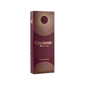
Celosome Aqua
The Celosome Aqua packaging indicates a 24 mg/mL non-crosslinked hyaluronic acid concentration, a parameter highly similar to the natural concentration of human synovial fluid. A 2.5 cc is filled with large polysaccharide molecules weighing between 1 million and 1.5 million Daltons. After undergoing an 8-step cross-flow filtration process, residual bacterial endotoxins in the raw material are strictly kept below 0.01 EU/mL. The injection resistance is set by the device at 15 Newtons. Once these large polysaccharide molecules enter the mid-dermis, they powerfully absorb 500 to 1,000 times their own weight in water from surrounding tissues within 48 hours. Under a 20 MHz high-frequency dermatological ultrasound, the previously shriveled gaps between collagen fibers at a depth of 1.5 mm are seen expanding, forming a micro-reservoir approximately 0.2 mm thick. Normal dermal water content fluctuates around 20%; dropping below the critical 10% threshold causes fine dehydration lines to form on the epidermis. After a single full-face administration of a 5 cc dose, the transepidermal water loss (TEWL) rate rapidly drops from 15 g/m²·h to 8 g/m²·h. Under an osmotic pressure of 300 mOsm/kg, the dehydrated and fractured elastic fiber network regains hydrostatic support. Physicians typically select ultra-fine 34G with an outer diameter of merely 0.23 mm, executing a grid-like injection pattern with 1 cm spacing.
- The injection volume per point is precisely controlled between 0.01 mL and 0.02 mL.
- The bevel of the 4 mm tip is consistently kept facing upward at a 15 to 20-degree angle.
- The treatment area strictly avoids major arterial and venous branches larger than 1 mm in diameter.
- Between 100 and 120 micro-wheals, each 3 mm in diameter, are evenly distributed across a single cheek.
The rate at which these wheals subside serves as an indicator of the macrophage phagocytosis rate within the dermis. When non-crosslinked hyaluronic acid (HA) is injected at a depth of 1.0 mm, hyaluronidase in healthy skin typically metabolizes and flattens it within 24 to 48 hours. If accidentally injected into the dense 0.5 mm epidermal layer, the drug molecules cannot exchange with the bloodstream, extending the metabolic cycle to 5 to 7 days. The formulation deliberately includes 0.9% mannitol by mass. Mannitol molecules contain six hydroxyl groups, enabling them to neutralize hydroxyl radicals within the tissue at an extremely fast rate of 107 M ¹s ¹. Formulations containing mannitol extend the half-life of non-crosslinked HA in the dermis from the standard 72 hours to between 10 and 14 days. During their degradation phase, free HA molecules continuously release trace amounts of lactic acid and 18 amino acids, fully participating in the 28-day metabolic cycle of basal cells. Ultrasound pachymeter data records show that after three consecutive overlapping treatment sessions, the density of the collagen network in the mid-dermis exhibits a subtle 15% increase after three months. HA molecules naturally permeate and diffuse through interstitial spaces with a radius of about 0.5 cm. Because the total skin thickness around the eyes is only 0.5 mm, the injection volume per point must be compressed to a strict limit of 0.005 mL. The injection depth must also be adjusted back to 0.8 mm to prevent persistent eyelid edema that can last for up to two weeks. Automated mesotherapy and manual injections differ in delivery precision at the micrometer level. Vacuum-assisted devices utilize a 32G 9-module and maintain a constant vacuum negative pressure of 250 mmHg, ensuring every drop of the solution is precisely deposited at a depth of 1.2 mm.
- The mechanical vacuum suction action significantly reduces the epidermal leakage rate at the puncture site to below 5%.
- The constant injection motor pressure drastically lowers the probability of forming 0.1 cm localized hard nodules.
- The operation time for a single full-face 600-shot session is strictly controlled within 20 minutes.
- The vertical puncture trajectory reduces the lateral mechanical tearing and wear on pain nerve endings.
The temperature and blood flow velocity in different facial regions invisibly interfere with the retention time of HA. The T-zone is rich in sebaceous glands, with capillary network blood flow velocities reaching up to 5 cm/s and tissue temperatures 0.5°C higher than the U-zone. Consequently, localized hyaluronidase activity is stronger, causing the solution to degrade 20% faster than along the jawline. Maintaining high hydration levels in the dermis structurally defends against photoaging. During severe dehydration, it takes only 3 minutes for ultraviolet rays with wavelengths between 320 and 400 nm to penetrate the epidermis and reach the dermis. Sufficient interstitial water refracts and scatters the light, cutting UV penetration by 30% and protecting fragile elastin. At a room temperature of 25°C, the Celosome Aqua solution exhibits a fluid viscosity of 100,000 cP, retaining a jelly-like yet non-sticky spreadability. The minimal plunger resistance of the significantly alleviates physician hand fatigue, enhancing the absolute stability of the tip depth during hundreds of facial punctures. During the critical 72-hour post-treatment recovery window, approximately 15% of transepidermal water is lost through the microscopic punctures. Applying a sterile, ceramide-infused medical dressing for a 20-minute occlusive wet compress utilizes a powerful osmotic pressure gradient of 50 mOsm/L to force lost dermal moisture back into the basal layer. Measuring the stratum corneum moisture content of 50 test subjects, Corneometer readings reached their peak on day 14 post-treatment. The moisture retention rate in the superficial dermis increased by 42%, and the skin surface gloss value jumped from a baseline of 35 to 58. After 30 days, the HA molecules, degraded into water and carbon dioxide, are completely flushed out through the lymphatic microcirculation at a flow rate of 2 mL per day, smoothly returning the skin to its pre-injection state. In clinical practice, this product is frequently used as a base solution, mixing 2.5 cc of the formulation with 10 to 15 units of Type A botulinum toxin. This mixed formulation is injected in a grid pattern at a depth of 1.0 mm, allowing the drug molecules to intervene in tissue microcirculation:
- Reduces acetylcholine release at nerve synapses by 60%, decreasing excessive sebum secretion.
- Blocks superficial neurotransmitter transmission to smooth out 0.5 mm deep facial expression lines.
- Delays hyaluronidase activity, increasing the reticular water retention duration in the dermis by 30%.
- Downregulates full-face pro-inflammatory IL-6 activity, reducing the incidence of epidermal erythema.
Pure HA solutions tend to be slightly acidic at a pH of 6.8. After adding the botulinum toxin saline, the pH of the compounded mixture slightly adjusts toward a neutral 7.2, subsequently reducing the localized burning pain during injection by 40%. The liquid’s surface tension changes, accelerating the interstitial diffusion rate by 10% and advancing the onset of visible hydration effects to just 24 hours post-procedure.
2XSOME
Unboxing 2XSOME reveals two separate. Agent 1 is a lyophilized powder containing 10 billion exosome particles, while Agent 2 is a 3 mL diluent blended with 59 skin nutrients. On a sterile workspace, a nurse slowly injects Agent 2 into the Agent 1 glass, leaving it to sit for 3 minutes until the powder dissolves into a clear, pale yellow liquid. The exosome particles measure a mere 30 to 150 nm in size. A single strand of human hair is roughly 80,000 nm thick, making an exosome less than one-thousandth of its size. This microscopic scale allows them to effortlessly slip straight into the skin channels opened by microneedles.
These intercellular communication vesicles, carrying over 1,000 types of proteins and more than 600 miRNAs, precisely settle at the dermal-epidermal junction.
The physician swaps the for a Microneedle Therapy System (MTS) device equipped with 16 gold-plated. The penetration depth is firmly locked between 0.5 mm and 0.8 mm via a dial. Oscillating at a high frequency of 6,000 times per minute, the motor creates nearly 200,000 absorption channels across the 400 cm² surface area of the face. Basal melanocytes and stem cells are tucked away at a skin depth of 0.8 mm. Because exosomes possess a lipid bilayer structure, they fuse with the stem cell membranes within 30 seconds of contact. The anti-inflammatory molecules packaged inside then migrate toward the nucleus to begin their work. On compromised cheeks with severe telangiectasia, performing 0.8 mm microneedling triggers pinpoint bleeding. Platelets exuding from the microvessels release Platelet-Derived Growth Factor (PDGF). The miRNAs carried by the exosomes, synergizing with the PDGF, forcefully shut down 80% of the internal tissue inflammatory response within 24 hours.
- Interleukin-6 (IL-6) markers plummet by 65% on day 3.
- Pro-inflammatory Tumor Necrosis Factor (TNF-α) is heavily suppressed below 10 pg/mL.
- Vascular Endothelial Growth Factor (VEGF) secretion doubles to repair ruptured capillaries.
- The rate at which mast cells expel histamine is slowed by a stark 40%.
Erythema clearance speed is a reliable metric for judging delivery depth accuracy. When the 0.8 mm “golden depth” micro-trauma meets exosome anti-inflammatory action, full-face flushing completely fades within 12 to 24 hours.
A heavy-handed operator adjusting the to 1.5 mm will puncture through the papillary dermis, allowing the tips to laterally tear the larger capillaries in the reticular layer. The resulting severe subcutaneous hematomas take an agonizing 7 to 10 days to fully absorb. Furthermore, the exosome solution is washed backward out onto the skin surface alongside the blood. Agent 2 (the diluent) is blended with 10 mg/mL of non-crosslinked HA and four types of biomimetic peptides. Acting as a lubricant, the HA coats the exosome particles, helping them tunnel down the 0.5 mm channels. Along the penetration route, the biomimetic peptides awaken Tissue Inhibitors of Metalloproteinases (TIMPs). Agent 1’s lyophilized powder is dehydrated by 98% in a -40°C environment. This vacuum freeze-drying technology preserves the long-chain mRNA structures inside the vesicles. Once reconstituted at a 25°C room temperature, the exosomes retain activity for only 4 hours, meaning the opened must be completely applied to the face within 10 minutes. Laboratory stained-section tracking data reveals that basal cell division activity increases by 35% on day 7. Receiving synthetic commands from the exosomes, fibroblasts push Type I collagen secretion to its peak by day 14, yielding a total synthesis volume 40% higher than normal.
Newly generated collagen fibers grow outward at 2 μm per day, filling in the collapsed areas surrounding the pores. The spacing between stratum corneum cells is pulled tight, dropping from 15 μm to 8 μm.
Changes in pore size exhibit clear differences on a high-definition facial imaging system at day 30. Following a single 0.8 mm depth infusion, the cross-sectional area of large pores around the nasal alae shrinks by an average of 18%. After completing 5 consecutive standard sessions, three-dimensional pore depth data shallows by 25%, and the roughness (Ra value) drops from 3.2 to 1.8. The total skin and tissue thickness on the forehead is under 1.2 mm, lying flush against the frontalis muscle fascia. When the device glides over the forehead, the microneedle depth must be dialed back to between 0.3 mm and 0.5 mm. Because the epidermal stratum corneum is only 0.02 mm thick, a 0.3 mm easily pierces the stratum lucidum to deliver the solution to the stratum spinosum.
- The 0.3 mm puncture on the forehead severs irregularly growing superficial keratin.
- The ultra-shallow 0.25 mm pricks under the eyes awaken dormant orbicularis oculi muscle cells.
- A 0.6 mm depth on the highest point of the cheekbones perfectly dodges the pain points of zygomatic nerve branches.
- An 0.8 mm depth along the jawline relies on mechanical tugging to stimulate local lymphatic microcirculation.
The gliding speed is strictly controlled at 2 cm per second. Moving too quickly causes the 16-array to carve continuous 0.5 cm long bloody scratches across the cheeks. A slow, deliberate “stamping” motion restricts the diameter of individual puncture lesions to a micro-trauma limit of 0.1 mm. Breaching the stratum corneum barrier triggers inflammation, accompanied by a transepidermal water loss of up to 30 g/m²·h. However, ceramide synthase directives within the exosome vesicles activate after 48 hours. The stratum granulosum aggressively secretes natural lipids, reweaving a 0.01 mm thick sebum membrane within 5 days.
The rebuilt sebum membrane rapidly pulls the epidermal pH from a slightly alkaline 6.5 back to a weakly acidic defense line of 5.5. On this acidic epidermis, the survival rate of external Staphylococcus aureus drops to less than one-tenth.
Topical numbing cream is applied to the face for 30 minutes before being wiped completely clean with saline. The lidocaine in the anesthetic cream penetrates to a maximum depth of 0.4 mm. When the 0.8 mm microneedles penetrate, a slight, dull, compressive prickling sensation is still felt; this is the true reaction of the tips hitting the free nerve endings in the papillary dermis. The areas rolled over by the microneedles exhibit a visible, dense, white-frost-like array of micro-lesions. The physician divides the reconstituted 3 mL solution into 4 parts, dripping it evenly over these lesions. The application must follow within 2 seconds of the microneedles leaving the skin, racing to flow down the channels into the deep basal layer before the exudate coagulates.
Parameter Comparison & Delivery Indications
Because large HA molecules and nano-scale exosome vesicles differ vastly in size, the injection tools and parameters must be rigidly calibrated to specific skin layer thicknesses. Grabbing the wrong or injecting at the wrong depth means thousands of dollars’ worth of serum will be flushed out as waste within 48 hours. Lying on the treatment bed, a few millimeters of penetration difference is virtually impossible to spot with the naked eye.
| Delivery Parameter Metrics | Celosome Aqua HA | 2XSOME Exosomes |
|---|---|---|
| Standard Delivery Device | 34G Ultra-Fine Single / 9-Vacuum Device | 16-Gold MTS Device |
| Set Penetration Depth | 1.0 mm – 2.0 mm | 0.5 mm – 0.8 mm |
| Insertion Angle | 15 to 20-Degree Bevel Up | 90-Degree Vertical Stamping |
| Single Face Dosage | 5.0 cc (Two Standard ) | 3.0 mL (Reconstituted Mixture) |
| Pain Score (VAS) | 2 to 3 (Mild Aching Pressure) | 4 to 5 (Dense Prickling) |
| Macroscopic Tissue Response | Raised 3 mm Diameter Micro-Wheals | Dense Frost-Like Erythema with Micro-Bleeding |
A 34G ultra-fine single has an outer diameter of 0.23 mm. Targeting the mid-dermis at a 1.5 mm thickness, the physician’s wrist must press at a 15-degree angle to drive the in. Given the immense viscosity of the 24 mg/mL gel-like polysaccharide, a vertical injection would cause the gel to leak entirely out onto the epidermis. The operational technique for exosomes completely flips. A densely packed array of 16 microneedles is pressed down vertically at 90 degrees, relying on the 6,000 RPM high-frequency motor vibration to punch through the stratum lucidum. A 0.5 mm length requires no angling; the watery 3 mL solution instantly glides down the vertical channels right next to the basal cells. The sensation of pain is entirely different. Pushing 0.02 mL of gel-like HA into a single point forcefully stretches open the dehydrated subcutaneous fiber network. The localized tissue endures an osmotic pressure of 300 mOsm/kg, causing nerve endings to feed back a strong, aching pressure. Microneedling exosomes avoids that swelling ache of liquid tearing tissue apart. The pain stems entirely from the tips repeatedly tracking across the papillary dermis at a depth of 0.8 mm. Sustaining 500 micro-punctures per square centimeter, the brain registers a burning, prickling pain akin to sanding the epidermis, resulting in a pain score 2 levels higher. The tactics to alleviate pain diverge sharply. Slowing the HA injection speed to 0.01 mL per second gives the subcutaneous tissue a 3-second adaptation window to stretch, cutting the aching pain in half. Microneedling relies on applying a 2 mm thick layer of compound lidocaine cream beforehand, occluding it with plastic wrap for 40 minutes to thoroughly numb the epidermis. Facial reactions during the first 24 hours serve as an indicator of the absorption rate. Post-HA cheeks display neat rows of 120 micro-wheals, each 3 mm in diameter. Because the mannitol component is phagocytized very slowly by macrophages, it takes 48 hours for the wheals to gradually flatten out; during this time, the skin feels like it has tiny, bumpy granules. The 0.8 mm length ruptures capillary loops at the top of the dermis, covering the face with pinpoint bleeding that merges into erythema resembling a severe sunburn. Under the anti-inflammatory intervention of the 59 nutrients, this redness fades completely within 12 hours. At a depth of 1.5 mm, non-crosslinked HA cannot withstand the degradation from the body’s own hyaluronidase. Within 30 days, the 5 cc of serum is broken down into water and carbon dioxide and excreted via urine, causing the hydrated sensation to experience a steep cliff-drop exactly at week 4. Vesicle particles carrying mRNA don’t just passively resist subcutaneous breakdown. They fuse with stem cells in 30 seconds, issuing synthetic directives that continue to take effect for 28 days. The physical structural changes of newly generated collagen filling the pores cannot be reversed; after 3 to 5 consecutive sessions, the pore-shrinking effect is firmly maintained for 8 months without rebound. Mixing the two formulations creates a mutually complementary effect. Blending 1 cc of HA solution into 3 mL of raw exosome fluid and pressing them together into the facial skin using a 1.0 mm microneedle roller allows the polysaccharide polymers to coat the fragile vesicles. This forcefully extends their active viability window at room temperature from 4 hours to 12 hours. The 200,000 channels punctured by the microneedles are accompanied by a TEWL rate of 30 g/m²·h. The infused 1 cc of HA absorbs 500 times its weight in water at a shallow depth of 0.8 mm, becoming a liquid artificial sebum membrane that compresses the scabbing and shedding cycle of micro-wounds from 5 days down to 3 days.