Rejuran I (White Box) is specifically designed for the fragile periocular skin with a thickness of only about 0.6mm. With a lower viscosity texture, it is the safe first choice for fading dark circles and fine lines around the eyes. The small papules (recovery period) generated after injection usually subside within only 1-2 days. While Rejuran HB (Hydro Booster) adds extra Hyaluronic Acid (HA) and 0.3% Lidocaine on the basis of the core ingredient Polynucleotide (PN). It focuses on deep full-face hydration and barrier repair. The built-in anesthetic significantly reduces injection pain, and the recovery period is approximately 1-3 days.
Under Eye Use
The skin thickness around the eyes is only 0.3–0.5mm, and there are almost no sebaceous glands. A blinking frequency of about 22,000 times per day makes it the earliest area to age. The PN concentration of Rejuran I is 1%, specifically designed for the dermis layer with a thickness of less than 1mm; While Rejuran HB combines 1% PN + 1% HA + 0.3% Lidocaine. Due to the high osmotic pressure of HA, HB can increase water content by more than 20%, but it may also increase postoperative swelling for 24-48 hours.
Molecular Viscosity Differences
The dynamic viscosity design of Rejuran I is approximately 1.0–1.2 Pa·s. This rheological property, which is close to a liquid state, gives it extremely strong dispersive power in the superficial dermis at a depth of 0.3mm around the eyes. In an experimental environment of 25°C, the shear rate of its PN (Polynucleotide) molecules is much higher than that of Rejuran HB, which ensures that the drug solution can quickly diffuse to the surroundings after entering the subcutaneous tissue, rather than aggregating into a spherical shape. This ultra-low viscosity is particularly friendly to subjects with extremely thin fat layers under the eyes because, under the physical stimulation of a 1.5mm, low-viscosity liquids can reduce tissue injection resistance by about 30%, thereby reducing bruising caused by capillary rupture. In contrast, because Rejuran HB contains 1% non-cross-linked hyaluronic acid, its intermolecular forces are stronger, and it tends to form a tiny water reservoir at the injection point.
- Diffusion Radius: After a single-point injection of 0.02ml of Rejuran I, its lateral diffusion radius can reach 2.5-3.0mm, covering more damaged fibroblasts.
- Tissue Compatibility: PN chains with a molecular weight distribution of around 1000kDa can slide more smoothly into the gaps of the collagen scaffold in a low-viscosity state.
- No Pressure Sensation: Experimental data shows that the increase in internal tissue pressure at the eyelid from low-viscosity preparations is lower than 5mmHg, avoiding discomfort caused by a squeezing sensation.
- Residual Rate: In ultrasound observations 4 hours after surgery, the height of local protrusions of Rejuran I typically drops by more than 70%, showing a flattened distribution.
When these highly mobile PN molecules encounter fine lines under the eyes, they exhibit a very low sense of physical filling, relying more on biological signaling to induce endogenous collagen regeneration. This “non-occupying” repair logic is completely different from traditional HA filling; it does not change the anatomical contours of the eye area but gradually guides the skin thickness from 0.4mm back to a healthy level of around 0.55mm. Since the lymphatic drainage rate of the skin under the eyes is only 60% of that in other areas of the face, any substance with too high a viscosity will increase the burden on lymphatic vessels, leading to weeks of morning puffiness. The low-viscosity formula of Rejuran I, assisted by a 34G nanoneedle, can achieve precise “dot-matrix” coverage. A single treatment usually involves 20-40 tiny injection points around the eyes. This high-frequency, micro-quantity operation is extremely dependent on the compliance of the drug solution.
- 34G Compatibility: The internal diameter of the tip is only 0.12mm. Only low-viscosity PN can pass through smoothly without increasing injection pressure.
- Tyndall Effect Defense: Reduced viscosity means molecules will not aggregate abnormally, and there will be no wavelength shift when reflecting light, avoiding skin blueness.
- Internal Pressure Balance: The exchange rate between low-viscosity liquid and extracellular fluid (ECF) is increased by 25%, accelerating the elimination of metabolic waste.
- Immediate Socializing: Within 60 minutes after injection, the visibility of local papules will decrease to 1/3 of the initial state, making them almost invisible to the naked eye.
Rejuran I can penetrate skin textures like a lubricant. Through a 14-21 days cell cycle, it increases the reflectivity of the skin under the eyes by about 12%. The source of this gloss is not the physical reflection of hyaluronic acid, but the optimization of diffuse reflection brought about by the improvement of skin surface smoothness after the increase in dermal thickness. For subjects over 35 years old, the loss rate of elastic fibers around the eyes usually exceeds 1.5% per year, which causes the skin to lose its ability to wrap high-viscosity substances. Using a watery preparation like Rejuran I can effectively prevent the drug from shifting under the influence of gravity. This physical stability is manifested as continuous improvement in the smoothness under the eyes in clinical observations of 6-9 months, rather than a temporary volume filling.
- Dynamic Performance: When blinking, low-viscosity PN deforms naturally with muscle contraction, without creating a stiff sense of layering or foreign body sensation.
- Absorption Dynamics: Under thermal imaging detection, the blood flow velocity in low-viscosity areas is 15% faster than in high-viscosity areas, which is conducive to ingredient uptake by cells.
- Nodule Incidence: Clinical statistics show that the probability of Rejuran I producing long-term nodules around the eyes is close to 0%, much lower than cross-linked products.
- Layer Precision: Doctors can utilize its fluidity to perform “feather-sweep” injections at a depth of 0.5mm, covering a wider area.
Osmotic Pressure and Edema
The standard osmotic pressure of human extracellular fluid is usually maintained at 280-310 mOsm/L. The osmotic pressure of Rejuran I’s pure polynucleotide (PN) formula is in an isotonic state, with a measured value of about 300 mOsm/L. When the drug solution is injected into the dermis under the eye with a thickness of less than 0.5mm, the water exchange inside and outside the cells remains balanced, and the incidence of physical tissue expansion is lower than 5%. For Rejuran HB with 1% non-cross-linked hyaluronic acid (HA) added, the local microenvironment osmotic pressure can instantly jump to over 400 mOsm/L. HA molecules carry a large number of hydrophilic groups; 1 gram of free HA has the chemical property of capturing 1000 times its own weight in water.
- Water Capture Ratio: 1mg of non-cross-linked HA can adsorb about 1ml of tissue fluid within 4 hours.
- Volume Expansion: The local volume at the injection point usually increases by 15%-25% within 12 hours after surgery.
- Maintenance Time: The hypertonic liquefied state gradually subsides after 48 hours with monomer metabolism.
- Muscle Compression: The swollen papules will exert a slight pressure of about 2mmHg on the underlying orbicularis oculi muscle.
The lymphatic capillary network around the orbit is extremely fine, with a pipe diameter of only 10-50 microns. The lymphatic drainage rate of the eye area is in a low-load operation all year round, only being able to process about 0.1-0.2ml of excess fluid per hour. When the hypertonic water absorption rate of Rejuran HB exceeds the maximum discharge capacity of the local lymphatic system, the retained tissue fluid will form visible edema in the superficial fascia layer. During 8 hours of flat-lying sleep at night, gravity no longer assists the venous and lymphatic drainage of the face. Venous flow rate drops by about 30%, making it very easy for moisture to stagnate in the loose connective tissue of the eyelids. Since the single-point injection volume of Rejuran HB is usually 0.05ml, the strong water-absorbing property will temporarily increase the skin thickness around the eyes in the morning by 0.2-0.3mm.
| Osmotic Pressure Parameters | Pure PN (Isotonic) | PN + 1% HA (Hypertonic) |
|---|---|---|
| Solute Concentration | 10 mg/ml PN | 10 mg/ml PN + 10 mg/ml HA |
| Microenvironment Water Increase | 2% – 5% | 20% – 35% |
| Lymphatic Metabolic Cycle | 12 – 24 hours | 48 – 72 hours |
| Morning Swelling Incidence | Below 10% | About 60% – 80% |
When doctors inject hypertonic liquid, the depth must be strictly controlled in the dermal reticular layer at 1.0-1.5mm subcutaneous. Once the HA solution mistakenly enters the epidermal basal layer with a thickness of less than 0.2mm, intense water accumulation will push up the extremely thin stratum corneum. This not only extends the subsistence period of the papules to 7 days but also easily triggers a visual blue-tinted transparency due to light refraction. The metabolism of pure PN preparations in tissue fluid relies entirely on macrophage phagocytosis and enzymatic hydrolysis, a process not accompanied by intense hydration reactions. Under a standard dose of 1ml for the entire eye area, the pressure change in the tissue space does not exceed 1.5mmHg. After subjects complete ice packing for 2 hours after the procedure, 80% of the slight redness and swelling caused by microvascular expansion can subside.
- Hypersensitive Constitution: For those whose eye bags protrude more than 3mm before surgery, pure PN is preferred to prevent worsening fluid accumulation.
- Extremely Dry Skin: When the stratum corneum water content is below 10%, hypertonic HA can establish a micro-reservoir within 24 hours.
- Social Interval: If it is necessary to appear in public within 48 hours, avoid the peak period of hypertonic water absorption.
- Overlapping Operations: When combined with Botox injections, hypertonic edema will increase the physical diffusion radius of the toxin by 1-2mm.
Within 48 hours of the dermis being forcibly stretched by moisture, mechanical tension receptors of fibroblasts are activated. After the Integrin protein on the cell membrane surface perceives a stretching deformation of more than 15%, it will increase the synthesis rate of Type I collagen precursor protein. The non-cross-linked HA free molecules introduced by Rejuran HB will be degraded into water and carbon dioxide by hyaluronidase in the tissue within 14-21 days. The hypertonic environment subsequently collapses, and the physical edema completely subsides. The remaining PN molecules have simultaneously completed the early release of Vascular Endothelial Growth Factor (VEGF), increasing local capillary density by about 12%, taking over to maintain the natural water content of the periocular tissue. To artificially intervene in the early hypertonic expansion, cold compresses at 15°C are often used within 24 hours after surgery to reduce capillary permeability. By reducing capillary filtrate extravasation by 40%, it effectively offsets the absolute volume increase brought about by HA water capture. It is forbidden to perform radiofrequency or hot compresses at temperatures higher than 40°C within 72 hours, as local high temperature will double the tissue fluid exudation rate.
Comfort and
The density of trigeminal nerve endings in the dermal layer around the eyes is as high as 200-300 per square centimeter, which is 3 times that of the cheek area. The extremely high density of mechanical pain receptors means that any entry of a foreign body with a diameter exceeding 0.2mm will trigger strong neural signal transmission. In order to reduce the stinging sensation brought by physical piercing, the clinical standard operation of Rejuran I requires being equipped with 34G or 33G ultra-fine nanoneedles.
- Outer Diameter Size: The outer diameter of a 34G is only 0.18mm, about half that of a traditional 30G hyaluronic acid injection.
- Bevel Angle: The tip usually adopts a multi-bevel cut of 15-20 degrees to reduce tissue cutting resistance by 40%.
- Injection Depth: The standard length is 4mm, making it easy to accurately pierce into the superficial dermis at 1.0-1.5mm at a 15-degree angle.
- Discharge Thrust: Combined with extremely low viscosity solutions, the physical finger force required for a single injection of 0.02ml of drug solution drops by 35%.
When the 0.18mm micro-tip penetrates the stratum corneum with a thickness of only 0.05mm, the extremely small cross-sectional area effectively avoids 80% of the superficial capillary network. Since no large-scale tissue tearing is triggered, the rate at which mast cells release histamine is significantly reduced. When subjects receive dense injections of up to 40 points, physical pain scores can usually be controlled between 3-4 on the Visual Analogue Scale (VAS).
The ultra-fine 34G not only reduces the irritation frequency of pain nerves but also compresses the probability of postoperative bruising larger than 2mm in diameter to below 5%.
Upgrading the specifications of physical instruments cannot completely eliminate the chemical soreness caused by liquid expansion of the dermis. The pH value of the preparation is about 6.8, which is slightly different from the weak alkaline environment of 7.35-7.45 in human tissue fluid. When 1ml of pure solution is pushed subcutaneously into the eyelid in a grid-like distribution, the tiny pH difference will stimulate free nerve endings, producing a burning sensation that lasts for 2-3 minutes. Rejuran HB intervenes from a pharmacological level, with 0.3% lidocaine hydrochloride added to the formula. As an amide local anesthetic, lidocaine molecules can quickly block sodium ion channels on nerve cell membranes within 30-60 seconds after contacting tissue. After the depolarization process is inhibited, the transmission efficiency of pain electrical signals to the central nervous system falls off a cliff.
- Onset Time: After the first point is injected, lidocaine diffuses within a 2mm radius along the tissue gaps within 45 seconds.
- Pain Reduction Range: The pain from subsequent adjacent injections and liquid expansion decreases simultaneously by 40%-60%.
- Metabolic Half-life: Tiny amounts of anesthetic ingredients will be completely metabolized by the liver into monoethylglycinexylidide within 90-120 minutes.
- Soreness Offset: The hypertonic expansion pain brought by 1% hyaluronic acid is blocked by the anesthetic, and the VAS score drops to around 2 points.
Preparations containing lidocaine will still produce extremely short-lived stinging when the first 0.01ml contacts the tissue during the initial entry. Since anesthetic ingredients are slightly acidic, the drug solution needs to undergo a physiological buffer period of about 10 seconds at the moment it enters the dermal reticular layer at a depth of 1.5mm. Once the sodium ion channels are successfully locked, the subsequent 0.04ml injection will produce almost no sensation.
The addition of 0.3% lidocaine shortens the operation time for 20 injections per eye from the usual 8 minutes to 5 minutes, reducing the frequency of defensive muscle contractions.
Intense blinking and muscle tension not only increase the doctor’s resistance when injecting to 1.5mm but also easily lead to tip slippage and rupture of micro-veins with a diameter of 0.5mm. The establishment of a chemical painless environment allows the muscle tension around the eyes to remain in a relaxed state of 15mmHg throughout the 10-minute treatment cycle. 30 minutes after applying 5% prilocaine compound cream to the eyelid surface, the maximum depth of anesthetic ingredient penetration is about 1.0mm. When the tip breaks through 1.2mm to find deep fibroblasts, the effect of surface anesthesia decays sharply, and the peeling pain of the liquid pushing apart subcutaneous tissue will still be clearly transmitted.
- Surface Anesthesia Limitations: The stratum corneum barrier makes the effective molecular absorption rate of 5% cream less than 10%.
- Layer Decay: Beyond 1.0mm depth, the surface anesthesia effect decreases rapidly at a rate of 20% per 0.1mm.
- Physical Analgesia: Ice packing for 3 minutes before surgery can slow down local nerve conduction speed by 15%, assisting the operation with pure preparations.
- Emotional Index: 65% of first-time subjects show a heart rate change of less than 5 bpm after receiving anesthetic-containing preparations.
To compensate for the lack of chemical analgesia in non-anesthetic preparations, physical control of the flow rate is precise to the second. Operators usually extend the release time of 0.02ml of liquid to 2-3 seconds, slowly increasing the subcutaneous tissue internal pressure to 5-8mmHg. Reducing the flow rate by 50% can provide sufficient mechanical extension time for the dermal fiber network, preventing connective tissue tearing pain caused by rapid hydraulic impact. The internal diameter of a 34G nanoneedle is only 0.12mm. If a high-viscosity liquid is forcibly pushed, it will cause the internal pressure of the tube to break through 100kPa, triggering uncontrollable hand tremors. The watery low-viscosity property allows it to be discharged at a uniform speed under a steady pressure of 40kPa, ensuring that the tip’s hover deviation in the extremely thin 0.5mm skin does not exceed 0.1mm.
The reduction in the hover deviation rate allows more than 90% of the injection points to accurately hit the middle dermis, maximizing the mechanical activation rate of biological cells.
The hypertonic pain caused by 1% hyaluronic acid is effectively offset by 0.3% lidocaine, while pure physiological soreness relies on the tiny wound of the 34G ultra-fine and the 50% deceleration of the injection to be relieved. Every micro-drop release, precise to 0.02ml, completes the avoidance of dense nerve endings in the 0.3mm thick under-eye dermis. 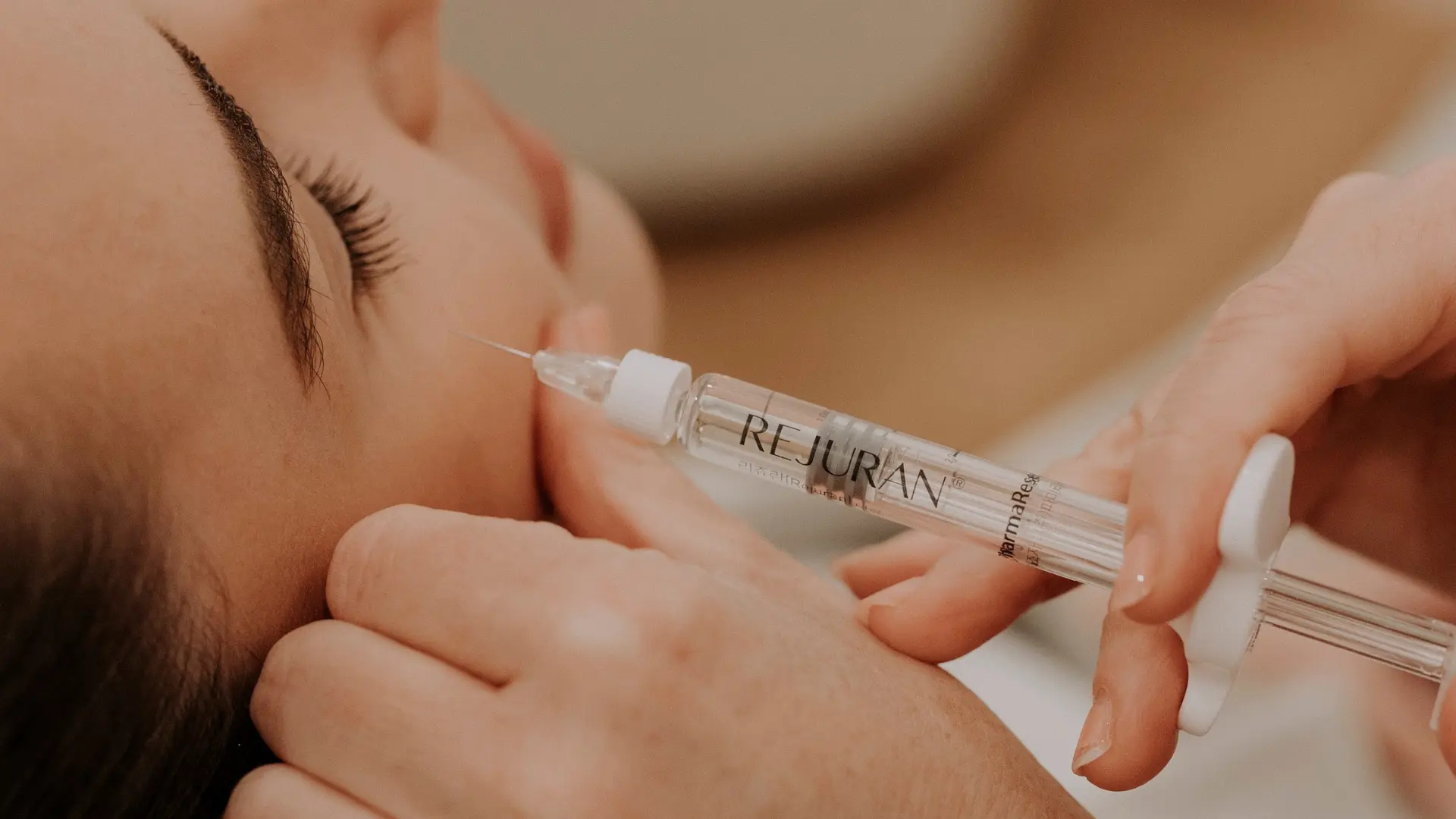
Hydration
The difference in hydration between Rejuran HB and Rejuran I primarily lies in the Hyaluronic Acid (HA). HB contains 1% Polynucleotide (PN) and 1% HA. After injection, the moisture level in the skin layer increases by about 20%-30% within 2-4 weeks, making it suitable for deep facial hydration. I contains only 1% PN and is completely free of HA. It does not provide a direct physical hydrating effect; instead, it improves fine lines around the eyes mainly by stimulating collagen production (increasing dermal thickness by about 20%). Choose HB for overall facial radiance; choose I for the under-eye area to prevent chronic puffiness caused by excessive water absorption by HA.
Differences in Ingredient Hydration
The Rejuran HB formula contains 10mg/ml of Polynucleotide (PN), 10mg/ml of non-cross-linked Hyaluronic Acid (HA), and is equipped with 0.3% Lidocaine. The pH value of the solution is stabilized between 6.8 and 7.6, and the osmotic pressure is set at 280-300 mOsm/kg, which is highly close to human tissue fluid. The molecular weight of the HA is fixed at the 1000 kDa level, presenting a three-dimensional network distribution within the liquid. After the non-cross-linked hyaluronic acid is injected into the mid-dermis at a depth of 1.5 to 2.0 mm, it rapidly resides in a sponge-like physical form. 1 gram of non-cross-linked HA can bind approximately 6 liters of free water in biochemical reactions. The fluid dynamics of the subcutaneous interstitial fluid change accordingly, and the volume of local tissue fluid expands by 15% to 20% within 48 hours. The monomer concentration of Rejuran I is 10mg/ml of Polynucleotide, with any form of hyaluronic acid component removed. To adapt to the extremely thin anatomical structure around the eyes, its kinematic viscosity is strictly controlled between 0.8 and 1.2 Pa·s. The DNA fragments extracted from deep-sea salmon range in length between 50 and 1500 base pairs. There are quantitative differences in the water metabolism rates of the two solutions within the extracellular matrix (ECM):
- 72 hours after HB administration, the water content of the stratum corneum increases by 25% to 30%
- The half-life of non-cross-linked HA in HB is measured to be 14 to 21 days
- Skin impedance tests in the early stage after I administration show no significant increase or decrease in moisture
- I requires more than 28 days to trigger changes in endogenous water metabolism
Polynucleotides primarily act on the A2A purinergic receptors of fibroblasts in the dermis. Once binding occurs, the rate of DNA synthesis within the nucleus increases by 30% within 72 hours. The metabolic activity of fibroblasts increases, releasing Type I and Type III collagen. The density of the newly formed collagen fiber network increases by 15%, filling the structural gaps in aged tissue. Since it does not contain exogenous HA, the biochemical pathway of PN alone relies on the remodeling of the epidermal barrier. Histological observations show that for every 0.1 mm increase in dermal thickness, the Transepidermal Water Loss (TEWL) rate decreases by 5% to 8%. Moisture is tightly locked within the dense collagen matrix, making it difficult to evaporate outward through capillary microcirculation. Clinical data testing indicates that in skin tissue receiving only the PN component, the lipid synthesis rate of the sebum film increases by 12.5% after 12 weeks. Physical hydration is highly dependent on the input volume of exogenous ingredients, while biological-level hydration relies on the internal synthesis mechanisms of the tissue. Rheological properties profoundly affect the diffusion radius and water retention morphology of the solution in the subcutaneous reticular layer:
- The high viscoelasticity of HB limits the diffusion radius of a single-point injection to within 3 mm
- The absorption and degradation time of a single HB papule is maintained at 3 to 5 days
- The low viscosity of I’s liquid state increases its diffusion radius in loose connective tissue by 40%
- The total absorption time for a single point of I is usually shorter than 24 hours
- The Newtonian fluid characteristics of I avoid obstruction of lymphatic drainage under the eyes
Once non-cross-linked HA is exposed subcutaneously, it is continuously subjected to degradation and cleavage by enzymes in the body. Approximately 3% to 5% of exogenous hyaluronic acid is decomposed into monosaccharides and enters the blood daily. The plump sensation of hydration will follow a linear decay curve along with the enzymatic hydrolysis process. The degradation pathway of polynucleotides in the body differs from that of hyaluronic acid. PN is slowly hydrolyzed into nucleotide monomers by nucleases secreted by macrophages, with a half-life of up to 30 days. After fibroblasts are stimulated by A2A receptors, they not only secrete collagen but also synthesize endogenous hyaluronic acid themselves. The absolute volume of endogenously synthesized HA is not as massive as that of exogenous injections, but its biocompatibility is as high as 99%. In areas injected with pure PN, the distribution density of endogenous HA increases by 18% by the 8th week. The endogenous moisture retained by structural thickening has an actual duration of effect exceeding 16 weeks. Small changes in the osmotic pressure gradient control the flow of water across cell membranes and the probability of local edema:
- The hypertonicity of HB leads to a rapid accumulation of extracellular fluid in the short term
- If the reticular dermis in the infraorbital area absorbs more than 15% water, it easily forms bulging eye bags
- The isotonic formula of I maintains a pressure difference balance of 285 mOsm/L across cell membranes
- Pure PN solution does not break the homeostasis of fragile tissue fluid
Local blood perfusion shows a short-term decrease of about 5% within the first 48 hours. As HA binds moisture and diffuses to the surroundings, tissue pressure is released, and microcirculation flow rate recovers to 0.5 mm per second. Pure polynucleotide solution is dedicated to repairing the water-storage microenvironment at the cellular base. The expression level of Aquaporin 3 (AQP3) in the basal layer of the epidermis is raised by 15% under the action of polynucleotides. The transport efficiency of water molecules across cell membranes increases by 20%, resulting in substantial replenishment of intracellular fluid. The 10mg/ml HA injected by HB pulls the water-holding capacity of tissue gaps to a physical peak within 14 days. The average thickness of the reticular dermis in the cheek reaches 1.8 mm, which is sufficient to accommodate a 20% volume expansion. Within 42 days, I increases the dermal thickness from 0.5 mm to 0.6 mm, completing the iteration of natural moisturizing power.
Injection Area Selection
The total skin thickness in the cheek and forehead areas usually fluctuates between 1.5 mm and 2.5 mm. The epidermal layer accounts for 0.1 mm, while the reticular dermis and papillary layer occupy the remaining physical space. Collagen fibers are arranged in a dense cross-pattern, forming a microscopic three-dimensional support network with high tension. When up to 10mg/ml of non-cross-linked hyaluronic acid is injected at a depth of 1.5 mm into the cheek, local tissue pressure will instantly soar to 15 mmHg. The dense collagen network of the face can withstand an additional 20% volume expansion without structural tearing. Rich capillaries and facial venous plexuses can adjust the interstitial fluid osmotic pressure to a balanced state of 285 mOsm/L within 48 hours after the initial injection. The gaps between the malar fat pad and the buccal fat pad allow 2.0 cc to 4.0 cc of Rejuran HB solution to diffuse evenly within a 3 mm radius subcutaneously. The ample physical space within the tissue blocks the physical path for high-viscoelasticity liquid to squeeze backward into the epidermal layer. The total thickness of the eyelid skin is measured by instruments to be 0.4 mm to 0.6 mm, which is only one-third of the cheek area. The epidermal layer thickness here drops sharply to 0.04 mm, and the density of sebaceous glands is lower than 10 per square centimeter, resulting in an extremely weak natural lipid barrier. The subcutaneous area around the eyes lacks solid fat pad support, and the dermal layer is attached to the surface of the orbicularis oculi muscle. Rejuran I solution, containing only 1% polynucleotide monomer, can reside without pressure in the extremely shallow dermis at a depth of 0.5 mm. Low-viscosity liquid without hyaluronic acid generates a local physical pressure of less than 5 mmHg after entering the periorbital area. The metabolic load limit of the infraorbital capillary network and lymphatic drainage system is extremely low. Once water-absorbing HA is injected, the accumulation of water molecules in the reticular tissue will exceed the maximum drainage limit of the lymphatic vessels within 12 hours. Free water stagnates in the loose connective tissue above the orbicularis oculi muscle, forming iatrogenic edematous eye bags that persist for 3 to 6 months.
| Anatomical Parameters | Cheek/Forehead Target Area | Infraorbital/Periocular Target Area |
|---|---|---|
| Average Epidermal Thickness | 0.10 mm | 0.04 mm |
| Average Dermal Thickness | 1.40 – 2.40 mm | 0.36 – 0.56 mm |
| Safe Injection Depth | 1.00 – 1.50 mm | 0.50 – 0.80 mm |
| Tissue Tension Tolerance Peak | 20% Volume Expansion | 3% Volume Expansion |
| Local Capillary Density | Moderately Dense | Extremely Dense and Superficial |
If 1000 kDa molecular weight HA gel is pushed into the under-eye dermis as thin as 0.5 mm, Rayleigh scattering occurs after light penetrates the extremely thin epidermis. A persistent light-blue reflective band appears subcutaneously; clinical science defines this physical optical phenomenon as the Tyndall effect. The kinematic viscosity of 0.8 to 1.2 Pa·s of Rejuran I is highly compatible with the fluid mechanics of periocular tissue. After the solution enters the 0.5 mm skin layer, it penetrates into tiny pores in a flat two-dimensional physical form. The pure nucleotide component does not change the local optical refractive index, eliminating the physical hazard of blue light scattering caused by shallow injection. Based on anatomical data from two different regions, the mechanical parameters of insertion and instrument specifications are strictly divided:
- The cheek often uses 33G or 34G that are 4 mm long
- The insertion angle for facial papules is maintained at 15 to 30 degrees
- The single-point injection volume for HB is set at 0.02 to 0.05 cc
- The infraorbital area mandatorily uses 34G 2 mm ultra-short nanoneedles
- The insertion angle for intradermal injection around the eyes must be lowered to 5 to 10 degrees
- The single-point micro-injection of I is controlled within 0.01 cc
The thicker dermis of the face contains a denser population of immune cells, with a phagocytosis efficiency of free nucleotides as high as 2.5% per day. In the infraorbital dermis, the extracellular matrix volume is smaller, and the macrophage phagocytosis rate drops to about 1.5% per day. A Rejuran I dose of 1.0 cc each time can be completely integrated by the 0.5 mm infraorbital dermal network within 24 hours. The osmotic pressure inside and outside the cell membrane quickly returns to the baseline value of 280 mOsm/kg within 60 minutes after the injection is completed. The ratio of dermal matrix components in the target area intervenes in the rate of subsequent collagen regeneration. The cheek area, with a reserve of Type I collagen as high as 70%, can make a strong fibroblast proliferation response to solutions containing HA and PN. Within 72 hours, the collagen synthesis rate subcutaneously in the face shows a steep upward curve of 30%. The originally scarce collagen reserve under the eye lengthens the physical cycle of cell remodeling. The biochemical reaction path of pure PN components in the infraorbital target area is different from facial operations:
- Targeted activation of weak A2A purinergic receptors takes longer
- Only microscopic reorganization of the extracellular matrix occurs in the first 14 days
- The synthesis of Type III collagen begins to climb slowly after 21 days
- The proliferation and repair period of infraorbital vascular endothelial cells lasts up to 28 days
The healing trajectories of physical trauma caused by differ across skin layers of different thicknesses. Around the 1.5 mm deep track in the face, platelet-derived growth factors can seal microvascular bleeding points within 12 hours. For the 0.5 mm extremely shallow wound around the eyes, due to the lack of sebaceous gland secretions, the physical closure of the stratum corneum usually requires 24 to 36 hours.
Comparison of Onset Time
After Rejuran HB is injected into the dermis, 10mg/ml of non-cross-linked hyaluronic acid initiates the physical water absorption mechanism within 24 hours. The three-dimensional network structure of HA molecules begins to capture free water molecules, and the monomer volume expands by 15% to 20% within 48 hours. Data from the stratum corneum moisture tester shows that the epidermal water content in the administered area rises sharply by 25% on the 3rd day. Dry desquamation on the skin surface is relieved within 72 hours. In contrast, the Rejuran I solution, which does not contain hyaluronic acid, follows a flat curve within the same timeline. After 10mg/ml of pure polynucleotide enters the 0.5 mm under-eye dermis, it does not trigger a change in osmotic pressure. The under-eye moisture test data for the first 72 hours stays at the baseline level, with a fluctuation rate lower than 2%. At this time, PN molecules are being slowly uptaken by A2A receptors on the surface of fibroblasts at a rate of 1% per hour.
The biochemical response of pure PN occurs at the microscopic level. The DNA polymerase activity within fibroblasts increases by 15% after 72 hours, starting to output repair instructions from the cell nucleus.
Advancing to days 7 to 14, the exogenous hydration performance of HB reaches the peak of the parabola. The Transepidermal Water Loss (TEWL) rate decreases by 30%, and the face presents a highly reflective, hydrated texture. Hyaluronidase in the body begins to intervene at this time, cutting the glycosidic bonds of non-cross-linked HA at a constant rate of 3% to 5% per day. The sense of exogenous hydration shows a slight decline after the 14th day. Rejuran I shows an angiogenesis response at the histological level on the 14th day. The under-eye area, lacking the physical support of HA, instead relies on microcirculation reconstruction to improve the hollowed state.
- The secretion of Vascular Endothelial Growth Factor (VEGF) increases by 12%
- Under-eye microcirculation blood flow velocity increases to 0.6 mm per second
- The fibroblast proliferation rate records a steady climb of 20%
- Early Type I procollagen chains begin to polymerize in the extracellular matrix
- Pigment cell metabolism speeds up, and periocular pigmentation fades by 10%
Between the 21st and 28th day after surgery, the non-cross-linked hyaluronic acid in HB passes its 21-day half-life. Free HA molecules are degraded into monosaccharides and drained into the micro-venous network below 0.5 mm. At this point, the 1% PN component in the HB formula fully takes over the skin state. The density of newly formed reticular collagen fibers in the dermal matrix increases by 12%, locking in the remaining 15% moisture by thickening the barrier. Rejuran I, composed of pure polynucleotides, welcomes the first wave of tissue remodeling climax on the 28th day. High-frequency ultrasound probes show that the physical thickness of the under-eye dermis slowly thickens from 0.5 mm to 0.55 mm. The increased 0.05 mm of solid tissue blocks the outward evaporation of deep moisture. The depth of static dry lines around the eyes measures a 15% reduction under a 3D skin scanner.
Along with an 18% increase in dermal structural density, TEWL data for the under-eye area records a 15% natural decrease in the 4th week. The ceramide synthesis rate of the sebum film climbs by 10% simultaneously.
Entering the long-term maintenance stage from 4 to 12 weeks, the physical hydration period of HB completely ends. The remaining 10mg/ml PN molecules are hydrolyzed into oligonucleotides by macrophages at a rate of 2% per day. Skin elasticity indicators for the cheek area increase by 15% to 18% compared to before surgery. Rejuran I, lacking HA assistance, exhibits a lag and persistence highly dependent on the cell cycle. Micro-remodeling continues to operate at high intensity after 60 days.
- At the 8th week, the ratio of Type III collagen to Type I reaches an optimal steady state of 1:3
- Physical extensibility test values for the eyelid skin increase by 22%
- The expression of endogenous hyaluronic acid synthase is upregulated by 18%
- The total dermal thickness reaches a peak of 0.6 mm in the 12th week
- The endogenous moisturizing efficacy brought by the thickened tissue remains stable for up to 24 weeks
Since HB contains fast-degrading HA, a second injection is often required after 3 to 4 weeks to continue covering moisture loss. Each superposition of 2.0 cc doses allows the dermis to complete 3 alternating cycles of physical hydration and biochemical repair within 12 weeks. Rejuran I, with only 1.0 cc of pure PN, follows the complete 28-day metabolic cycle of fibroblasts. The interval between two adjacent under-eye administrations is set at 4 to 6 weeks. This gives the extremely thin under-eye connective tissue sufficient time to synthesize 20% of the collagen matrix. The accumulation of tissue structural thickening from 3 treatments constructs a physical dam that blocks water loss. Long chains of deoxyribonucleic acid require 6 months to be completely metabolized and excreted. Half-life tests in the blood confirm that free nucleotide concentrations drop to the initial baseline of 0.1 μg/mL on the 180th day. By the 60th day after HB injection, 3D facial volume scans show that 95% of the exogenous hydrating volume has subsided. The remaining 5% of volume enhancement comes from the thickening of the fiber scaffold induced by polynucleotides. The high-purity I solution continues to restructure the extracellular matrix in the absence of exogenous moisture. Confocal microscopy observations on the 90th day point out that the wavy structure fluctuation of the dermal papillary layer around the eyes increased by 25%. The density of anchoring fibers at the dermo-epidermal junction increases by 30%. 
Downtime
Rejuran I (containing 1% polynucleotide), due to its lack of cross-linking and low viscosity, typically has its blebs (papules) fully absorbed by the dermis within 24 to 36 hours after injection. Rejuran HB (containing 1% polynucleotide + 1% hyaluronic acid), due to the water-absorbing properties of hyaluronic acid, requires 24 to 48 hours for the blebs to flatten. Eyelid capillaries are dense, and the tiny bruises caused by Rejuran I holes require an average of 3 to 7 days for metabolism; The redness from HB injections on the cheeks can subside in 1 to 3 days. The normal social recovery period for both is within 1 to 2 days.
Bleb Subsidence Period
After the Rejuran series is administered via multi-point micro-droplet injection into the reticular matrix of the dermis, small hemispherical bumps with a diameter between 2 to 4 mm will form on the local skin surface due to liquid filling. Clinically, ultra-fine 33G or 34G are usually used, with the single-point volume strictly controlled between 0.01 to 0.02 ml, precisely injected at the junction of the superficial and middle dermis. The high concentration of polynucleotide (PN) polymers instantaneously enters the tissue gaps, disrupting the original osmotic balance of the area. Before the macromolecular gel is fully uptaken by surrounding fibroblasts and deconstructed by the reticular fiber network, it physically expands the dermal space, which is only 1.5 to 2.5 mm thick. The rheological parameters of Rejuran I are specifically adjusted for the extremely thin 0.5 mm skin around the orbits, and its PN molecular weight is lower than the standard version. Its low-viscosity, water-like fluid state allows it to rapidly diffuse radially along the pressure gradient of the interstitial fluid once it leaves the tip. The initial phagocytosis by macrophages and liquid exchange in local capillaries together accelerate the rapid degradation of the single low-concentration PN component. When the single-point injection volume is within 0.01 ml, the micro-bumps formed by Rejuran I typically undergo a significant reduction in volume within 12 to 24 hours.
- Single-point injection volume (strictly <0.01ml)
- Anatomical depth of insertion (superficial dermis 0.5-1.0mm)
- Local lymphatic microcirculation rate around the orbit
- Natural evaporation rate of moisture from the epidermal stratum corneum
- Phagocytic activity of fibroblasts in the reticular layer
The ingredient list of Rejuran HB includes 1% PN, along with 1% non-cross-linked hyaluronic acid (HA) and 0.3% lidocaine hydrochloride. Non-cross-linked HA molecules possess strong physical properties in three-dimensional space, binding water molecules up to 1000 times their own weight. This compound formula inevitably creates a biphasic physiological cycle of absorption after the liquid is injected into the dermis. The initial stage involves the rapid metabolism of lidocaine within 2 to 4 hours, followed by a powerful water-binding reaction between the hyaluronic acid molecules and the tissue fluid. Within 6 to 12 hours after injection, the HA component actively absorbs moisture from the surrounding interstitial spaces, causing the bump volume to visually undergo a compensatory micro-expansion of 5% to 15%. The sharp rise in water content irreversibly prolongs the timeline of physical space occupation. Micro-blebs containing HA are highly dependent on the slow enzymatic reaction of the body’s inherent hyaluronidase. The half-life of hyaluronidase in the facial dermis of a normal adult is maintained approximately between 24 to 48 hours. The complete flattening of Rejuran HB micro-bumps until they are invisible to the naked eye usually requires a full physiological metabolic cycle of 36 to 72 hours. If the injection layer is incorrect and the product mistakenly stays 0.2 to 0.3 mm below the epidermis, the degradation rate of HA molecules lacking sufficient tissue fluid coverage will significantly slow down.
- 0-4h: Lidocaine metabolism is complete, local numbness subsides
- 6-12h: HA hydration expansion reaches its peak value
- 24h: The outlines of the physical micro-bumps begin to blur
- 48h: The absolute height of the overall bumps decreases by over 70%
- 72h: The subcutaneous granular sensation completely disappears
The anatomical structure around the orbit is extremely unique, with the subcutaneous fat layer thickness generally less than 1 mm and a significant lack of sebaceous glands. If Rejuran HB containing non-cross-linked HA is mistakenly injected into the periorbital area, the excessive accumulation of water molecules will inevitably compress the fragile microvascular network of the orbicularis oculi muscle. Compression of microvessels will cause the flow rate of local venous return to drop by 20% to 30%, leading to persistent non-inflammatory edema lasting 7 to 14 days. Long-term imbalance of subcutaneous osmotic pressure will trigger the Rayleigh scattering effect, causing light to refract into an abnormal light-blue tone in superficial tissues rich in free moisture. The dermal thickness of the cheeks and jawline can generally reach above 2 mm, and the underlying microvascular and lymphatic networks are extremely rich and dense. The multi-point array flat injection method allows Rejuran HB with a single-point volume of 0.05 ml to achieve high histocompatibility and degradation rates.
- 15-20°C sterile cold compress intervention (10 minutes per session)
- Maintain a supine position with the head slightly elevated (at a 15-30 degree angle)
- Maintain a daily intake of over 2500ml of pure water
- Strictly block ethanol (alcohol) intake within 72 hours
The physical low-temperature cold compress applied to the epidermis can cause the cross-sectional diameter of local capillaries to contract rapidly by 10% to 15%, effectively reducing the cellular permeability of the vascular walls. Temperature intervention inhibits the release of histamine molecules, blocking the excessive exudation of tissue fluid into the bumps during the first 24 hours after injection. The complete epithelialization and closure of the tiny wounds left by 33G are typically completed within 24 hours through the division and proliferation of basal cells. Once the physical barrier of the stratum corneum is rebuilt, the osmotic pressure gradient within the dermis will fully take over and lead the degradation and integration process of the remaining PN and HA.
Edema and Bruising
When a 33G or 34G sharp is inserted into the reticular dermis at a depth of 0.5 to 2.0 mm at an angle of 15 to 30 degrees, the physical cutting of the body will sever the microvascular networks interwoven along the way. The hydrostatic pressure within the extracellular matrix will soar to 2.5 times the normal physiological value within the first 30 minutes. The gaps between vascular endothelial cells are subsequently forced open to 10 to 50 nanometers, and moisture and macromolecular proteins in the blood are squeezed out, forming initial edema. The Rejuran I formula contains only a low-viscosity polynucleotide (PN) at a concentration of 10mg/mL, with no hydrophilic polysaccharide macromolecules added. Free-state PN chains do not possess the osmological ability to actively capture surrounding free water molecules in the tissue gaps. The skin thickness below the eyelids constantly hovers between 0.2 to 0.6 mm, and the reticular fiber arrangement is extremely loose, making the capacity for liquid severely limited. When the total injection volume is controlled within 1.0mL, the micro-swelling from tissue fluid exudation typically subsides on its own within 12 to 24 hours along with lymphatic microcirculation. Rejuran HB is mixed with 10mg/mL of non-cross-linked hyaluronic acid (HA), which has the physical property of binding 1000 times its own weight in water molecules. After 1.0mL of the mixed solution enters the subcutaneous layer, it will actively draw 2.0 to 3.0mL of moisture from the surrounding microcirculation system within 4 to 8 hours. Free water molecules are forced to bind within the high-molecular-weight polysaccharide network, prompting the local tissue volume to expand by 15% to 25% compared to before the injection. The natural hyaluronidase in the facial dermis requires 48 to 72 hours to completely cleave the exogenous HA long chains into single molecules that can be metabolized via the lymph. If the formula containing HA is mistakenly injected into a high position in the tear trough less than 5 mm from the lower eyelid margin, the water-absorbing expansion of hyaluronic acid will severely compress the orbicularis oculi muscle. The capillary lymphatic return rate in the compressed area will drop sharply by 40% to 50%, causing a large amount of tissue fluid to stagnate in the loose connective tissue for a long time. Light refracting in the superficial subcutaneous tissue rich in free moisture will trigger a visible light-blue tone, which is difficult to dissipate for two to four weeks.
| Rheology and Pathological Parameters | Rejuran I (1.0mL) | Rejuran HB (1.0mL) |
|---|---|---|
| Peak Osmotic Pressure Change | Within 30 minutes after injection | Within 6-12 hours after injection |
| Local Volume Expansion Rate | Tiny physical volume only (< 5%) | Water-absorbing expansion up to 15% – 25% |
| Edema Subsidence Half-life | 12 – 24 hours | 48 – 72 hours |
| Histamine Release Intervention Period | 0 – 2 hours | 0 – 6 hours |
Extremely dense branches of the angular vein and terminals of the transverse facial artery are distributed in the periorbital area, with a total microvascular length exceeding 150 cm per square centimeter. When a 34G ultra-fine penetrates the superficial dermis, there is still a 20% to 30% anatomical probability of puncturing micro-arteries or micro-veins with a diameter between 0.1 to 0.3 mm. Blood spills from the damaged vessel walls into the tissue gaps, and hemoglobin deoxygenates within 12 hours, presenting as a purple-red ecchymosis. The subcutaneous fat thickness around the eyes is less than 1 mm, lacking a buffer layer, which causes the spilled blood to quickly spread and stain outwards along the loose fascia layer. Heme within red blood cells is engulfed by macrophages and converted into biliverdin and bilirubin, with the biochemical degradation cycle strictly limited by the individual’s metabolic rate. A second-degree bruise around the eye larger than 5 mm in diameter requires an average of 7 to 14 days for a full evolutionary cycle to fade to pale yellow and be absorbed by the body.
- The area 1.0 to 1.5 cm horizontally outward from the outer canthus
- The area above the infraorbital foramen where the 2.0 mm microvascular network is dense
- The intersection point of the zygomatic-mandibular cutaneous nerve branch and its accompanying vein
- The superficial arterial plexus 0.5 cm below the inferior tarsal plate margin
The dermal thickness in the cheeks, forehead, and jawline areas reaches 1.5 to 2.5 mm, with rich subcutaneous fat cells acting as a physical buffer zone. When Rejuran HB is administered via grid-like flat injection on the cheeks, the insertion depth is usually set at 1.2 to 1.5 mm in the relatively avascular middle dermis. Facial vessels are mostly distributed in a vertical perforator structure, and the multi-point injection method parallel to the dermal plane can maximize the avoidance of trunk vessels with diameters exceeding 0.5 mm. Clinical data shows that with a full-face injection of 2.0mL of Rejuran HB, the probability of significant bruising larger than 3 mm in diameter is only 5% to 8%. The pKa value of lidocaine hydrochloride is 7.9. Once injected into the dermis, it blocks sodium channels on nerve cell membranes and triggers transient smooth muscle paralysis. Smooth muscle paralysis causes the microvascular diameter to passively enlarge by 10% to 15% within the first 90 minutes after injection, leading to a temporary compensatory surge in blood flow. The drastic fluctuation in local hemodynamics increases the physical probability of red blood cells leaking through the gaps of damaged endothelial cells, requiring immediate post-operative physical intervention.
- Maintain 15°C physical cold compress for 10 minutes per session to constrict vessels
- Prohibit aerobic exercise with a heart rate exceeding 120 bpm for 48 hours
- Elevate the head of the bed to a 30-degree angle during sleep to prevent congestion
- Stop taking non-steroidal anti-inflammatory drugs that inhibit platelet aggregation 7 days in advance
- Apply gel containing 20% Arnica extract to accelerate bilirubin metabolism
After the dermal matrix encounters forced filling by exogenous liquid, the local micro-acidic environment (pH drops to around 6.5) slightly inhibits the activity of endogenous thrombin. A single 1.0mL injection typically requires a 24 to 48-hour macrophage phagocytosis cycle to complete the preliminary cleanup of free red blood cells and macromolecular debris. The pH value of the local microenvironment will rise back to the physiological standard range of 7.35 to 7.45 within 24 to 36 hours after the injection ends, relying on the continuous replacement of tissue fluid.
Daily Social Period
Lattice-style puncturing with ultra-fine 33G to 34G in the dermis will leave hundreds of tiny physical wounds with diameters between 0.1 to 0.2 mm. The migration and division of the basal cell layer toward the stratum corneum to complete the epithelialization and closure of these tiny wounds strictly requires 24 to 36 hours. During the first 24 hours when the holes are open, the reticular dermis is exposed to the external aerobic bacterial environment. Foundation liquids and concealers commonly contain iron oxide and titanium dioxide powders with particle sizes of 0.5 to 5.0 microns. Once macromolecular mineral powders and synthetic esters seep into the subcutaneous tissue as deep as 1.0 mm along unclosed micro-channels, they will trigger a macrophage rejection reaction.
Strictly adhere to the principle of zero makeup contact within 24 hours to prevent mineral powders from triggering delayed foreign body granulomas or Staphylococcus aureus infections.
Rejuran I applied to the periorbital area with a thickness of only 0.5 mm has its 10mg/mL low-viscosity polynucleotide (PN) degraded extremely quickly. Under ideal anatomical conditions without touching the subcutaneous microvascular network, the histamine release flushing triggered by sticks will subside within 12 to 18 hours. For tiny blebs with a single-point volume controlled at 0.01mL, their absolute height will drop below 0.5 mm after 24 hours, making them difficult to detect with the naked eye. If a 34G tip severs a 0.2 mm diameter micro-vein at 2.0 mm below the lower eyelid margin, the blood spill will form a purple-red ecchymosis with a diameter of 3 to 5 mm. The physiological metabolic cycle of converting hemoglobin to bilirubin lasts 7 to 14 days, greatly extending the visual social awkwardness period. After the epidermis is completely closed for 48 hours, chemical concealer products with yellow or orange tones must be used to neutralize blue-violet light reflections with wavelengths of 400 to 450 nm.
- Confirm that the wound has been closed for a full 48 hours
- Choose high-pigment orange-based products to cover pigmentation
- Apply 0.1 to 0.2 grams of the product using a dabbing method
- Control the blending range within 2 mm of the bruise extension
- Layer 10-micron level transparent loose powder for physical setting
After full-face injection of Rejuran HB containing 1% non-cross-linked hyaluronic acid (HA), the benchmark for assessing the social period shifts significantly. Within 12 to 24 hours after injection, HA molecules will draw water molecules equivalent to 1000 times their own weight from the surrounding microcirculation. The overall visual presentation of the face shows a mild edema state of 15% to 25%, and the surge in water content within the dermal matrix forces a temporary distortion of local contours. The hemispherical bumps formed in the superficial reticular structure of the cheek dermis, forced open by 0.02mL of the mixed solution, subside extremely slowly. Limited by the cleavage rate of endogenous hyaluronidase, the high-molecular-weight polysaccharide network injected once requires 48 to 72 hours to complete preliminary degradation.
Under fluorescent lamps with a color temperature above 4000K or strong overhead directional light sources, the 2 to 3 mm micro-bumps on the face will produce noticeable physical shadows during the first 72 hours.
Melanocytes in the basal layer of the epidermis exhibit extremely high photosensitivity to UVA ultraviolet light with wavelengths of 320 to 400 nm within 72 hours of sharp mechanical puncture. Premature exposure to sunlight will cause tyrosinase activity to jump by 30% to 40%, significantly increasing the incidence of post-inflammatory hyperpigmentation (PIH). From the 2nd to the 7th day after wound closure, physical sunscreen must be applied to the face at a thickness of 2.0 mg/cm². Zinc oxide and titanium dioxide particles with sizes greater than 100 nm can construct a reflective shield on the surface of the stratum corneum, blocking over 98% of UVB ultraviolet rays from penetrating the dermis.
- Prohibit entering saunas with temperatures above 45°C within 72 hours
- Avoid high-intensity aerobic exercise with a heart rate exceeding 120 bpm for 48 hours
- Block oral ethanol metabolism within 72 hours
- Avoid eating vasodilating foods containing high amounts of capsaicin
- Strictly prohibit mechanical massage with a pressure exceeding 5 Newtons on the injection area
After lidocaine metabolism is complete, the capillary cross-sectional area will still maintain a passive expansion state of 10% to 15% for 24 to 36 hours. The compensatory surge in blood flow causes the skin tone to present a persistent pink hue, and the basal body temperature will rise slightly by 0.2 to 0.5 degrees Celsius above normal. For a large-area flat injection of 2.0mL of Rejuran HB on the face, the ideal state for complete recovery without social traces is typically set at the 3rd to 4th day after injection. At this time, 90% of free moisture has been metabolized and excreted by the lymphatic system, and non-cross-linked HA and PN macromolecules are fully integrated into the extracellular matrix, restoring the light refractive index to a healthy physiological baseline of 1.34.